Cardiac Examination I
1/114
There's no tags or description
Looks like no tags are added yet.
Name | Mastery | Learn | Test | Matching | Spaced | Call with Kai |
|---|
No analytics yet
Send a link to your students to track their progress
115 Terms
indications for a cardiac exam
- chest pain
- palpitations
- shortness of breath
- orthopnea/paroxysmal nocturnal dyspnea
- swelling/edema
- syncope
- *systemic complaint
important HPI information for a complaint of chest pain
exertional, radiation, associated symptoms (diaphoresis, nausea, vomiting, palpitations, SOB, lightheadedness, syncope, edema), treatment/medications
important HPI information for a complaint of palpitations
racing, irregular, skipping
important HPI information for a complaint of dyspnea (SOB)
sudden vs progressive, exertional, orthopnea, paroxysmal nocturnal dyspnea
orthopnea
difficulty breathing when lying down
How do you document orthopnea?
by quantifying the number of pillows used for sleeping
paroxysmal nocturnal dyspnea
SOB awakening from sleep, improved by sitting upright
important HPI information for a complaint of swelling
dependent edema, anasarca (severe generalized edema)
What cardiac pathology commonly causes fatigue?
common in chronic decreased cardiac output
What should you consider if syncope is sudden and without prodrome?
arrhythmia
What should you consider if syncope is exertional?
reduced cardiac output (CHF, valvular disease)
Cardiac PMHx
- cardiac surgery and hospitalization (catheterization, stenting, CABG)
- congenital heart disease
- rhythm disorder
- acute rheumatic fever, unexplained fever, swollen joints, inflammatory rheumatism
- Kawasaki disease
- chronic illness
Cardiac Family Hx
- long QT syndrome (watch medications!)
- Marfan syndrome
- Diabetes
- Heart disease
- Dyslipidemia
- Hypertension
- Congenital heart defects
- Family members with cardiac risk factors
Cardiac Personal/Social Hx
- employment (physical demands/stress, environmental hazards)
- nutritional status/diet/exercise
- weight
- alcohol consumption/illicit drugs/tobacco
- known hypercholesterolemia/triglycerides
- relaxation/hobbies
What are the main arteries to be evaluated during an exam?
carotid artery, internal carotid artery, external carotid artery
What are the two circulatory systems blood enters after it leaves the heart?
pulmonary and systemic
pulmonary circulation
right heart -> lungs -> left heart
purpose of pulmonary circulation
oxygenation
systemic circulation
left heart -> body -> right heart
purpose of systemic circulation
deliver oxygen/nutrients
Describe the pressures in pulmonary and systemic circulation.
Pulmonary circulation = lower pressure system
Systemic circulation = higher pressure system
How do veins change in response to significantly increased blood volume?
the veins can expand and act as a repository for extra blood
What are arterial pulses the result of?
ventricular systole (produces a pressure wave throughout the arterial system, arterial pulse)
How long does it take for the impact of the the pressure wave to be felt in the dorsalis pedis artery?
0.2 seconds
What can effect the characteristic of a pulse?
- volume of blood ejected
- distensibility of the aorta
- obstruction of blood flow
- peripheral arteriolar resistance
- viscosity of the blood
Intima of artery
lumen, regulates thrombosis and clotting
Media of artery
smooth muscle, can dilate/constrict to change blood flow
Adventitia of artery
connective tissue, contains nerve fibers
What must arteries be able to respond to?
variations that cardiac systole and diastole generate
How do anatomy and size of arteries vary?
varies according to distance from the heart (highly elastic arteries -> medium-sized arteries -> small arteries -> arterioles)
When are arterial pulses palpable?
when arteries lie close to body surface
brachial pulse
at bend of elbow, just medial to biceps tendon
radial pulse
lateral flexor surface at wrist
ulnar pulse
medial flexor surface (overlying tissues may obscure)
How many arterial arches are in the wrist/hand?
two, prevents arterial occlusion
What does the aorta branch into in the abdomen?
celiac trunk, superior mesenteric artery, inferior mesenteric artery (not palpable)
femoral pulse
below inguinal ligament, midway between ASIS and symphysis
popliteal pulse
passes medially behind the femur, palpable behind knee
dorsalis pedis
dorsum of foot; lateral to extensor tendon of big toe
posterior tibial
behind medial malleolus of ankle
Describe the structure of a vein
- thin-walled and highly distensible
- deep, superficial, and perforating veins have ONE-WAY VALVES
Purpose of one-way valves
propel blood toward heart, prevent pooling, venous stasis, and backward flow
Contraction of what muscle acts as a venous pump?
calf muscles (even if you aren't able to get up and walk around, doing calf raises in your seat cen be helpful to decrease swelling)
Deep veins carry ____% of venous return from lower extremities.
90%
Describe the support of deep veins.
well-supported by surrounding tissues
Where are superficial veins located, and how are they supported?
subcutaneously, with poor tissue support
What superficial veins are important to know?
greater saphenous and small saphenous veins (anastomotic veins connect two saphenous veins)
Perforating veins
connect superficial (saphenous) system with deep system
How should you examine the patient?
inspect the patient from the end of the bed whilist at rest, looking for clinical signs suggestive of underlying pathology
Cyanosis
a bluish discoloration of the skin due to poor circulation (i.e., peripheral vasoconstriction secondary to hypovolemia) or inadequate oxygenation of the blood
What might SOB indicate?
underlying cardiovascular (i.e., CHF, pericarditis) or respiratory disease (i.e., pneumonia, pulmonary embolism)
Pallor
a pale color of the skin that can suggest underlying anemia (i.e., hemorrhage, chronic disease) or poor perfusion (i.e., congestive cardiac failure)
What does pallor of the hands indicate?
poor peripheral perfusion (i.e., congestive heart failure)
What does cyanosis of the hands indicate?
underlying hypoxemia
What is tar staining a sign of?
smoking, risk factor
Xanthomata
raised yellow cholesterol-rich deposits that are often noted on the palm, tendons of the wrist and elbow, associated with hyperlipidemia
Arachnodactyly (spider fingers)
Marfan's - associated with mitral/aortic valve prolapse and aortic dissection
Finger clubbing
Congenital cyanotic heart disease
Endocarditis signs
splinter hemorrhages, Janeway lesions, Osler nodes
Splinter hemorrhages
longitudinal, red-brown hemorrhage under a nail that looks like a wood splinter
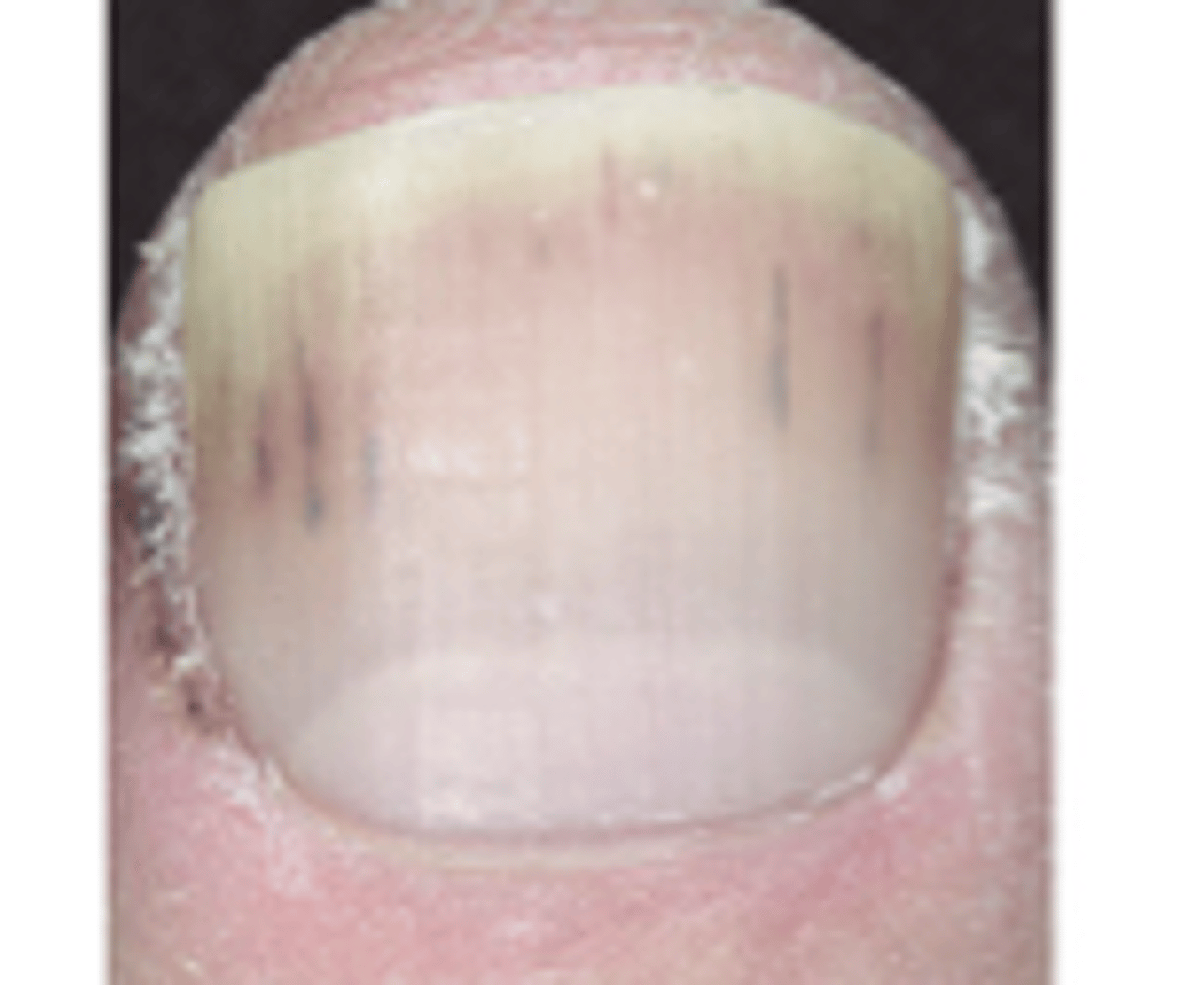
Causes of splinter hemorrhages
local trauma, infective endocarditis, sepsis, vasculitis, and psoriatic nail disease
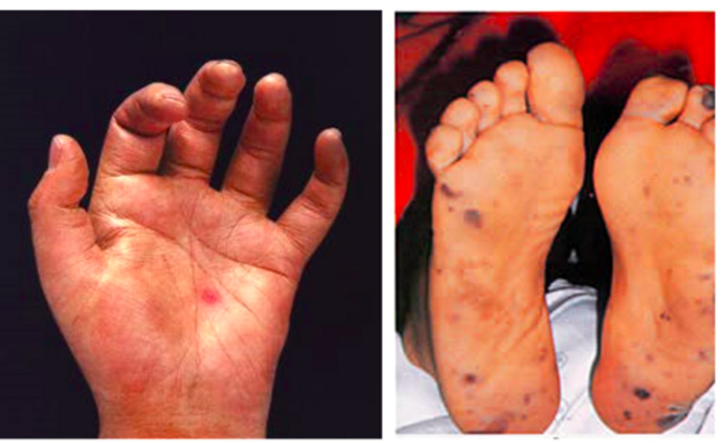
Janeway lesions
non-tender, hemorrhagic lesions that occur on the thenar and hypothenar eminences of the palsm (and soles)
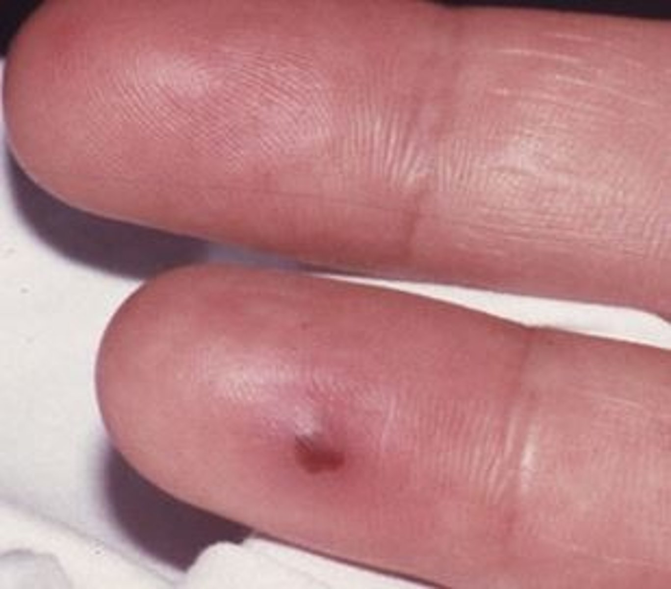
Osler nodes
red-purple, slightly raised, tender lumps, often with a pale center, typically found on the fingers or toes
Eye inspection
- conjunctival pallor (anemia)
- corneal arcus
- xanthelasma
- kayser-fleischer rings
Mouth inspection
- central cyanosis
- angular cheilitis
- high-arched palate
- dental hygiene
How to assess skin temperature
- assess at hands using the dorsal aspect of your hand
- the hands should be symmetrically warm, suggesting adequate perfusion
What do cool hands suggest?
poor peripheral perfusion (i.e., congestive cardiac failure, acute coronary syndrome)
What do cool and sweaty/clammy hands suggest?
acute coronary syndrome
How to perform a capillary refill test?
apply five seconds of pressure to the distal phalanx of one of a patient's fingers and then release
What is a normal capillary refill?
The initial pallor of the area you compressed should return to its normal color in less than two seconds
What does a capillary refill greater than 2 seconds suggest?
poor peripheral perfusion
Edema
accumulation of fluid in the interstitial space that occurs as the capillary filtration exceeds the limits of lymphatic drainage, producing noticeable clinical signs and symptoms
What type of edema are we looking for in the cardiac exam?
pitting edema
Where do we evaluate for edema?
- sacral edema
- peripheral edema (lower extremities, note pitting vs non pitting)
What type of edema is caused by right sided heart failure?
peripheral edema
What type of edema is caused by left sided heart failure?
pulmonary edema
SLIDE 34
What does amplitude of a pulse correlate with?
pulse pressure (difference between systolic and diastolic pressure)
Amplitude of pulse
- strength, force, or volume of the pulse
- often described as weak, thready, bounding, or normal
- you may have variations beat to beat
What might a high-amplitude (bounding) pulse indicate?
fever or anxiety
What might a low-amplitude (thready or hypokinetic) pulse indicate?
heart failure, shock, or severe dehydration
FINISH SLIDE 36
SLIDES 37-42
What is the diaphragm of the stethoscope used for?
high-pitched sounds (S1, S2, aortic and mitral regurgitation, pericardial friction rubs)
What is the bell of the stethoscope used for?
low-pitched sounds (S3, S4, mitral stenosis, bruits)
What type of pressure should you use when listening with the diaphragm?
firm, push down
What type of pressure should you use when listening with the bell?
light pressure
In a carotid examination, why do we auscultate before palpate?
assess for the presence of bruits
Bruit
a vascular sound, like a murmur, caused by turbulent blood flowing through an artery ("whooshing" sound)
What does a bruit indicate?
atherosclerosis, stenosis, or high-velocity flow
Where can a bruit be heard?
carotid arteries, abdomen, renal, or femoral arteries
What does an early systolic bruit indicate?
narrowing
What does a "pan-systolic" (throughout systole) bruit suggest?
more advanced narrowing (60% or more)
How do you differentiate a heart murmur radiating to the neck versus a bruit?
heart murmur radiating to the neck is usually louder below the clavicle, whereas a bruit is louder above it
Why should you never compress both carotids simultaneously?
avoid compromising cerebral blood flow
SLIDE 47
Why should you avoid palpation if you hear a bruit?
it may decrease the flow further or it may dislodge the plaque
Positioning for a carotid examination
supine with head of bed elevated to approx. 30-45 degrees
Palpation technique
- use the pads of your fingers
- use the index/middle finger in lower 1/3 of neck
Why is it important to avoid high neck palpation
to prevent stimulation of the carotid sinus, which can cause a sharp drop in heart rate or blood pressure