Lecture 2: Injury, Inflamation, and Repair
 Call Kai
 Learn
Learn Practice Test
 Spaced Repetition
 Match
Match Flashcards
 Knowt Play
1/76

There's no tags or description
Looks like no tags are added yet.
Name | Mastery | Learn | Test | Matching | Spaced | Call with Kai | Chat |
|---|
No analytics yet
Send a link to your students to track their progress
77 Terms
general pathology
A basic reactions or processes common to most tissues
specific pathology
A specific response or process in specific organs or tissues
etiology
cause of disease
pathogenesis
the manner in which a disease develops
hypoxia
decreased oxygen supply
anoxia
absence of oxygen due to the availability
asphyxia
absence of oxygen due to interruption of breathing (suffocation)
ischemia
decreased blood flow (loss of oxygen and nutrients)
What are six acquired causes of cell injury?
1)hypoxia/ischemia
2)physical damage
3)chemical damage
4)immunological
5)nutritional deficiency
6)infection
What are two intrinsic causes of cell injury?
1)genetic derangements (mutations)
2)normal aging process
What are four factors that are common in mechanisms of cell injury?
1)calcium (increased intracellular)
2)ATP (reduction)
3)membrane permeability
4)oxygen (reactive species present or decreased amount)
How does increased intracellular calcium promote cell injury?
Increased intracellular calcium activates phospholipase and protease, which break down cell membranes. It also activates endonucleases, which cause damage to nuclear/chromatin structures.
How do reactive oxygen species (ROS) promote cell injury?
Increased ROS leads to lipid peroxidation of membranes (breakdown), oxidative modification of proteins, and DNA damage.
reversible cell injury
impairs cell function but does not result in cell death
Ex/cellular swelling or fatty change
irreversible cell injury
autolysis and apoptosis
Ex/denaturation of proteins or membrane lipids
What are the five types of necrosis?
1)coagulative necrosis
2)liquefactive necrosis
3)caseous necrosis (special)
4)enzymatic fat necrosis (special)
5)gangrenous necrosis (special)
necrosis
irreversible cell death that is passive (does not require energy)
coagulative necrosis
A loss of nucleus, but cell shape and organ structures are preserved by the coagulation of proteins.
Ex/myocardial infarction

liquefactive necrosis
Influx of neutrophils releases catalytic enzymes that destroy cell outlines and tissue structure.
Ex/bacterial abscess (puss)

caseous necrosis
It has a cheese-like appearance and is typically seen in tuberculosis infections. It results from a combination of coagulative and liquefactive necrosis, forming granulomas with central necrotic debris.

enzymatic fat necrosis
Enzymatic fat necrosis occurs when pancreatic enzymes digest fat tissue, often seen in acute pancreatitis.

gangrenous necrosis
Gangrenous necrosis is tissue death due to loss of blood supply, commonly affecting limbs. It can be dry (coagulative), wet (with bacterial infection and liquefaction), or gas gangrene (caused by Clostridium species).

infarction
Process of coagulative necrosis that occurs distal to an occlusion in an artery feeding the tissue

apoptosis
programmed cell death that requires energy
What is the difference between apoptosis and necrosis?
Apoptosis is a controlled, energy-dependent process of programmed cell death that avoids inflammation and helps maintain tissue health.
Necrosis is an uncontrolled, often injury-induced cell death that leads to cell rupture and triggers inflammation in surrounding tissues.
How is apoptosis induced?
1)withdrawal of tropic factors (hormones or growth factors)
2)engagement of death receptors
Compare necrosis and apoptosis:
necrosis: non-physiological, unregulated, no energy required, cell membrane leakage, death of cell groups, and inflammatory response
apoptosis: physiologic and non-physiologic, highly regulated, energy dependent, cell membrane intact, death of an individual group of cells, and no inflammatory response
hyperplasia
increase in number of cells

hypertrophy
increase in cell size

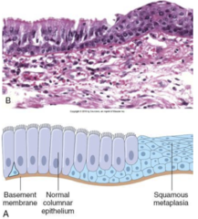
metaplasia
premalignant condition where a cell type is replaced with a different cell type
Ex/barrett's metaplasia of the esophagus
premalignant
A lesion or process that will probably transform into cancer
amyloidosis
the accumulation of the abnormal protein amyloid in the kidneys and other organs that can lead to organ damage, as well as renal failure
hemachromatosis
An inherited disorder with an abnormal excess of iron leads to deposition in other parts of the body

monocyte
a phagocytic WBC found in the blood
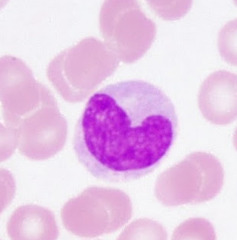
neutrophil
A type of white blood cell that engulfs invading microbes and contributes to the nonspecific defenses of the body against disease.

lymphocyte
A type of white blood cell that make antibodies to fight off infections

inflamation
localized response to infection that is designed to protect the host, but excessiveness can be damaging to the host
What WBCs are found in acute inflammation?
neutrophils
What WBC are found in chronic inflammation?
lymphocytes and monocytes
Describe acute inflammation
-infiltration of tissue w/neutrophils
-rapid onset (minute to hour) and transient
-phagocytosis of bacteria and necrotic tissue
-innate immune response

Describe chronic inflammation
-infiltration of tissue w/monocytes and lymphocytes
-slow persistent onset (24 hours)
-phagocytosis, secrete cytokines, kill infected cells, and reject foreign tissue
-adaptive immune response

What is the sequence of events for acute inflammation?
1)increased blood flow to the region (vasodilation)
2)deposition of fluid and plasma proteins
3)accumulation of neutrophils from the microcirculation
4)endothelial damage causes more edema

edema nasal polyp
swelling (edema) within the nasal sinus that is common with asthma, allergies, or chronic sinusitis

histamine
A chemical secreted by mast cells that causes vasodilation of arterioles and increased permeability of venules. This response causes a fluid leak and is a common allergic response.
serotonin
A chemical in platelets that has a similar activity to histamine
inflammatory complement system
A series of plasma proteins with proteolytic activity that, when contacted with bacteria or antibody-bound bacteria/antigen, initiates a cascade of activated components.

What is the function of C3a and C5a?
vasodilation and permeability
What is the function of C3b and C3bi?
act as an opsonin (attach to bacterial cell wall and promote phagocytosis by neutrophils)
What is the function of C5a?
chemotactic (attractor) agent for neutrophils
What are two arachidonic acid metabolites that affect acute inflammation?
1)prostaglandins (inhibited by aspirin)
2)leukotrienes (allergic response)
All leukocyte traffic requires adhesion between surface of __________ _________ _______ and _________ _______ _______
apposing extracellular cells and extracellular matrix proteins
How do leukocytes transmigrate between endothelial cells and cross the basement membrane?
They are initially tethered to endothelial cells, which causes them to slowly roll until they firmly adhere and cross the basement membrane
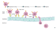
What are the three types of cell adherence molecules?
1)selectins
2)ICAMs
3)integrins
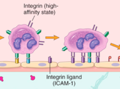
integrins
Transmembrane heterodimers (alpha-beta) that are important for cell-cell and cell-membrane interactions. They are expressed on all leukocytes.
Ex/Mac-1 (neutrophil) and LFA-1 (lymphocyte)

ICAM-1
Expressed at low levels on endothelial cells that become super-induced in the presence of cytokines, which are released by lymphocytes and macrophages. They bind to LFA-1 and Mac-1
L-selectin
Expressed on monocytes, neutrophils, and lymphocytes to mediate adhesion to carbohydrate ligands induced on endothelial cells that are inflamed.
How does expression of integrins increase?
activation of leukocytes (ICAM-1 on endothelial cell)
-neutrophils (MAC-1) within minutes
-lymphocytes (LFA-1) couple days
How does the affinity of integrins increase?
chemokines (change conformation to increase affinity)
selectin-mediated adhesion
rapid-rate, low binding affinity, too weak to stop cells, and loose rolling interaction

integrin-mediated adhesion
slow-rate, high bonding affinity, and firm adherence
What is responsible for loose rolling interaction?
selectins
What is responsible for firm adherence?
integrins
What are three outcomes after inflammation?
1)complete resolution (regain function)
2)tissue replacement (fibrosis)
3)chronic inflammation (persistent damage)
angiogensis
formation of new blood vessels
fibrogenesis
migration and proliferation of fibroblasts that cause the development of fibrous tissue
granulation tissue
new tissue that is pink/red in color and composed of fibroblasts and small blood vessels that fill an open wound when it starts to heal
What is the major regulatory molecule for angiogenesis?
vascular endothelial growth factor (VEGF)
What is the major regulatory molecule for fibrogenesis?
transforming growth factor-beta (TGF-beta)
In ACUTE inflammation, which of the following often causes damage to the tissue?
A. Toxic enzymes secreted by infiltrating T lymphocytes
B. Hydrolytic enzymes and reactive oxygen species released from neutrophils
C. Overactive and persistent monocyte phagocytosis
D. Caspase activation in tissue-resident macrophages
E. The secretion of endonucleases by infiltrating neutrophils, which breaks down cells' DNA5
B. Hydrolytic enzymes and reactive oxygen species released from neutrophils
Damage to epithelial cells lining the alveoli (microscopic air
sacs in the lung) by bacteria often results in which of the following?
A. Complete resolution, with return of normal function.
B. Scar formation, with thickening of alveolar walls by collagen.
C. Pulmonary edema.
D. Decreased gas exchange.
E. All of the above are correct.
E. All of the above are correct.
The cellular events that occur in apoptosis include which of the following?
A. The secretion of Ca+2 ions from the cell
B. Suppression of integrin expression
C. Hyperoxidation of plasma membrane lipids
D. Rapid decrease in cell's energy stores
E. Release of cytochrome C from mitochondria into the cytosol
A. The secretion of Ca+2 ions from the cell
What key feature of L-selectin renders it so uniquely effective in its particular role in leukocyte-endothelial adhesion?
A. Their expression on the surface of all leukocytes
B. They adhere especially tightly (high affinity) to ligand on endothelium
C. It does not have to be induced by cytokines
D. It is a target for inside-out signaling
E. It binds to its ligand with a very high on-rate
E. It binds to its ligand with a very high on-rate
In tissue repair, fibroblast proliferation and collagen
secretion is stimulated by which one of the following?
A. complement component C5a
B. transforming growth factor-beta (TGF-beta)
C. interferon
D. tumor necrosis factor alpha (TNF-alpha)
E. macrophage chemokines
B. transforming growth factor-beta (TGF-beta)
Stimuli that initiate an inflammatory response must induce the
secretion of a cytokine such as IL-1 or gamma-interferon for which
one of the following reasons:
A. To diffuse toward blood vessels and cause their increased permeability
B. To activate macrophages to become more phagocytic
C. To induce the rapid translocation of cytoplasmic integrins to the surface of circulating leukocytes
D. To induce the increased expression of ICAM-1 and other adhesion molecules on endothelial cells
E. To attract leukocytes to the source of the cytokine in the tissue.
D. To induce the increased expression of ICAM-1 and other adhesion molecules on endothelial cells
MOST bacterial infections give rise to what type of
response?
A. a neutrophil-rich infiltrate
B. tissue edema
C. complement activation
D. local vasodilation
E. all of the above
E. all of the above
Which one of the following statements about metaplasia is NOT true?
A. Metaplastic tissues have a high risk of developing into cancer.
B. Metaplasia results from chronic tissue injury.
C. Only cells that are not capable of proliferation undergo metaplasia.
D. It is a common result of gastric reflux (reflux of stomach acid into the
esophagus)
E. Metaplasia usually results in a decrease in normal tissue function.
C. Only cells that are not capable of proliferation undergo metaplasia.
A myocardial infarct can result from which of the following?
A. A thrombus (blood clot) in a coronary artery, leading to tissue
ischemia
B. Asphyxia
C. An acute viral infection in cardiac myocytes
D. Excessive iron deposition in the heart
E. Leakage of cardiac enzymes from the myocytes
A. A thrombus (blood clot) in a coronary artery, leading to tissue ischemia