BIOCHEM Exam 1
 Call Kai
 Learn
Learn Practice Test
 Spaced Repetition
 Match
Match Flashcards
 Knowt Play
1/243

There's no tags or description
Looks like no tags are added yet.
Name | Mastery | Learn | Test | Matching | Spaced | Call with Kai | Chat |
|---|
No analytics yet
Send a link to your students to track their progress
244 Terms
Prokaryote
Nucleus: Absent Cytoplasmic organelles: Absent
Eukaryote
Nucleus: Present Cytoplasmic organelles: Present
Eukaryote cells Possess Organelles
such as mitochondria, plasma membrane, nucleus, Endoplasmic Reticulum, Golgi apparatus, Lysosomes, and Peroxisomes
Plasma membrane
encloses body of cell, limits transport of molecules across
Nucleus
contains the genetic information
Mitochondria
Responsible for aspects of metabolism
Endoplasmic Reticulum
synthesis and storage
Golgi apparatus
processing and sorting
Lysosomes
removal of old organelles, processing of ingested material
Peroxisomes
breakdown of catabolites
The Phylogenetic Tree of Life
All organisms can be placed in one of three domains: Eukarya, Bacteria, or Archaea
– Groups based on their biochemical characteristics
– Branches are points of divergence
Covalent Bonds
– formed by electron sharing between two adjacent atoms
– the strongest bonds
Resonance
• Some molecules, such as adenine, exhibit multiple covalent structures called resonance structures.
Ionic Interactions
Noncovalent interactions that occur between fully charged atoms or molecules
Electric Dipoles
Molecules with no overall charge can have regions where electron distribution is uneven.
– leads to electric dipoles (dipoles)
• Dipoles can interact with ions or with other dipoles.
Hydrogen bonds are a specific example of a
dipole-dipole interaction
Hydrogen-Bond Donors
The group that includes both the atom to which the hydrogen atom is covalently bonded and the hydrogen atom itself
Hydrogen-Bond Acceptor
The lone pair of electrons that is on the atom less tightly linked to the hydrogen atom
van der Waals interactions:
–occur when two atoms are sufficiently close.
–occur when transient asymmetry in electron distribution in one atom induces complementary asymmetry in a neighboring atom.
–involve neighboring atoms attracting each other.
–are relatively weak.
van der Waals Contact Distance
• Attraction increases as two atoms come closer to each other, until they are separated by the van der Waals distance.
• At distances shorter than the van der Waals contact distance, strong repulsive forces become dominant.
Properties of Water
• Water is a polar molecule with a partial positive and partial negative end.
• Water is highly cohesive.
• A large # of hydrogen bonds are formed in liquid water, and the maximum # of hydrogen bonds are formed in crystalline ice
The Hydrophobic Effect
• Nonpolar molecules in water can be driven together by the hydrophobic effect.
– powered by the increase in entropy of water
Hydrophobic interactions
• Nonpolar molecules are not soluble in water
• Water interacts with itself and excludes the nonpolar molecules
• Phospholipids form biological membranes in water because of the hydrophobicity of the fatty acids
Oxidation-Reduction reactions
• Gain of electron-reduction
• Loss of electron- oxidation
Oxidation-Reduction example

Acyl Linkages
Condensation of carboxylic acids with other electronegative functional groups
• Drugs are designed as “pro-drugs” that we intake and then are hydrolyzed to their active forms
• An example of acyl linkage: the condensation of a carboxylic acid with an amine forms a peptide bond; hydrolysis can break that bond
First Law of Thermodynamics
The total energy of a system and its surroundings is constant
Second Law of Thermodynamics
The total entropy of a system plus that of its surroundings always increases
Buffers Regulate pH
• Buffers resist changes in the pH of a solution.
• Buffers are most effective at a pH near its pKa.
What are Proteins?
Linear polymers made of monomers called amino acids:
– can interact with one another and other macromolecules to form complex assemblies.
- contain a diverse range of functional groups that contribute to the protein’s structure and function.
– can be rigid or flexible.
– Responsible for physical structures within cells as well as catalysts for reactions (i.e. enzymes).
An α-amino acid consists of the α carbon linked to:
– an amino group.
– a carboxylic acid group.
– a hydrogen atom.
– a specific R group (or side chain).
Chiral amino acids:
– have four different groups bonded to the α carbon.
– exist as two mirror-image forms called the Lisomer and the D isomer.
– Proteins only contain L isomers.
Peptide bond (or amide bond) formation involves:
– the linking of the α-carboxyl group of one amino acid to the α-amino group of another amino acid through an acyl linkage.
– the loss of a water molecule.
Polypeptide chains have directionality because its ends are different:
– α-amino group at the beginning (often referred to as the ”N terminus”)
– α-carboxyl group at the end (often referred to as the “C terminus”)

Fatty Acids Are Key Constituents of Lipids
• Lipids are water-insoluble biomolecules that are highly soluble in organic solvents
– most lipids are hydrophobic due to fatty acids
• Fatty acids are long hydrocarbon chains that terminate with carboxylic acid groups
Fatty Acid Names
• From the parent hydrocarbon by
substitution of oic for the final
• First number is the number of carbon
atoms, and the second number is the number of double bonds.
Fatty Acid Carbon Atoms Can Be Numbered in Two Ways
• 1: Carbons can be numbered starting at the carboxyl terminal carbon atom.
– Carbon atoms 2 and 3 are often referred to as α and β.
– Position of a double bond can be represented by the symbol ∆ followed by a superscript number
• 2: The methyl carbon atom at the distal end of the chain is called the omega (ω) carbon.
– Position of a double bond can be represented by counting from the distal end.

• Fatty acids in biological systems contain:
– an even number of carbon atoms between 14 and 24 (16 and 18 are most common).
– an unbranched hydrocarbon chain in animals.
– a saturated(has double bonds) or unsaturated
• Short chain length and the unsaturation enhance the fluidity of fatty acids and their derivatives.
• Lipids function as:
– fuel molecules.
– highly concentrated energy stores.
– signal molecules and messengers in signal-transduction pathways.
– the essential component of biological membranes.
Principal lipids in eukaryotic membranes are
phospholipids, glycolipids, and cholesterol.
• Membranes
– are sheetlike structures, two molecules thick, that form closed boundaries.
– consist mainly of lipids and proteins with linked carbohydrates.
– contain lipids, small molecules with hydrophobic and hydrophilic that form lipid bilayers.
– proteins embedded in lipid bilayers with distinct functions.
– are asymmetric, non-covalent assemblies.
– are fluid structures.
– tend to be electrically polarized.
Phospholipids are composed of:
– one or more fatty acids.
– a platform to which the fatty acids are attached (ex: glycerol, sphingosine).
– a phosphate.
– an alcohol attached to the phosphate.
Phosphatidate Is the Simplest Phosphoglyceride
• phosphoglycerides = phospholipids derived from glycerol
– The –OH groups at C-1 and C-2 of glycerol are esterified to the carboxyl groups of the two fatty acid chains.

Common Alcohol Groups of The Phosphoglycerides
• Major phosphoglycerides are derived from phosphatidate.
• An ester bond forms between the phosphate group of phosphatidate and the hydroxyl group of an alcohol.
Sphingomyelin Contains a Sphingosine Platform

Cholesterol is a steroid built from
4 linked hydrocarbon rings
– contains a linked hydrocarbon tail at one end and an –OH group at the other end
– oriented parallel to fatty acid chains of phospholipids in membranes
– The –OH group interacts with phospholipid head groups
A Membrane Lipid Is an Amphipathic Molecule Containing a Hydrophilic and a Hydrophobic Moiety
• Moiety= part or portion
• Membrane lipids are amphipathic molecules.
– hydrophobic moiety: fatty acid tails
– hydrophilic moiety: phosphorylcholine
How do phospholipids and glycolipids form structures in aqueous media?
Membrane formation results from their amphipathic nature.
A micelle is a globular structure with polar head groups on the outside and hydrocarbon tails sequestered inside.
Lipid Bilayers
consists of two lipid sheets
– hydrophobic tails of each sheet interacting with one another, forming a permeability barrier
– Hydrophilic head groups interact with the aqueous medium
Q: How do phospholipids and glycolipids form lipid bilayers?
Their two tails take up too much space, so they do not form small micelles like single-tailed fatty acid salts.
They spontaneously form lipid bilayers in water, stabilized by
Hydrophobic interactions
Van der Waals interactions between hydrocarbon tails
Electrostatic + hydrogen-bonding attractions between polar heads and water
Biological Consequences of Hydrophobic Interactions
• Lipid bilayers have an inherent tendency to be extensive.
• Lipid bilayers will tend to close on themselves so that there are no edges with exposed hydrocarbon chains( forms compartments).
• Lipid bilayers are self-sealing. (A hole in a bilayer is energetically unfavorable.)
Lipid Bilayers Are Highly Impermeable to Ions and Most Polar Molecules
• Have a very low permeability for ions and most polar molecules.
• Permeability of small molecules is correlated with their solubility in a nonpolar solvent relative to their solubility in water.
• Water is an exception due to its:
– low molecular weight.
– high concentration.
– lack of complete charge.
Proteins Carry Out Most Membrane Processes
• Membrane proteins allow transport of molecules and information across a membrane.
Q: How do integral and peripheral membrane proteins interact with membranes?
Integral membrane proteins interact extensively with the hydrocarbon chains of membrane lipids.
Released by agents that compete with these nonpolar interactions
Most completely span the lipid bilayer
Peripheral membrane proteins are bound mainly by electrostatic and hydrogen-bond interactions with lipid head groups.
Disrupted by salts or changes in pH
Often bound to the surfaces of integral proteins
May be anchored to the bilayer by a covalently attached hydrophobic chain
How do lipids and many membrane proteins move within biological membranes?
Biological membranes are not rigid or static.
Lateral diffusion = the constant lateral movement of lipids and many membrane proteins within the membrane.
FRAP (fluorescence recovery after photobleaching) can be used to visualize protein movement.
Q: How does Fluorescence Recovery After Photobleaching (FRAP) work?
A fluorescent label is put on a cell-surface component.
A laser bleaches a small area.
Scientists watch how the fluorescence comes back over time.
This shows that membrane molecules are moving.
Q: What does FRAP recovery show?
Fluorescence increases in the bleached area when molecules move into it.
Faster recovery = more mobile molecules.
Slower recovery = less mobile molecules.
Lateral Diffusion of Lipids in Membranes Is Much More Rapid Than Transverse Diffusion
• Flip-flop of a protein molecule has not been observed.
– preserves membrane asymmetry

Q: How does unsaturation affect the melting temperature (Tm) of a membrane?
Saturated fatty acid chains are straight → pack tightly → higher Tm and more rigid.
Cis double bonds create bends → prevent tight packing → lower Tm and more fluid.
Q: How does fatty acid chain length affect membrane melting temperature (Tm)?
Longer hydrocarbon chains → stronger interactions → higher Tm and more rigid.
Q: How does cholesterol affect membrane fluidity?
Cholesterol’s bulky steroid structure disrupts tight packing of fatty acid chains.
What are lipid rafts and what do they do?
Lipid rafts = membrane areas where cholesterol + specific lipids form complexes.
They have reduced fluidity.
They can:
Change the shape and activity of membrane proteins
Help with cell signaling by bringing proteins together.
Monosaccharides Are the Simplest Carbohydrates
Carbohydrates are carbon-based molecules high in hydroxyl groups
• Monosaccharides are aldehydes or ketones that contain two or more hydroxyl groups.
• They are three to seven carbons in length
• Monosaccharides exist in many isomeric forms and are also called simple sugars.
Monosaccharides
Examples

Constitutional isomers
Molecules with identical molecular formulas that differ in how the atoms are ordered
Stereoisomers
Molecules that differ in spatial arrangement but not bonding order
stereoisomers can be
enantiomers (mirror images of each other) or diastereoisomers (not mirror images of each other)
Common Monosaccharides

Most Monosaccharides Exist as Interchanging Cyclic Forms
• An aldehyde can react with with an alcohol to form a hemiacetal
• A ketone can react with an alcohol to form a hemiketal
Anomers of Glucose
Anomer is a diastereoisomeric form of sugars that forms when a cyclic hemiacetal is formed and an additional asymmetric center is created
Q: What conformations can pyranose rings have?
Boat or chair forms.
Chair form is preferred because it has less steric hindrance.
In the chair form:
Axial = nearly perpendicular
Equatorial = nearly parallel
Axial groups on the same side sterically hinder each other.
Why does β-D-glucose prefer the chair form?
The chair form predominates because all axial positions are occupied by hydrogens.
The boat form is disfavored because of steric hindrance.
Q: What are glycosidic linkages and why are they important?
Monosaccharides can react with alcohols, amines, and phosphates.
These modifications increase biochemical versatility and can help with signaling and metabolism.
O-glycosidic linkage = anomeric carbon + oxygen of an alcohol.
N-glycosidic linkage = anomeric carbon + nitrogen of an amine.
Q: What are disaccharides?
Disaccharide = 2 sugars joined by an O-glycosidic linkage.
Sucrose, lactose, and maltose are common disaccharides.
They can be broken down into sugars that provide energy for ATP production.
What is sucrose?
Glucose + fructose
Glucose: α; fructose: β
Anomeric carbons of both sugars are linked.
Not a reducing sugar
Broken down by sucrase (invertase).
Q: What is lactose?
Galactose + glucose
Joined by a β-1,4-glycosidic linkage
Broken down by lactase in humans.
Lack of lactase → lactose intolerance.
What is maltose?
Glucose + glucose
Joined by an α-1,4-glycosidic linkage
Produced from hydrolysis of larger oligosaccharides.
Broken down by maltase (α-glucosidase).
Q: What is glycogen and how is it structured?
Storage form of glucose in animals
Large, branched glucose polymer
α-1,4 linkages = main chains
α-1,6 linkages = branches
Branching allows enzymes to rapidly break down glucose.
Q: What is starch and what are its two forms?
Storage form of glucose in plants
Amylose = unbranched, α-1,4 linkages
Amylopectin = branched, mostly α-1,4 with some α-1,6
Amylopectin is similar to glycogen but less branched.
Both are broken down by α-amylase.
What are DNA and RNA made of?
DNA and RNA = linear polymers that carry genetic information.
Nucleotide = monomer.
Each nucleotide contains:
Sugar
Phosphate
1 of 4 bases
Q: How do DNA and RNA differ?
RNA → ribose sugar → has 2′-OH
DNA → deoxyribose sugar → has 2′-H
The lack of 2′-OH makes DNA more resistant to hydrolysis.
RNA has uracil (U) instead of thymine (T).
What makes up the DNA/RNA backbone?
Sugars + phosphates linked by phosphodiester bridges.
3′-OH of one sugar connects to the 5′-OH of the next through phosphate.
Backbone is constant within a nucleic acid.
Each phosphodiester bridge has a negative charge, which helps resist hydrolysis.
Q: What are the bases in DNA and RNA?
Bases attach to the sugar’s 1′ carbon.
Purines: Adenine (A) and Guanine (G)
Pyrimidines: Cytosine (C), Thymine (T), and Uracil (U)
DNA: A, G, C, T
RNA: A, G, C, U

Type and structure
Purines:Purine
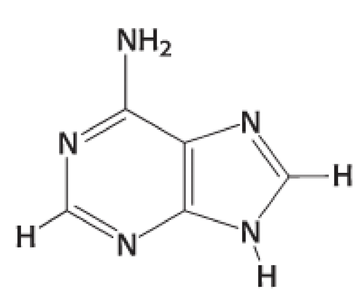
Type and structure
Purines: Adenine

Type and structure
Purines: Guanine

Type and structure
Pyrimidines: Pyrimidine

Type and structure
Pyrimidines: Cytosine

Type and structure
Pyrimidines: Uracil

Type and structure
Pyrimidines: Thymine
Q: What is the difference between a nucleoside and a nucleotide?
Nucleoside = base + sugar
Nucleotide = nucleoside + 1 or more phosphoryl groups
Nucleoside triphosphates are the precursors of DNA and RNA.
How are bases attached to the sugar in N-β-Glycosidic Linkage?
y an N-β-glycosidic linkage.
Purine → sugar C-1′ attaches to N-9
Pyrimidine → sugar C-1′ attaches to N-1
Q: What is the structure of RNA?
RNA is usually single-stranded.
Base pairing can still occur, giving some RNA molecules a 3D structure.
Q: How are nucleoside triphosphates related to energy?
They contain high-energy anhydride bonds between phosphates.
ATP is an example.
Mg²⁺ is often the divalent cation associated with them
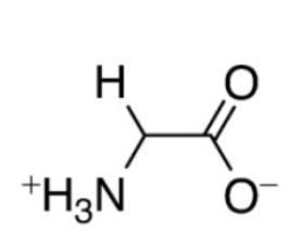
Amino Acid: Single letter designation, Three letter designation, Grouping of the amino acids
Glycine, Gly, G, Simple (Ambivalent)
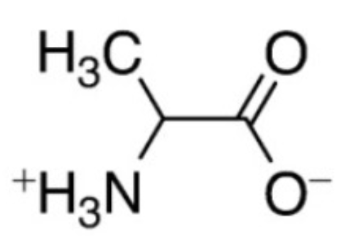
Amino Acid: Single letter designation, Three letter designation, Grouping of the amino acids
Alanine, Ala, A, Simple (Ambivalent)
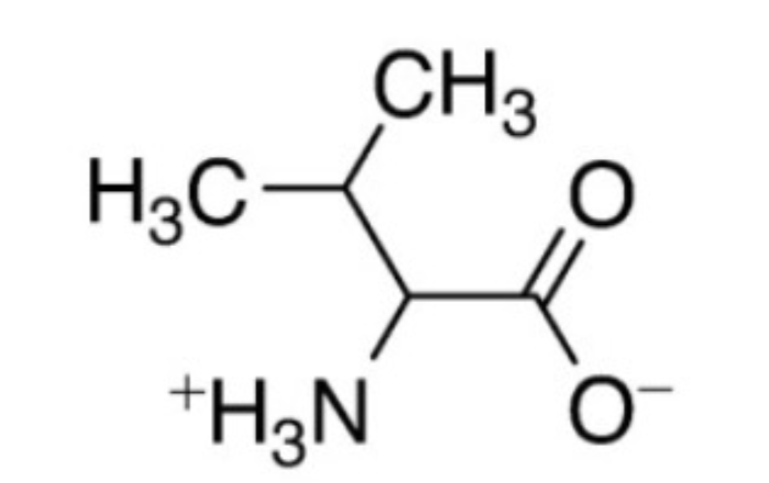
Amino Acid: Single letter designation, Three letter designation, Grouping of the amino acids
Valine, Val, V, Simple (Ambivalent)

Amino Acid: Single letter designation, Three letter designation, Grouping of the amino acids
Leucine, Leu, L, Simple (Ambivalent)

Amino Acid: Single letter designation, Three letter designation, Grouping of the amino acids
Isoleucine, Ile, I, Simple (Ambivalent)
Simple (Ambivalent) Amino Acids
Small, somewhat hydrophobic R groups with no hydrophilic components
Can orient facing inward or outward on an overall protein structure