Lecture 8: Dx & Management of Equine Ligament & Tendon Disorders
 Call Kai
 Learn
Learn Practice Test
 Spaced Repetition
 Match
Match Flashcards
 Knowt Play
1/55

There's no tags or description
Looks like no tags are added yet.
Name | Mastery | Learn | Test | Matching | Spaced | Call with Kai | Chat |
|---|
No analytics yet
Send a link to your students to track their progress
56 Terms
What are the general clinical signs of equine soft tissue injuries?
Swelling: localized to a site of injury
Heat: warm to touch due to inflammation
Tenderness: palpation may elicit pain and discomfort
Thickening: fibrosis causes firmer or thicker tissues
Altered posture: refusing to bear weight on the affected limb
Lameness: when asked to move or flex the affected limb
Joint instability: in severe cases, joint subluxation or luxation
What are the differential diagnoses for soft tissue injuries?
muscles - strain, contusion, laceration
tendon - tendonitis
ligament - desmitis (sprain)
synovial structures - synovitis
joint capsule - effusion
tendon sheath - tenosynovitis
bursa - bursitis
skin and fascia: edeam, cellulitis, seroma, hematoma
What is the most common soft tissue injury in racing Thoroughbreds?
tendon and ligament injuries because of their essential role in movement, load distribution, and joint stability
What type of equine soft tissue injury has a 43-94% risk of re-injury?
tendon and ligament injuries
What are the two most commonly affected structures in equine soft tissue injuries?
SDFT tendonitis: most freq. injured soft tissue structure of the lower limb associated with high-impact injuries
suspensory ligament desmitis: chronic repetitive use injuries common in show jumpers and dressage
Why do SDFT tendonitis and suspensory ligament desmitis have high recurrence rates?
they are bilateral conditions → unable to fully heal both at same time due to weight
What are the functional differences between tendons and ligaments?
tendons: connect muscle to bone and transfer muscle force to induce joint mobility
ligaments: connect bone to bone to provide joint stability
Describe the microscopic anatomy of tendons and ligaments.
composed mostly of collagen fibers: type 1 collagen to provide tensile strength and proteoglycans to provide viscoelasticity
fibers are arranged in parallel bundles that create the linear pattern seen on ultrasonography
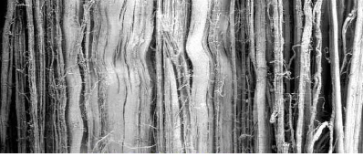
What has happened to these collagen fibers seen on this ultrasound image?
crimping/relaxation of collagen fibers when not under tension - normal
What are the three connective tissue coverings of tendons/ligaments?
endotenon: surrounds bundles of collagen fibers providing a path for blood
paratenon: surrounds entire tendon and contains synovial cells for movement and or gliding
synovial sheath: present in select areas to reduce friction (ex. digital flexor tendon sheath)
Why do most injuries occur where muscle meets tendon?
it is the weakest spot - tendon is 2x stronger than muscle → causes muscle to tear at musculotendinous junction
What is the purpose of the golgi tendon organ?
proprioception: detect stretch and tension → changes in joint position; feedback to golgi is needed to relax muscles
What is the purpose of the free nerve endings in tendons/ligaments?
nociception - detect noxious stimuli
Describe the biomechanics of tendons.
transmit forces from muscle to bones - induces joint movement → locomotion
resists applied forces: maintains joint alignment, provides joint stability
stores and releases elastic energy: act as shock absorbers (jumping)
How does tendon stiffness affect movement?
tendons exhibit degrees of stiffness that affects the ability to store and release elastic energy
stiff tendons and ligaments: supportive, controlled joint movement (ex. coffin jt)
compliant (stretchy) tendons: store elastic energy, aid in locomotion efficiency for running or jumping → want stretch for mobility (ex. fetlock or nuchal lig)
What tendons are critical for shock absorption and elastic energy storage and undergo high strain during galloping, making them prone to injury?
SDFT and DDFT
What ligament prevents hyperextension of the fetlock?
suspensory ligament
What are the 4 most common etiologies of tendon and ligament injuries?
overuse: accumulated microtrauma (chronic use without rest and repair)
acute overloading: sudden onset during high-impact activity
conformation: bone and joint anomalies may increase stress (foot and hoof issues like poor trimming or balance)
genetic disorders: collage defect → increased extensibility, reduced tissue healing
What part of the tendon do injuries tend to occur?
within the center of the tendon (core lesion) due to being a site of increased tissue loading and limited blood supply - hypoxia
What are the three phases of tendon healing?
acute inflammation (1-2wks): inflammatory products, blood, debris, fibrin
proliferation (3-6wks): type 3 collagen synthesis, thicken tendon - scar tissue
remodeling and maturation (6wks-12 mos): matrix remodels = type 1 collagen, fibrosis alters local tissue strain → risk of reinjury
Why do ligamentous injuries tend to recur?
torn ligament → injured nerves → neuromuscular dysfunction and altered proprioception → poor muscle activation and coordination, pain and ligamentous instability → reinjury
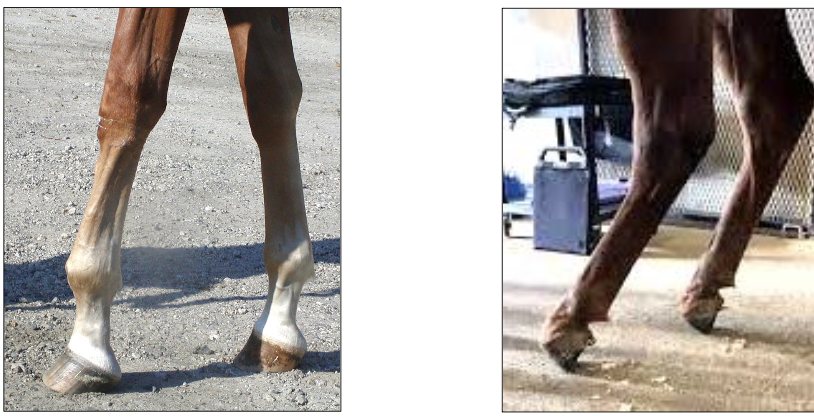
What contracted tendons are causing these two limb deformities?
left: SDFT - hoof is on the ground
right: DDFT - hoof is off the ground
What tendon has likely ruptured if a horse is has the toe of their hoof up off the ground and is walking on their heel?
DDFT
Why are tendon and ligament injuries common in athletic horses and difficult to treat successfully?
• Large forces applied – repetitive trauma
• Secondary joint instability
• High mechanical loading - inability to unload during healing
• Poor vascularity – limited healing
• Collagen density – scar tissue formation (fibrosis)
• Slow healing processes - remodeling
What are the three characteristic findings of tendon/ligament injuries upon palpation?
heat - acute inflammation
pain - localization of affected tissues
swelling
What is the preferred imaging modality for tendon and ligament injuries and how is it used to characterize tendon integrity?
ultrasound
• Location - structures affected
• Type of fiber disruption – diffuse, core lesion
• Extent of damage – area, length
What is located in each zone of the distal limb (distal to accessory carpal bone)?
• 1A, 1B – SDFT, DDFT, origin of suspensory ligament, origin of accessory ligament
• 2A, 2B – SDFT, DDFT, accessory ligament, suspensory ligament
• 3A, 3B – branches of suspensory ligament
• 3C – proximal sesamoid bones
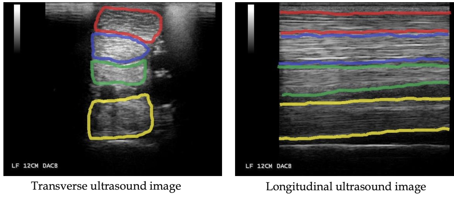
What are these structures?
red: SDFT
blue: DDFT
green: distal accessory (check) ligament
yellow: suspensory ligament
What characteristics of tendon integrity are evaluated on an ultrasound?
echogenicity
fiber alignment - linear and uniform
cross-section area: increased with edema, fibrosis, or chronic thickening
shape
margins
depth
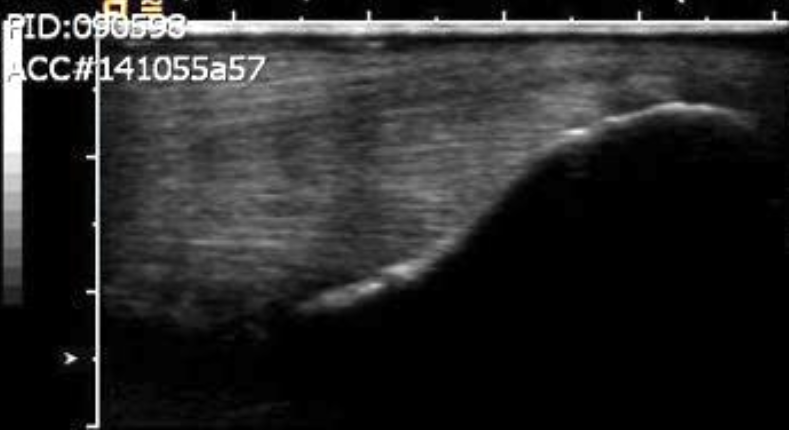
What is being shown here?
bone attachment site - entheses
What do hypoechoic vs hyperechoic mean on ultrasounds of tendons?
• Hypoechoic – fluid accumulation, fiber disruption
• Hyperechoic – fibrosis, mineralization
What do increases vs decreases of cross-sectional area mean on ultrasounds of tendons?
• Decrease – remodeling
• Increase - chronic thickening
Why would you choose to perform contrast rads on a distal limb injury?
to visualize if synovial structures are involved (joints, bursa, tendon sheath)
What is the most common tendon injury in horses, commonly caused by overuse, trauma, and high-intensity exercise?
bowed tendon aka superficial digital flexor tendonitis
How is SDF tendinitis diagnosed?
• Lame
• Characteristic swelling (if distal - need to r/o other causes like fetlock OA etc)
• Pain on palpation
• Ultrasound – definitive diagnosis
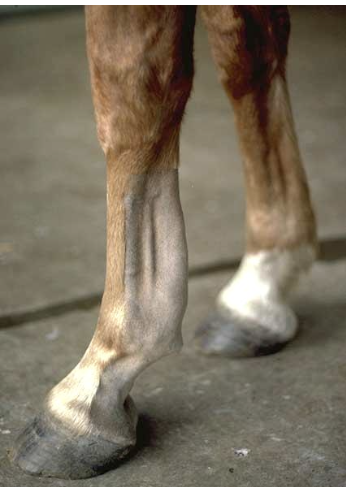
What injury is this swelling characteristic of ?
superficial digital flexor tendonitis
What is usually the cause of DDF tendinitis?
• High-speed injury
• Acute trauma - step in hole
• Wear and tear – distal lesion
Why might an MRI be recommended when diagnosing with DDF tendinitis?
DDFT goes all the way down to the coffin joint → may be associated with navicular syndrome - need MRI/CT to identify distal attachments
How does tenosynovitis of the digital flexor tendon sheath affect the distal limb?
constriction by palmar annular ligament (may need to surgically cut)
synovial fluid impedes tendon healing
prognosis decrease with chronicity
What causes suspensory ligament desmitis?
repetitive strain
high speed activities
poor conformation (upright)
inadequate warm-up
What is the impact of proximal suspensory ligament desmitis in the forelimb?
intermittent, subtle lameness: worse on soft ground and with limb on outside of circle
may be associated with splint because ligament lays on either side of medial/lateral splint bone
high recurrence rate → horse often does not return to former level of work
How is proximal suspensory ligament desmitis of the forelimb diagnosed?
• Pain on deep palpation of area
• Ultrasonography: Irregular fibers at attachment, Varying amount of muscle fibers
• Radiography – bone irregularity
• Nuclear scintigraphy, MRI
Perineural anesthesia ⇒ Lateral palmar nerve at the accessory carpal bone
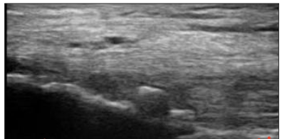
What is being seen on this ultrasound of a proximal suspensory ligament desmitis case and what can be done about it?
bone spur (enthesophyte) that constantly rubs against tendon/ligament → may tx with shockwave to help breakdown spur
What type of horse is proximal suspensory ligament desmitis in the hind limb commonly seen in?
sport horses: dressage and jumper, reiner
conformation: straight hocks, hyperextended fetlocks
Hpw is proximal suspensory ligament desmitis in the hind limb diagnosed?
• Insidious, chronic lameness - Shortened cranial phase of stride
• Can be positive to upper limb flexion: DDx - hock pain, Hock injections may improve short term
• Typically, worse on soft ground - With the affected limb on the outside of the circle
Perineural anesthesia ⇒ Deep branch of the lateral plantar nerve
What are the causes of suspensory ligament branch desmitis and how is it diagnosed?
cause: hyperextension or fetlock injury (galloping - forelimb, dressage & jumping - hind limb)
diagnosis: moderate lameness, thickened on palpation, ± positive to lower limb flexion, ultrasound, sesamoiditis on rads
What is usually the cause of degenerative suspensory ligament desmitis?
congenital collagen defect → progressive ligament degeneration that can progress to fetlock touching ground
While prognosis for degenerative suspensory ligament desmitis is guarded, what are the possible treatment options?
corrective shoeing or fetlock arthrodesis
What are the recommended treatments for tendonitis rehab in the acute inflammation stage?
• Compression wrap
• Confinement
• Cryotherapy
• NSAIDs
What are the recommended treatments for tendonitis rehab in the proliferative stage?
• Passive joint range of motion
• Controlled hand walking
• Tissue heating - if fibrosis, heat and stretch to help remodeling
• Laser therapy
• Orthobiological therapies
What are the recommended treatments for tendonitis rehab in the remodeling stage?
strengthening to avoid recurrence
lunge exercise
cavalletti work
ridden work
What are the surgical treatment options for soft tissue injuries?
• Tendon repair - laceration
• Ligament repair – joint subluxation
• Tendon sheath - debridement
• Tendon splitting – core lesion
• Tenotomy – tendon contractures (Accessory ligament desmotomy)
Why do extensor tendon lacerations have a very good prognosis?
because the tendon is just needed for extending the leg and not for “spring” mechanisms
Why is external coaptation commonly needed for flexor tendon lacerations?
because of weight bearing
How is collateral ligament rupture treated?
external coaptation for 4-6 weeks minimum
good prognosis if no underlying joint damage, but guarded for some joints b/c of difficulty in reducing the luxation
How can tendon and ligament injuries be prevented?
proper warm-up and cool-down to optimize MSK function and prevent tendon overload
rest and recovery
monitoring training load to avoid sudden increase in workload, intensity, or duration