L3 Chromatin Structure and Assembly in Eukaryotes Learning Objectives
 Call Kai
 Learn
Learn Practice Test
 Spaced Repetition
 Match
Match Flashcards
 Knowt Play
1/9

There's no tags or description
Looks like no tags are added yet.
Name | Mastery | Learn | Test | Matching | Spaced | Call with Kai | Chat |
|---|
No analytics yet
Send a link to your students to track their progress
10 Terms
Define histone variants, give one example, and say where in the genome the variants are found
The four core histones are the most common
Other histone variants can be incorporated into nucleosomes, and are found at special chromatin locations
For example, H2A.X is phosphorylated at sites of DNA double-stranded breaks and is thought to recruit repair machinery

Predict where along a gene nucleosomes are absent, and explain why promoters and terminators are relatively histone-free
Explain what a DNase I hypersensitive site indicates about the local chromatin state
They’re present in chromatin regions that lack nucleosomes
Describe how a nucleosome is built: which dimers associate with DNA first, how much DNA wraps the octamer, and the numbers that go with it
DNA wraps around a histone complex in a left handed manner to form a nucleosome - the histone complex at the center is the histone octamer
The histone octamer has two each of the four core histones H2A, H2B, H3, and H4 (histone variants exist)
The two H3-H4 dimers associate with DNA
The two H2A-H2B dimers then associate to form the octamer
About 146bp of DNA wrap around the octamer to form the nucleosome (1.76 turns)
Describe what H1 contributes to compaction, and what happens to chromatin when H1 is removed
H1 is a linker histone that binds to the linker DNA in between successive nucleosomes, helping compaction. Formation of the 30 nm fiber involves histone H1. The core histone tails are also involved in the formation of the 30 nm fiber, but it is not fully understood how. The 30 nm fiber is then compacted further into compact chromosomes in which large loops of chromatin are anchored to a central scaffold
Knocking out H1 leads to loss of chromatin structure and embryonic lethality
Explain how micrococcal nuclease digestion produced the ~200 bp ladder, and what that ladder established about how DNA is organized
Micrococcal nuclease is an extracellular enzyme (from Staphylococcus aureus) that digests both DNA and RNA by targeting the linker protein. Used on DNA it produced fragments of various lengths between 170-205 bp long. The 205 bp fragments were generated by one cut about half-way between the histone octamers, and the shorter 170 bo fragments resulted from “nibbling” up to the histone octamer. Longer fragments resulted from “multiples” - where the nuclease did not cut between one or more histone octamers
This showed that DNA is organized in a regular, repeating subunit structure and is not a random complex of DNA and protein
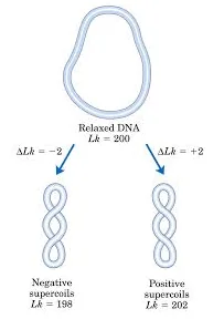
Explain why the left-handed wrap leaves negative supercoiling when the octamer is removed, and why that helps transcription and replication
The wrapping of the DNA is done in a left-handed position where, if the histone is removed, it will result in a linking number deficit that will create negative supercoiling making it slightly untwisted, and this is helpful because through this loop created the two loose strands can be used for DNA transcription and replication
Distinguish the histone-fold domain from the N-terminal tail by location and function, and say which one is required for 30 nm fiber formation
Histone-fold domain:
Structurally conserved motif near the C-terminus in every core histone
70 amino acids - 3 alpha helices separated by two loops
Responsible for the binding of histones into heterodimers
N-terminal tail:
Extends outwards of the core histone between the DNA coils
Tails are up to 25 amino acids, and have an undefined structure
Tails interact with other nucleosomes to help compact DNA further
Can also be chemically modified which are important for chromatin structure and function
The N-terminal tail is required for the 30 nm fiber formation
Compare the solenoid (one-start) and zig-zag (two-start) models, and explain why the question is still open
Similarities:
Both supported by experimental evidence
Both may exist in different areas of chromatin
Solenoid (one-start):
Nucleosome in a spiral shape
Flat slides of nucleosome disks next to each other
H1 linker bent inside center of the spiral to provide constant thickness of the spiral
Zig-Zag (two-start):
Presence of H1 linker favors this model (due to its binding geometry, physical interaction with linker DNA, and the energetic costs)
Zigzag histone pairs stack on each other
The question is still open because the in vitro results yielding a uniform 30 nm structure was different than the observed 10 nm disorganized structure in vivo

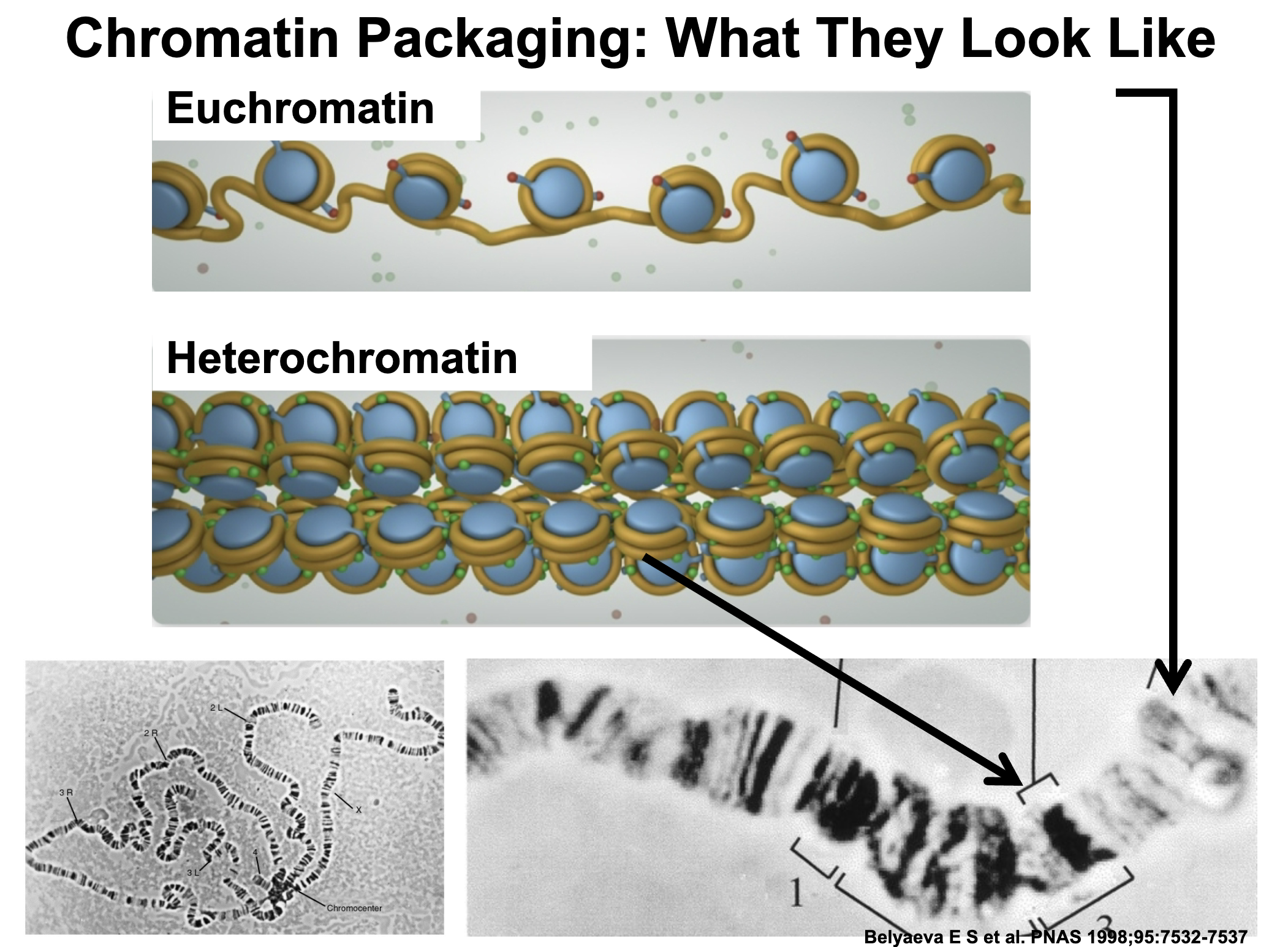
Distinguish euchromatin from heterochromatin, and give one consequence each for transcription, replication timing, and recombination
Euchromatin: Chromatin in relatively decondensed regions and stains lightly.
Heterochromatin: More compacted regions and stains more darkly. Some regions of chromosomes are particularly rich in heterochromatin. Telomeres (special DNA at the ends of chromosomes) and centromeres are heterochromatic. Regions with highly repetitive DNA
Chromatin structure affects transcription, but also replication, recombination, and chromosome transmission:
Rearrangements that place an origin of replication into heterochromatin result in late replication, possibly delaying cell division
Recombination involves breaking DNA and joining it to a different molecule. Heterochromatic regions experience less recombination, which can protect some parts of the genome, like the ribosomal RNA synthesis genes
The special histone CENP-A is needed to form a functional centromere, which is needed for proper chromosome separation
Some transcription does occur in heterochromatic regions, but translocation of a gene from a euchromatic to heterochromatic region can actively prevent transcription