radi 442 test 3 (noble course segment)
 Call Kai
 Learn
Learn Practice Test
 Spaced Repetition
 Match
Match Flashcards
 Knowt Play
1/309

There's no tags or description
Looks like no tags are added yet.
Name | Mastery | Learn | Test | Matching | Spaced | Call with Kai | Chat |
|---|
No analytics yet
Send a link to your students to track their progress
310 Terms
protons (+ charge, determines atomic # and element, inside the nucleus), neutrons (no charge, inside the nucleus), electrons (- charge, in valence shells, binding energy holds them in place, eV or keV electron volts where 1,000 eV = 1 keV)
atomic structure
radiation
energy; in our world, we equate it to x-rays
ionization
removal of an electron; losing an e- leaves you with an ion pair
ionizing radiation
radiation capable of removing electrons; x-rays have the ability to ionize atoms/knock out e-, causing biological impact from disrupting atoms
neutral elements have an equal amount of protons and electrons, so losing an electron induces a positive charge
how do neutral elements get ionized?
x-ray photon causes an electron to get ejected from orbit so the atom is left with a hole; the atom is no longer neutral, causing a biological impact
explain ionization
radiation protection
measures taken to protect patients, employees, and the public from unnecessary exposure to ionizing radiation
x-ray rooms/suites are designed with radiation protection in mind, so when you swap out equipment, you have to make sure the x-ray room can handle the unit being put in place; costs a lot of money
how are x-ray rooms designed with radiation protection in mind?
PREVENT tissue reactions and MINIMIZE risk of stochastic effects
what are the goals of radiation protection?
biological impact
ionized atoms cause…
we know exactly how much exposure it takes to cause different types of tissue reactions
why can we PREVENT tissue reactions from radiation?
we don’t know what the dose is or what exactly causes these effects
why can we only MINIMIZE the risk of stochastic effects and not prevent them?
example of a tissue reaction
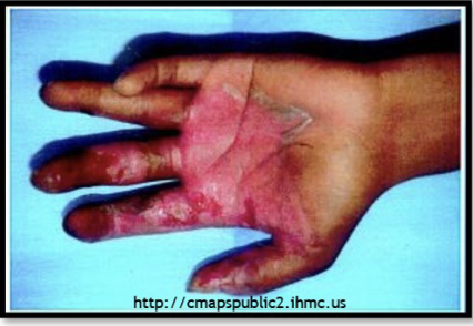
example of a stochastic effect
cancer
as low as reasonably achievable (exposures should be made with the least amount of radiation as possible)
occupational radiation protection concept; ALARA
optimization for radiation protection; same idea as ALARA, keep radiation doses to the minimum necessary to achieve the required image
occupational radiation protection concept; ORP
time, distance, shielding
what are the cardinal principles (rules)?
minimize time exposed to radiation, time in the x-ray room, etc.
explain the time cardinal principle
maximize distance from radiation
explain the distance cardinal principle
utilize shielding, protect yourself with protective wear and barriers
explain the shielding cardinal principle
weigh the risk vs. the benefit of the exam; this does NOT fall on the technologist, it is up to the ordering physician to decide how the risk of radiation compares to the benefit of the scan
patient radiation protection concept; justification
is this the appropriate procedure for the suspected condition or disease? also falls on the ordering physician’s shoulders
patient radiation protection concept; diagnostic efficacy
provided by the ACR (american college of radiology); evidence-based guidelines for selecting appropriate imaging procedures; based on patient condition
patient radiation protection concept; appropriateness criteria
time, distance, and shielding in regards to the patient; the cardinal principles apply to occupational workers (techs) AND patients
patient radiation protection concept; cardinal principles (rules)
background equivalent radiation time; risk perspective; puts patient radiation exposure into perspective
what’s the purpose of the BERT method?
BERT = background equivalent radiation time; compares radiation received by the patient to natural background radiation (which is about 3 mSv/year); emphasizes that radiation is part of our daily lives; does NOT imply risk, it is only a comparison; makes it easier for patients to understand their radiation exposure; just helps the patient understand, not tying a specific risk to the exam
explain the BERT method

3 mSv per year
what is the natural background radiation amount?
normal x-rays involve taking only 2-3 images, whereas CT involves longer exposure and more images
why does it make sense that x-rays have a lower approximate effective radiation dose compared to CT?
less than 3 hours (< 0.001 mSv)
how does extremity x-ray compare to natural background radiation?
6 months (1.4 mSv)
how does lumbar spine x-ray compare to natural background radiation?
10 days (0.1 mSv)
how does chest x-ray compare to natural background radiation?
2 years (6.1 mSv)
how does chest CT compare to natural background radiation?
2 years (6 mSv)
how does barium study (fluoroscopy, BE or UGI) compare to natural background radiation?
2.6 years (7.7 mSv)
how does abdomen & pelvis CT compare to natural background radiation?
DON’T; their bodies are more sensitive
peds (do/don’t) require the same amount of radiation exposure for an exam as adults do
image gently
what is the radiation protection campaign for pediatrics?
to change practice by raising awareness of the opportunities to lower radiation dose in the imaging of children
what is the goal of image gently?
started with CT, expanded to include fluoroscopy, interventional, nuclear medicine, digital imaging, dentistry
what is the focus of image gently?
provide information and free educational materials for medical professionals and parents
what is the strategy of image gently?
image wisely
what is the radiation protection campaign for older adults?
lower radiation dose in adults
what is the goal of image wisely?
provide resources for medical professionals and patients
what is the strategy of image wisely?
NEXT = nationwide evaluation of x-ray trends; fed. gov. agencies worked with state departments to collect dose information and establish dose reference levels (DRLs)
explain the radiation protection initiative - NEXT program
dose reference levels were brought about because of digital imaging and exposure; the NEXT program set the foundation for exam dose awareness; “for each exam, try for this target exposure level”; provided guidelines to help institutions adjust their exposures
explain DRLs and how they came about
there is a built-in system that tracks dose information and this information is saved and can become part of a patient’s medical record
explain digital imaging systems dose tracking software
requires patient dose monitoring - CT and fluoroscopic procedures have built-in monitoring; they have to have the software that traces patient exposure to radiation
to maintain accreditation status, the joint commission (TJC)…
annual education for staff that focuses on dose reduction, minimum qualifications for medical physicists, documentation of CT doses, mandated standardized CT protocols; the goal is always to be aware and minimize patient exposure to radiation!
what are the additional CT requirements for dose monitoring, as required by TJC?
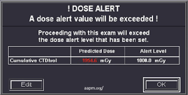
dose alert protocols
how do we handle situations where patients are overexposed or if there’s a risk of overexposure?
protocols for handling situations where patient doses are expected to exceed or have exceeded normal levels; radiographers and/or radiologist assistants may need to provide information to the medical physicist or radiologist for dose calculation; you may have to proceed; examples include pregnancy status, patient size, areas irradiated, technical factors, and dose data from imaging system
what are dose alert protocols?
the ability of machines or computer systems to learn, reason, make decisions, recognize patterns, and solve problems in ways that mimic or augment human cognitive abilities
what is artificial intelligence (AI)?
dose optimization, exposure settings, image processing, image interpretation (radiologist side of things); a lot of systems and equipment in the hospital incorporate AI; exposure settings can be given by AI after analyzing pt parameters and clinical implications, but you still need to think and know what’s correct because AI is not 100% accurate
how does AI play a role in medical imaging?
focuses on peds and ensures vendor software works for pediatric pts; promotes safe AI for pediatrics; AI systems must be specifically designed for peds and there must be standardized labeling for FDA-cleared medical devices that use AI for peds; the devices must clearly indicate that they’re safe and reliable for peds; remember that peds are different from adults so AI needs to be developed specifically for them; AI that uses adult data will not be accurate for peds, so models need to be based on peds
what does the image intelliGently campaign do?
energy
radiation is __
all types of radiation, different types of energy (because radiation is energy)
mechanical, thermal, chemical, electrical, sound, nuclear, electromagnetic (x-rays!)
radioactive decay
alpha particles are a product of __
the nucleus of an atom
where does radioactive decay occur?
unstable nuclei release charged particles to restabilize itself
why does radioactive decay occur?
uranium and radon (naturally occurring elements)
alpha particles are emitted from __
radioactive decay
beta particles are a product of __
iodine-131 (which is a radioisotope/unstable form of iodine)
beta particles are emitted from __
electromagnetic (EM) radiation; electric and magnetic properties
x-rays are an example of __

meters (m)
wavelength is measured in __
hertz (Hz)
frequency (f) is measured in __
frequency x wavelength
speed of light (c ) = ?
electron volts (eV)
energy (E) is measured in __
direct
wavelength and energy have a __ relationship
decreases
wavelength __ as you move to the right on the EM visible spectrum
increases
frequency __ as you move to the right on the EM visible spectrum
indirect/inverse
wavelength and frequency have a __ relationship
increases, direct
as frequency increases, energy __ because of the __ relationship between frequency and energy
lower
longer wavelength correlates with __ frequency
higher
shorter wavelength correlates with __ frequency
shorter, higher
x-rays have __ wavelength and __ frequency compared to visible light
the speed of light
radiowaves, microwaves, visible light, etc. all travel at __
x-rays have shorter wavelength, thus higher frequency, thus higher energy than visible light and are energetic enough to get into the body and cause ionization/disrupt atomic structure
why does the wavelength and frequency of x-rays matter?
electrical and magnetic disturbances traveling in space, no mass and no charge (they’re neutral and not a quantity of matter), can’t focus with a lens or be redirected, travel in straight lines in a vacuum (x-ray tube) at the speed of light (in a wave form/pattern but travels straight towards the patient), shorter wavelength and higher frequency means energy increases, interact with matter and ionize because they have high enough energy, exhibit wave and particle behavior (wave-particle duality)
list the properties of x-rays
bundle of energy (photon or quantum) traveling through space along a straight path but in an x-ray waveform; like a wave pattern as if it were contained in a particle/ball as a bundle of energy
explain wave-particle duality

the amount of energy deposited/transferred along the path of travel; how much energy is given up by a photon along its path of travel
what is linear energy transfer (LET) ?
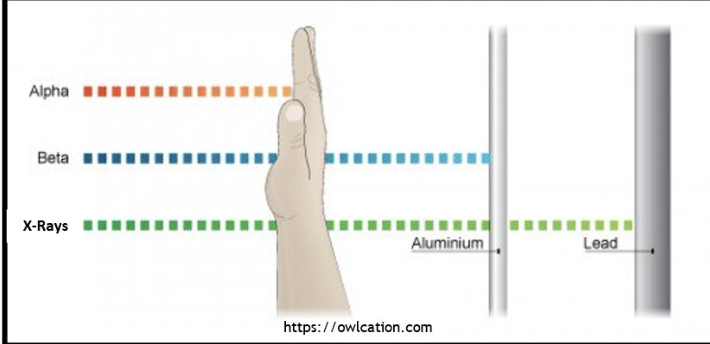
high
low penetrability = __ LET
the photon gives up all of its energy very readily; like a sprinter
what does a high LET mean?
low
high penetrability = __ LET
the photon gives up its energy over a longer period of time; like a marathon runner
what does a low LET mean?
high, low
alpha (uranium, radon) has __ LET meaning __ penetrability
high
because alpha is __ LET, it can be stopped by the skin
low, high
beta and x-rays are __ LET meaning __ penetrability
no, it just means you’re getting all the radiation at one spot
does alpha particles being high LET make them safe?
low
because beta is __ LET, it penetrates through the body but can be stopped by an aluminum sheet
the lowest
because x-rays are __ LET, they penetrate through the body and aluminum, needs lead to fully stop
alpha, x-rays
__ is the highest LET and __ is the lowest LET
on the electromagnetic (EM) spectrum
both non-ionizing and ionizing radiation are __
non-ionizing radiation
long wavelengths, short frequencies, low energy; not enough energy to ionize atoms
ionizing radiation
shorter wavelengths, higher frequencies, higher energy; enough energy to ionize atoms
non-ionizing
radio waves are __ radiation
non-ionizing
microwaves are __ radiation
non-ionizing
infrared light is __ radiation
non-ionizing
visible light is __ radiation
some is non-ionizing, some is ionizing, that’s why it’s important to protect yourself from the sun
ultraviolet (UV) light is __ radiation
vibrate or heat atoms
what is the result of non-ionizing radiation?
ionizing
x-rays (manmade) are __ radiation
ionizing
gamma rays (nucleus of atom, last on EMS) are __ radiation