Topic 2 - organisation
1/83
Earn XP
Description and Tags
Name | Mastery | Learn | Test | Matching | Spaced |
|---|
No study sessions yet.
84 Terms
Describe the relationship between cells and organisms.
Cells are the basic building blocks of all living organisms. A tissue is a group of cells with a similar structure and function. Organs are aggregations of tissues performing specific functions. Organs are organised into organ systems, which work together to form organisms.
Explain carbohydrates.
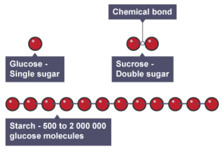
Carbohydrates in our diet include sugars and starches. The glucose molecule is small enough to be absorbed directly through the walls of the digestive system. Starch is a polymer of glucose. It must be broken down into glucose molecules - it is too large to pass through the gut. Cellulose is also made up of glucose molecules. It makes up plant cell walls. It is therefore a fundamental part of our diet. It cannot be broken down by the digestive system, so is egested from the gut. Once absorbed by the body, glucose molecules are transported to cells and: used for respiration reassembled into the storage form of carbohydrate in animals - glycogen In plant metabolism, the glucose produced by photosynthesis is converted into starch for storage, and cellulose, for cell wall synthesis. In humans and animals glucose is stored in glycogen. It is not converted into starch.
Explain the function of proteins.
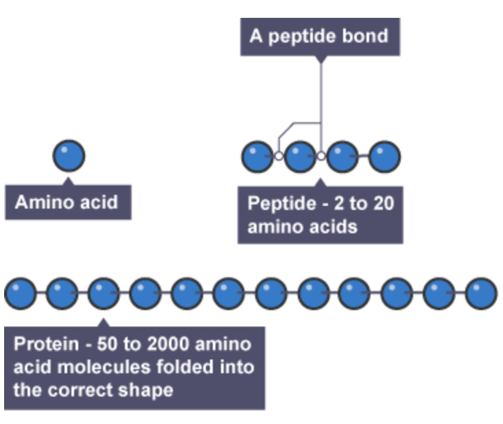
Proteins are made up of amino acids. Proteins are big molecules that are too large to pass through the gut wall. They must first be broken down into amino acids. Once inside the body, the amino acids are reassembled into the proteins the individual requires - the process of protein synthesis. Proteins are used for growth and repair. Excess amino acids are broken down in the liver.
Explain the function of lipids.
Lipids are used for energy, make up part of cell membranes so essential for normal growth. Lipids are esters of fatty acids and glycerol. Lipid molecules are too large to pass through the gut wall and must be digested first. In the body's cells, they are reassembled into the lipids the cell needs, for instance, for the cell membranes.

Explain the food test for sugars.
1. Create a solution in a test tube from the substance to be tested.
2. Add 5 drops of Benedict’s reagent to the substance to be tested.
3. Place the test tube in a hot water bath and leave for 5 minutes.
4. Record the colour change observed.
5. Bright blue - negative result, Green - little reducing sugar is present, Orange - some reducing sugar is present, Brick red - a lot of reducing sugar is present

Explain the food test for starch.
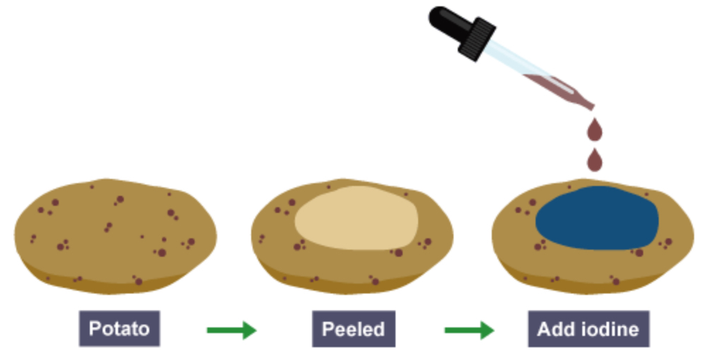
1. Place the substance to be tested in spotting tile.
2. Add two drops of iodine solution to the substance.
3. Record the colour observed.
4. If a red-brown colour remains the test for iodine is negative. If the solution turns blue-black the test for iodine is positive.
Explain the food test for protein.
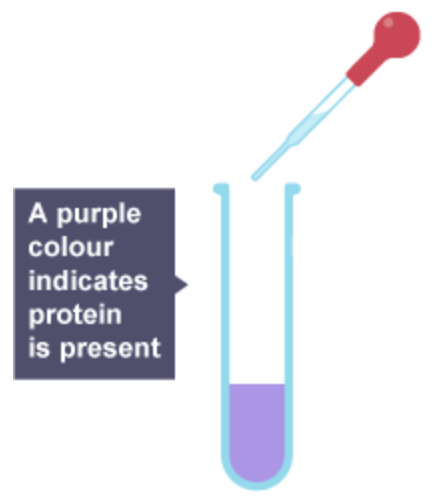
1. Create a solution in a test tube from the substance to be tested.
2. Add 5 drops of Biuret’s solution to the solution to be tested.
3. Record the colour observed.
4. If a light blue colour is observed, the result for protein is negative. If a lilac colour is observed, the result for protein is positive.
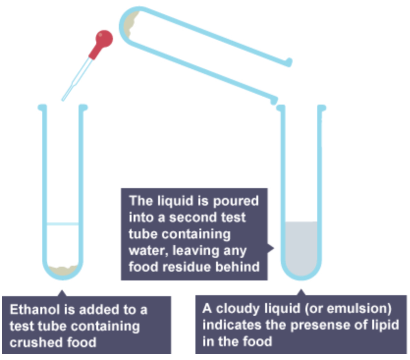
Explain the food test for fats.
1. Half fill a test tube with water.
2. Add the suspected fat to the water.
3. Move the test tube side to side to mix the water and fat thoroughly.
4. Add 5 drops of ethanol to the test tube mixture.
5. Place thumb over the top of the mixture and shake thoroughly.
6. As oils and fats do not dissolve in water, an emulsion will form. This makes the top layer cloudy. If there are no fats, the solution will be colourless.
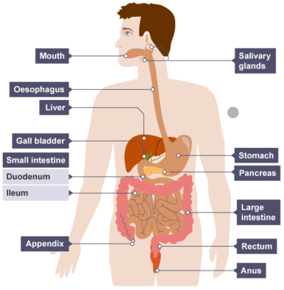
Explain the functions of different parts of the human digestive system.
Region | Function |
Mouth | Begins the digestion of carbohydrates |
Stomach | Begins the digestion of protein; small molecules such as alcohol absorbed |
Small intestine - Duodenum | Continues the digestion of carbohydrate and protein; begins the digestion of lipids |
Small intestine - Ileum | Completes the digestion of carbohydrates and proteins into single sugars and amino acids; absorption of single sugars, amino acids and fatty acids and glycerol |
Large intestine | Absorption of water; egestion of undigested food |
Define enzymes.
Enzymes are biological catalysts– they are proteins that speed up chemical reactions.
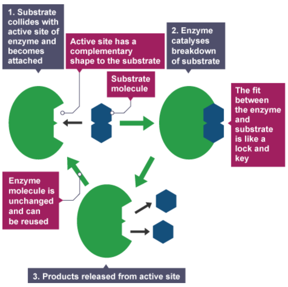
Explain the lock and key theory.
The lock and key theory shows how enzymes have an active site that is specific to its substrate so it can only speed up that specific reaction. It shows how enzymes are specific to its substrate and form an enzyme-substrate complex. The enzyme breaks its substrate down into products.
State the factors that affect enzyme action.
Temperature
pH
Explain how temperature affects enzyme action.
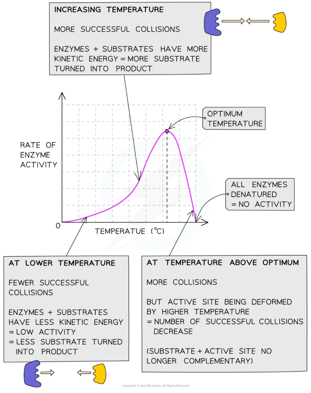
Increasing temperature from 0⁰C to the optimum increases the activity of enzymes as the more energy the molecules have the faster they move and the number of collisions with the substrate molecules increases, leading to a faster rate of reaction
This means that low temperatures do not denature enzymes, but at lower temperatures with less kinetic energy both enzymes and their substrates collide at a lower rate
The specific shape of an enzyme is determined by the amino acids that make the enzyme
Enzymes work fastest at their ‘optimum temperature’ – in the human body, the optimum temperature is around 37⁰C
Heating to high temperatures (beyond the optimum) will start to break the bonds that hold the enzyme together – the enzyme will start to distort and lose its shape – this reduces the effectiveness of substrate binding to the active site reducing the activity of the enzyme. Eventually, the shape of the active site is lost completely and the enzyme is described as being ‘denatured’
Substrates cannot fit into denatured enzymes as the specific shape of their active site has been lost
Explain how pH affects enzyme action.
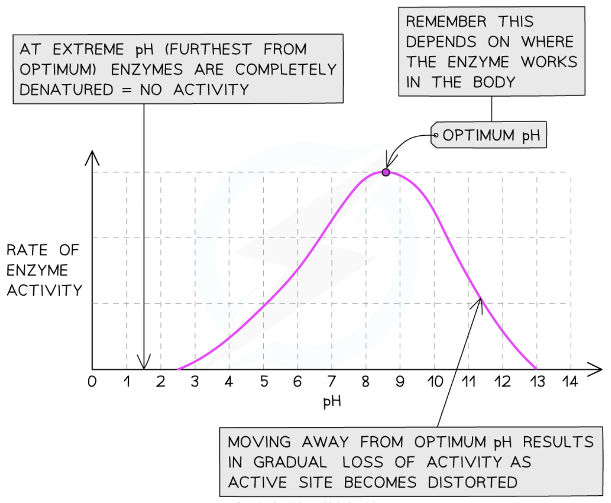
The optimum pH for most enzymes is 7 but some that are produced in acidic conditions, such as the stomach, have a lower optimum pH (pH 2) and some that are produced in alkaline conditions, such as the duodenum, have a higher optimum pH (pH 8 or 9)
If the pH is too high or too low, the bonds that hold the amino acid chain together to make up the protein can be destroyed
This will change the shape of the active site, so the substrate can no longer fit into it, reducing the rate of activity
Moving too far away from the optimum pH will cause the enzyme to denature and activity will stop
Explain carbohydrase.
Carbohydrases break down carbohydrates in several regions of the digestive system.
Most of the carbohydrate we eat is starch, so this will be the main substratein the early part of digestion for enzyme action.
Region of digestive system | Enzyme | Where produced | Substrate | Broken down into |
Mouth | Salivary amylase | Salivary glands | Starch | Maltose |
Small intestine - Duodenum | Pancreatic amylase | Pancreas | Starch | Maltose |
Small intestine - Ileum | Amylase | Wall of ileum | Maltose | Glucose |
Explain protease.
Proteases break down proteins in several regions of the digestive system.
Region of digestive system | Enzyme | Where produced | Substrate | Broken down into |
Stomach | Protease - pepsin | Gastric glands in stomach | Proteins | Begins the breakdown into amino acids |
Small intestine - Duodenum | Protease - trypsin | Pancreas | Proteins | Continues the breakdown into amino acids |
Small intestine - Ileum | Protease - peptidase | Wall of ileum | Peptides | Completes the breakdown into amino acids |
Explain lipase.
Lipases break down lipids in one region of the digestive system. In the duodenum of the small intestine, lipase made in the pancreas breaks down lipids into fatty acids and glycerol.
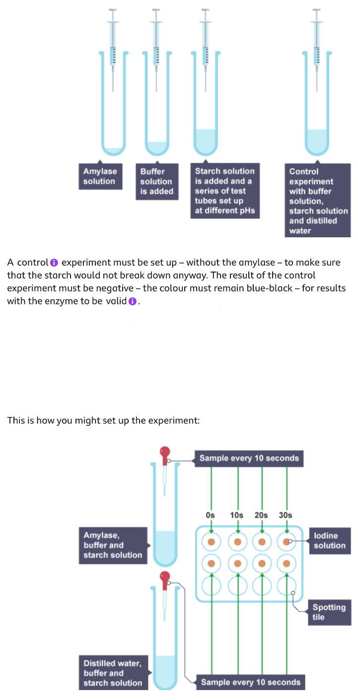
Describe a method to investigate the effect of pH on the rate of reaction of amylase enzyme.
1. Set up a water bath.
2. Place one drop of iodine into each well of a dimple tray.
3. Measure 2 cm3 of amylase solution into a test tube. Measure 1 cm3 of pH1 buffer solution into a different test tube. Put 1 cm3 of starch solution into the test tube with the buffer solution.
4. Allow the test tubes to acclimate in the water bath for about 5 minutes.
5. Use a pipette to drop one drop of starten/buffer solution into the dimple tray.
6. Thoroughly mix the test tube of starch and buffer solution with the test tube of amylase and start the stop clock.
7. Each minute, on the minute, use a dropper to drop one drop of the mixture onto a dimple well filled with iodine.
8. Record the time it takes for the iodine to no longer change colour and to stay red-brown.
9. Repeat the experiment with buffer solutions of pH 2,3,4,5,6,7,8,9,10,11 and 12.
Define bile.
Emulsifies lipids, breaking them up physically into tiny droplets. Tiny droplets have a much larger surface area, over which lipases can work, than larger pieces, or drops of lipid.
Contains sodium hydrogencarbonate, which is an alkali. It neutralises stomach acid and produces the optimumpH for pancreatic enzymes.
Is produced in the liver, but stored and concentrated in the gall bladder.
Explain why we need a transport system.
A transport system is needed to distribute essential molecules efficiently, as the diffusion distance is too large for molecules to diffuse from one cell to another.
Describe the structure of the heart.
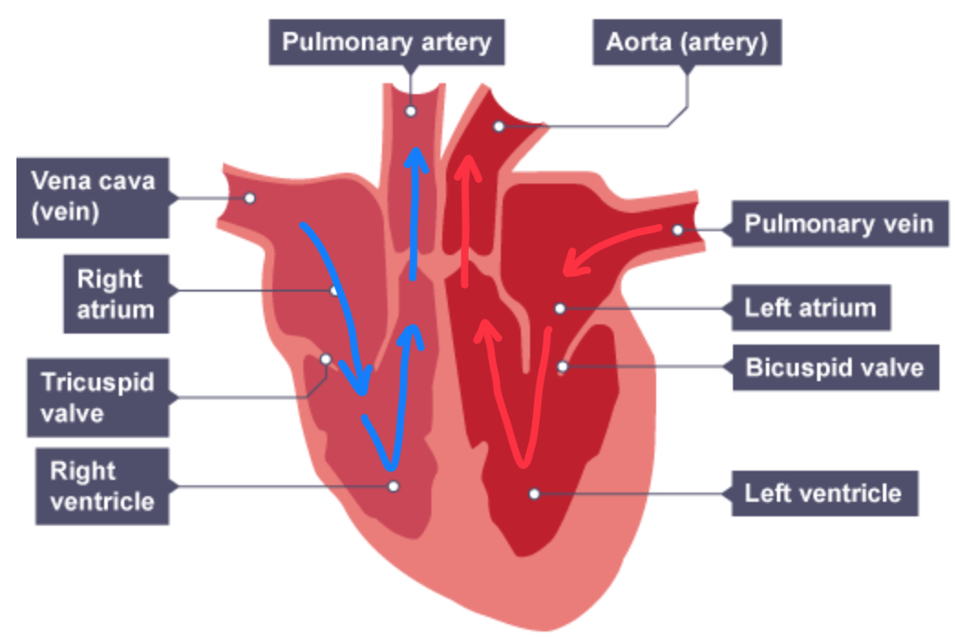
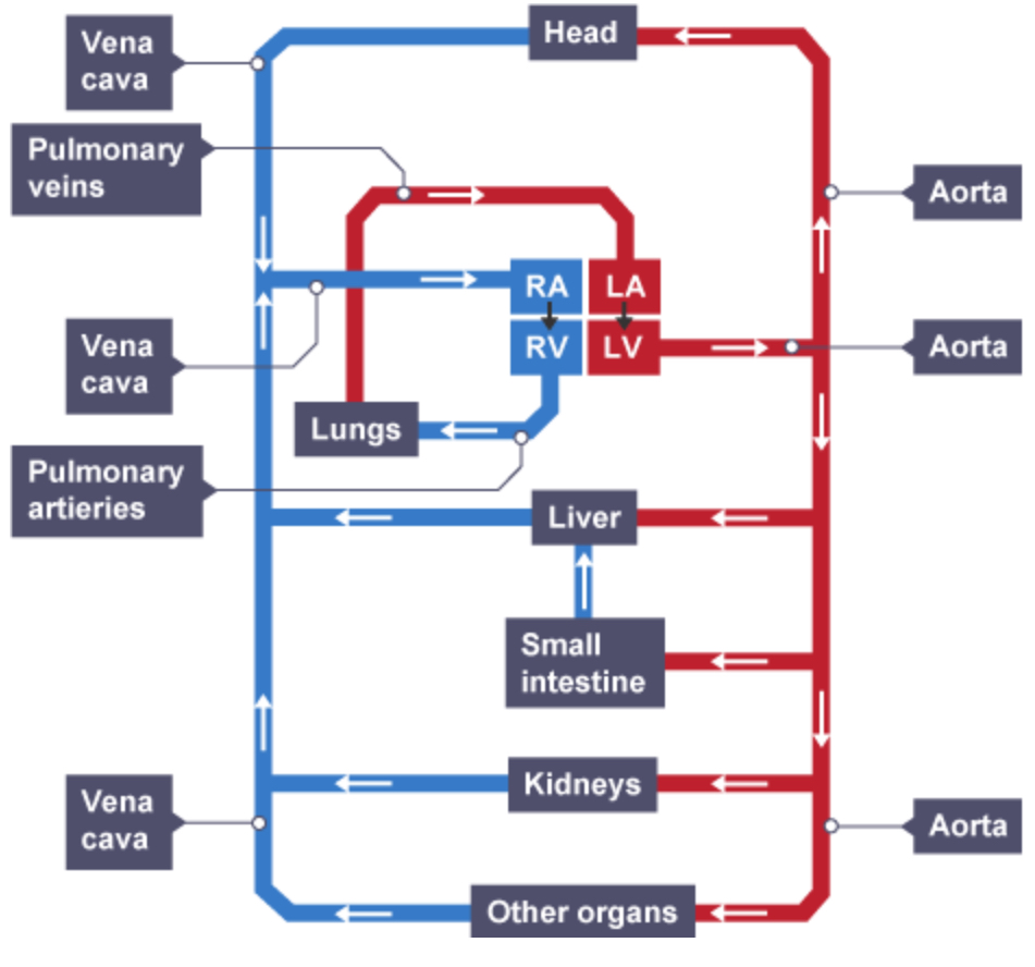
The right side of the heart receives deoxygenated blood from the body and pumps it to the lungs where oxygen diffuses in from the alveoli and carbon dioxide diffuses out
The left side of the heart receives oxygenated blood from the lungs and pumps it to the body
Blood is pumped towards the heart in veins and away from the heart in arteries
Describe the pathway of blood in the heart.
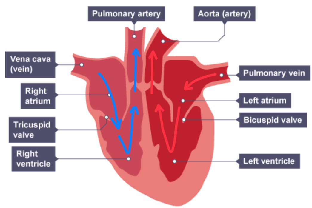
Deoxygenated blood enters the heart via the vena cava, emptying into the right atrium
Blood flows down through a set of atrioventricular valves into the right ventricle
When the ventricles contract, blood travels up through the pulmonary artery to the nearby lungs where gas exchange occurs (and the blood becomes oxygenated)
Oxygenated blood returns to the heart via the pulmonary vein, emptying into the left atrium
Blood flows down through a set of atrioventricular valves into the left ventricle
When the ventricles contract, blood travels up through the aorta, and to the rest of the body
Explain the double circulatory system.
The human heart is part of a double circulatory system
The circulatory system is a system of blood vessels with a pump (the heart) and valves that maintain a one-way flow of blood around the body
The heart has four chambers separated into two halves:
The right side of the heart pumps blood to the lungs for gas exchange (this is the pulmonary circuit)
The left side of the heart pumps blood under high pressure to the body (this is systemic circulation)
The benefits of a double circulatory system:
Blood travelling through the small capillaries in the lungs loses a lot of pressure which reduces the speed at which it can flow
By returning oxygenated blood to the heart from the lungs, the pressure can be raised before sending it to the body, meaning cells can be supplied with oxygenated blood more quickly
Explain adaptations of the human heart.
The walls of the ventricles are much thicker than those of the atria as they are responsible for pumping blood out of the heart and so need to generate a higher pressure
The wall of the left ventricle is much thicker than that of the right ventricle as it has to pump blood at high pressure around the entire body, whereas the right ventricle is pumping blood at lower pressure to the lungs (which are close to the heart)
There are two sets of valves inside the heart which function to prevent the backflow of blood in the heart
The two sides of the heart are separated by the septum which prevents the mixing of deoxygenated and oxygenated blood inside the heart so more oxygenated blood is pumped around the body
The heart is made of a special type of cardiac muscle tissue which does not fatigue like skeletal muscle
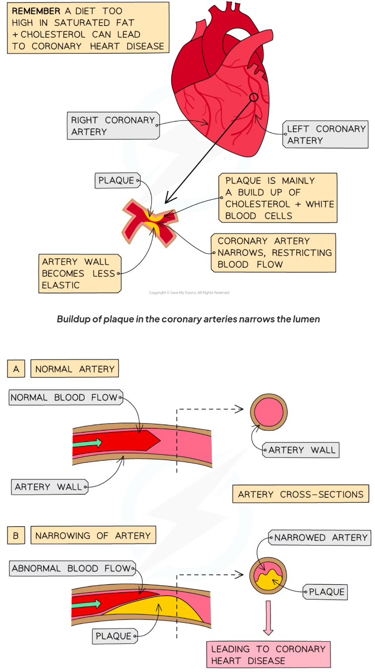
Explain CHD.
Coronary heart disease.
How can CHD be treated?
Treatment of CHD involves either:
increasing the width of the lumen of the coronary arteries using a stent
prescribing statins to lower blood cholesterol
Explain how stents work.
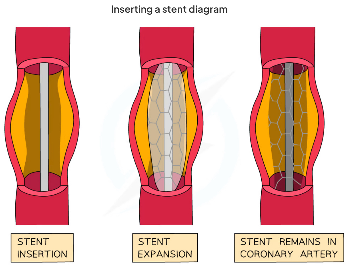
Stents can be used to keep the coronary arteries open
A narrow tube is threaded up through the groin up to the blocked vessel
A tiny balloon is then inflated
The balloon pushes the metal or plastic stent against the wall of the artery, increasing the width of the lumen
The balloon and tube are then removed
Stents are very effective at reducing the risk of a heart attack as they widen the lumen to increase blood flow to the coronary arteries, and the procedure is relatively simple
Stents also last a long time, which is a positive, however, there is a risk of blood clots(thrombosis) occurring
Evaluate the use of stents.
Advantages of stents | Disadvantages of stents |
Effective at reducing the risk of a heart attack as they widen the lumen and increase blood flow | Risk of blood clots occurring |
Last a long time | Risk of infection during surgery |
Simple minor surgery to insert a stent | Risk of damage to the blood vessel during surgery |
Explain how statins work.
Statins are drugs that are widely used to reduce the levels of fatty deposits(cholesterol) in the blood
They block an enzyme in the liver which is needed to make cholesterol
This slows down the rate of fatty material building up in the blood, reducing the risk of CHD occurring
There are many advantages and disadvantages of statins:
Evaluate the use of statins.
Advantages of statins | Disadvantages of statins |
Reduce the levels of 'bad' cholesterol (LDL) in the blood which reduces the risk of fatty deposits building up - this reduces the risk of CHD | Need to be taken regularly and long-term, otherwise, they are not as effective |
Increase levels of 'good' cholesterol (HDL) in the blood which remove 'bad' (LDL) cholesterol circulating in the blood | Take some time to have an effect |
Side effects of statins include muscle and joint pain, kidney problems and neurological issues |
Explain faulty valves.
The heart valves play a vital role in ensuring blood is pumped from the ventricles to the arteries, in a one-way direction
In some people, heart valves may become faulty as a result of illness, old age or a heart attack
Valves can stiffen which can prevent them from opening fully to let blood flow through
This reduces the volume of blood pumped by the heart
Sometimes a faulty heart valve might develop a leak
This allows blood to flow from the ventricles back into the atria or the arteries back into the ventricles
Both issues reduce the effectiveness of the heart in pumping oxygenated blood around the body
How are faulty valves treated?
Faulty heart valves can be replaced via surgery using:
biological valves from cows or pigs
mechanical valves
Evaluate the use of replacement valves.
Valve replacement type | Advantages | Disadvantages |
Biological valve | Highly effective Less likely to leak | Need to be replaced after 12-15 years Risk of immune rejection |
Mechanical valve | Long-lasting Less need to replace | Can increase the likelihood of blood clots Lifelong need to take anticoagulant (anti-blood clotting) medication |
How can heart failure be treated?
In the case of heart failure a heart can be transplanted from a donor who has recently died
Waiting lists for organ transplants are often long, so an immediate solution can involve replacing the heart with an artificial heart made from plastic and metal
Artificial hearts may be used to:
keep patients alive while waiting for a heart transplant,
allow the heart to rest as an aid to recovery
Evaluate the use of artificial hearts.
Advantages | Disadvantages |
Shorter waiting times | Do not work as well as real hearts |
Less chance of the patient's immune system rejecting it | Increased risk of blood clots, leading to increased risk of str |
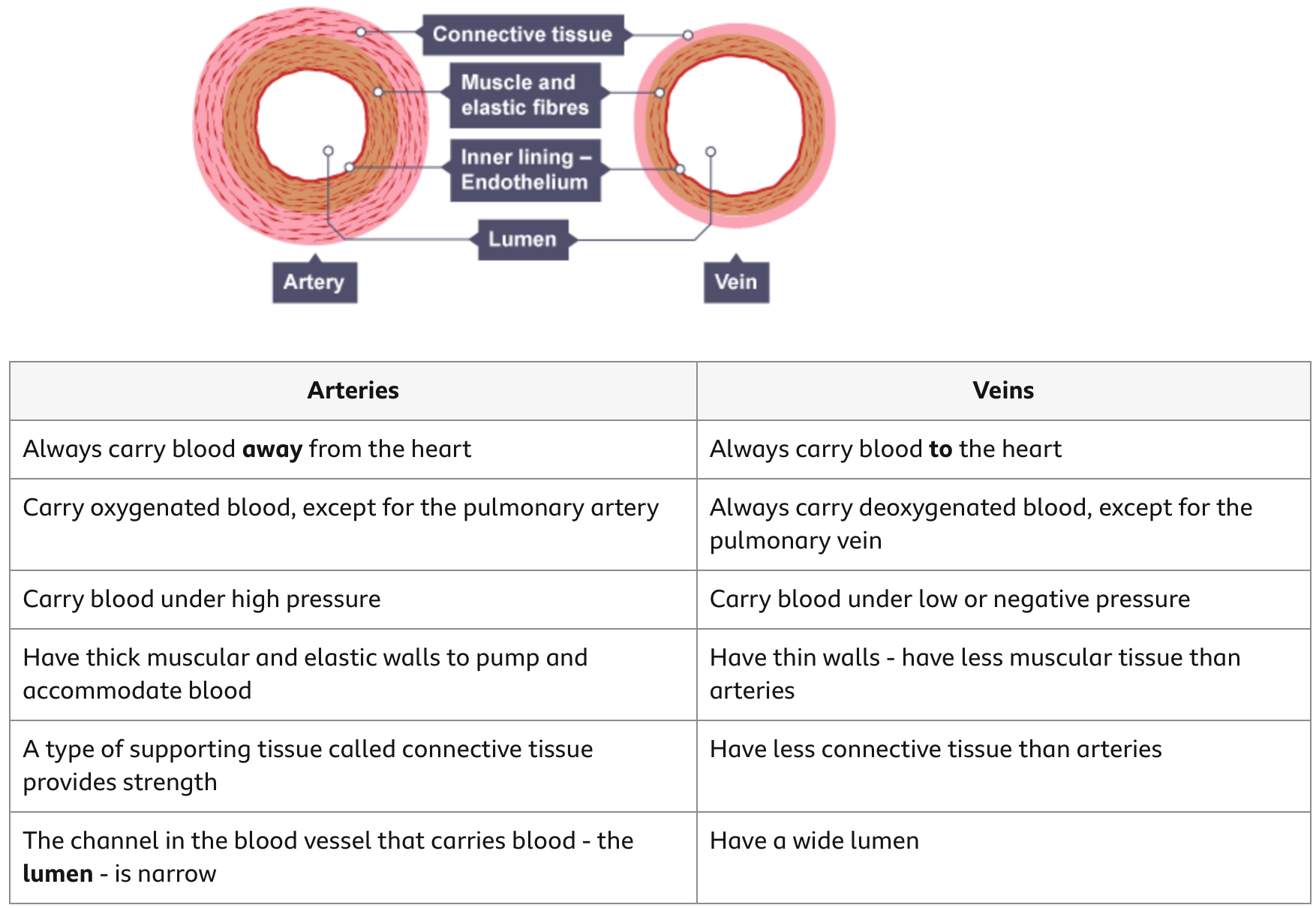
Compare the structures of an artery and a vein.
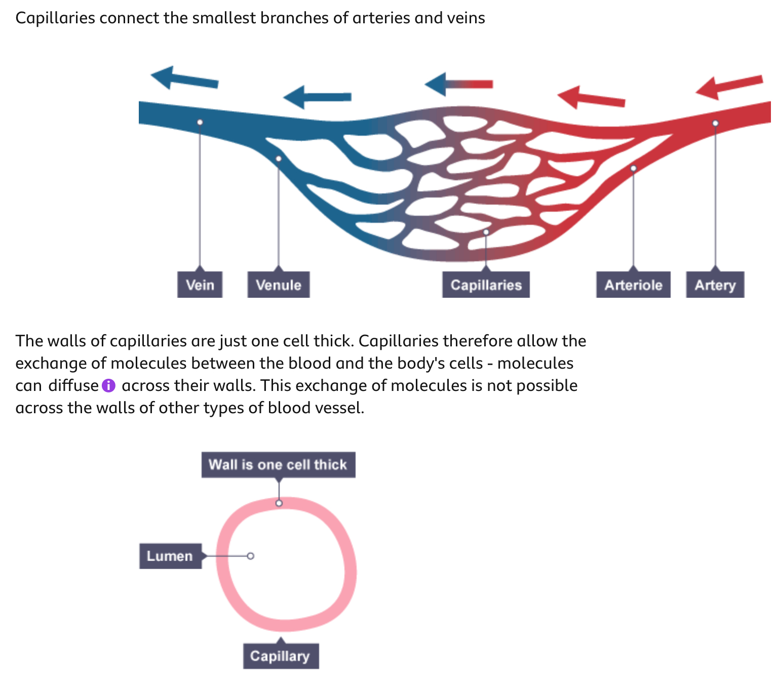
Describe the structure of a capillary and how it is adapted.
Explain the function of a pacemaker.
The natural resting heart rate is controlled by a group of cells located in the right atrium
These cells form a structure called the pacemaker
The role of the pacemaker is to coordinate the contraction of the heart muscle, therefore it regulates the heart rate
Up to a point, the faster the heart contracts, the more quickly oxygenated blood can be delivered around the body
When a person is at rest, the oxygen demand of their cells is relatively low and so a lower heart rate is maintained
When a person is exercising, the oxygen demand of their muscle cells increases and so a higher heart rate is necessary
The pacemaker sends out an electrical impulse which spreads to the surrounding muscle cells, causing them to contract
The pacemaker does this every time the heart needs to “beat”, so if a person has a resting heart rate of 60 beats per minute (bpm), then the SAN will be sending out electrical impulses on average once every second
Sometimes, the pacemaker of the heart stops functioning properly (this can cause an irregular heartbeat)
Artificial pacemakers are electrical devices used to correct irregularities in the heart rate
Name the components of the blood.
Plasma
Red blood cells
White blood cells
Platelets
Explain the function of plasma.
Plasma is a straw-coloured liquid that makes up 55% the volume of blood. Plasma transports carbon dioxide, digested food molecules, urea and hormones and distributes heat.
Explain the function and adaptations of red blood cells.
Red blood cells transport the oxygen required for aerobic respiration in body cells.
Red blood cells have adaptations that enable them to carry a maximum amount of oxygen. They contain the protein haemoglobin, which gives them their red colour.
Haemoglobin can combine reversibly with oxygen. This is important - it means that it can combine with oxygen as blood passes through the lungs, and release the oxygen when it reaches the cells.
red blood cells:
have no nucleus - they lose it during their development - so they can pack in more haemoglobin.
are small and flexible so that they can fit through narrow blood capillaries.
have a biconcave shape - they are the shape of a disc that is curved inwards on both sides - to maximise their surface area for oxygen absorption.
are thin, so there is only a short distance for the oxygen to diffuse to reach the centre of the cell.
Explain the function of different types of white blood cells.
There are several main types of white blood cell.
Phagocytes
About 70 per cent of white blood cells are phagocytes. Phagocytes engulf and destroy unwanted microorganisms that enter the blood, by the process of phagocytosis. They are part of the body's immune system.
Lymphocytes
Lymphocytes make up about 25 per cent of white blood cells. They are also part of the body's immune system. Lymphocytes produce soluble proteins called antibodies when a foreign body such as a microorganism enters the body.
Explain the function of platelets.
Platelets are cell fragments produced by giant cells in the bone marrow.
Platelets stop bleeding in two main ways:
they have proteins on their surface that enable them to stick to breaks in a blood vessel and clump together
they secrete proteins that result in a series of chemical reactions that make blood clot, which plugs a wound.
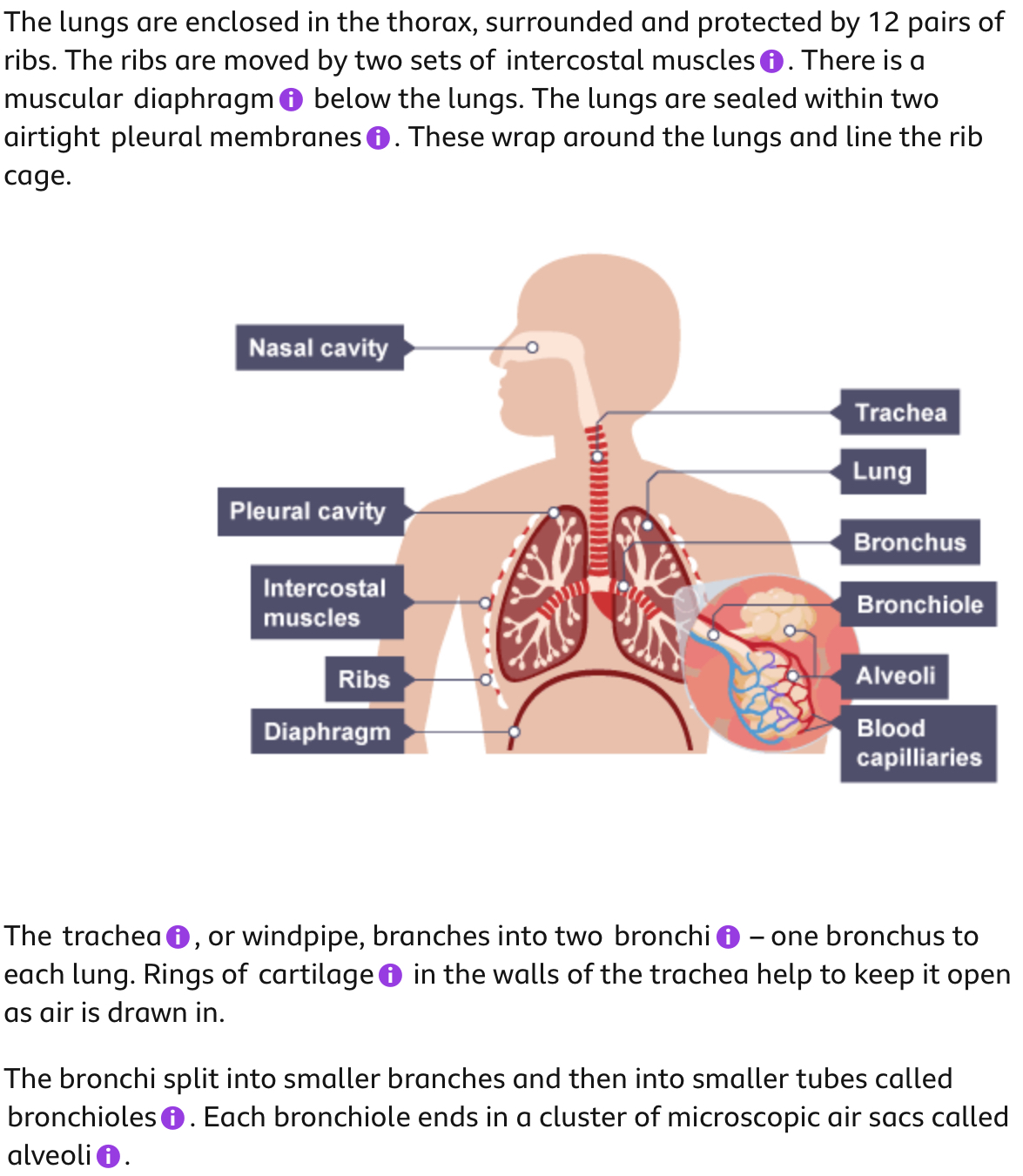
Describe the structure of the lungs.
How is the lungs adapted for gas exchange?
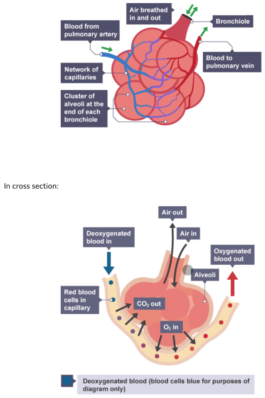
The exchange of gases occurs between the alveoli and blood in the capillaries that supply the lungs. Capillaries cover 70% of the outside of alveoli, providing a large surface area for gases to diffuse across.
The alveoli are adapted to provide a very large surface area for gaseous exchange:
small size - each alveolus is a small sphere about 300 μm in diameter, giving it a larger surface area to volume ratio than larger structures
number - there are around 700 million alveoli
The total surface area of the alveoli is around 70 square metres.
There is also a short diffusion path - the walls of blood capillaries and alveoli are just one cell thick. The alveoli are also lined with a thin film of moisture. Gases dissolve in this water, making the diffusion path even smaller.
The ventilation of the lungs and the blood flow through the surrounding capillaries mean gases are being removed continually, and steep concentration gradients are set up for gases to diffuse.
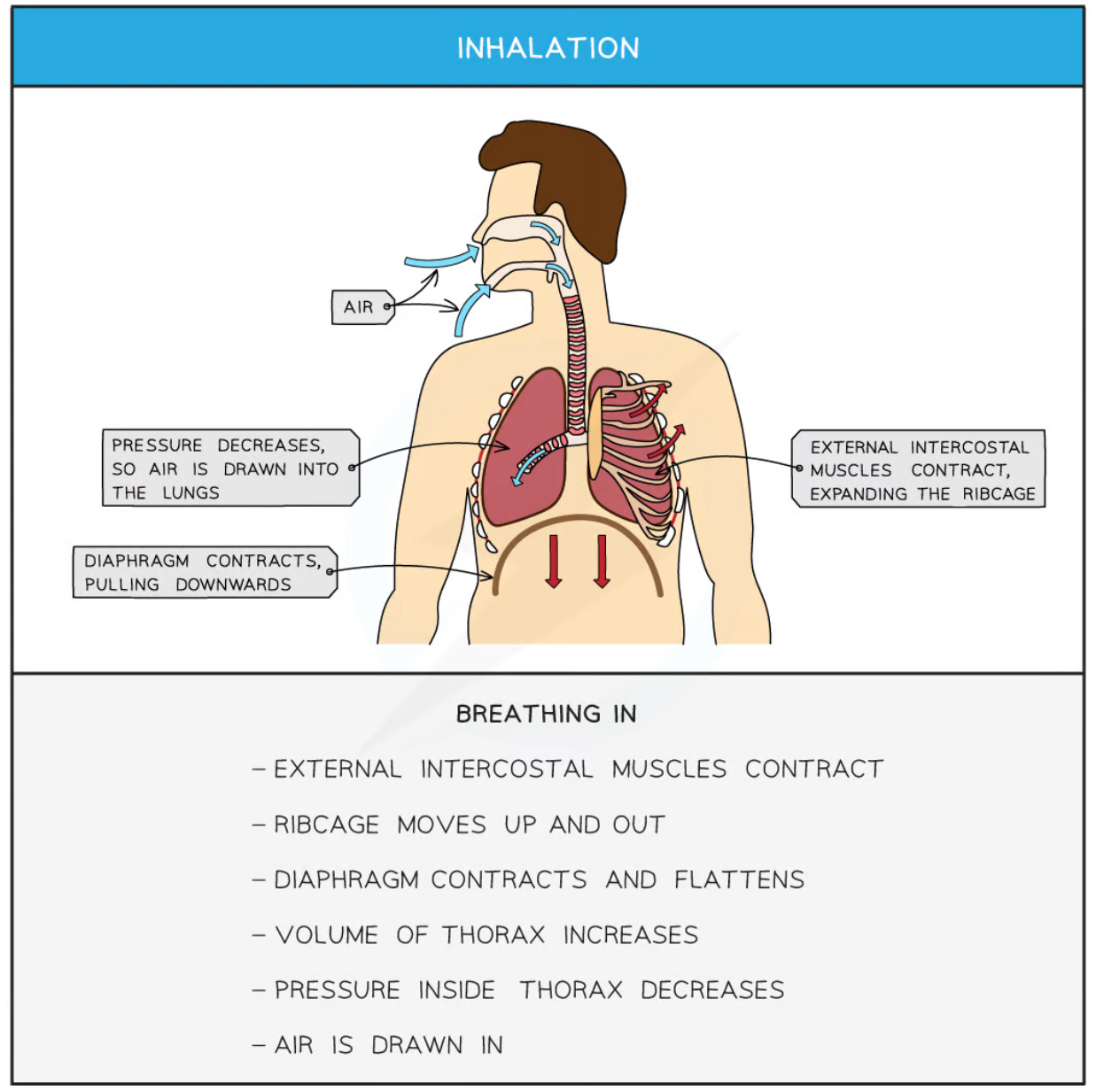
Describe the process of inhalation.
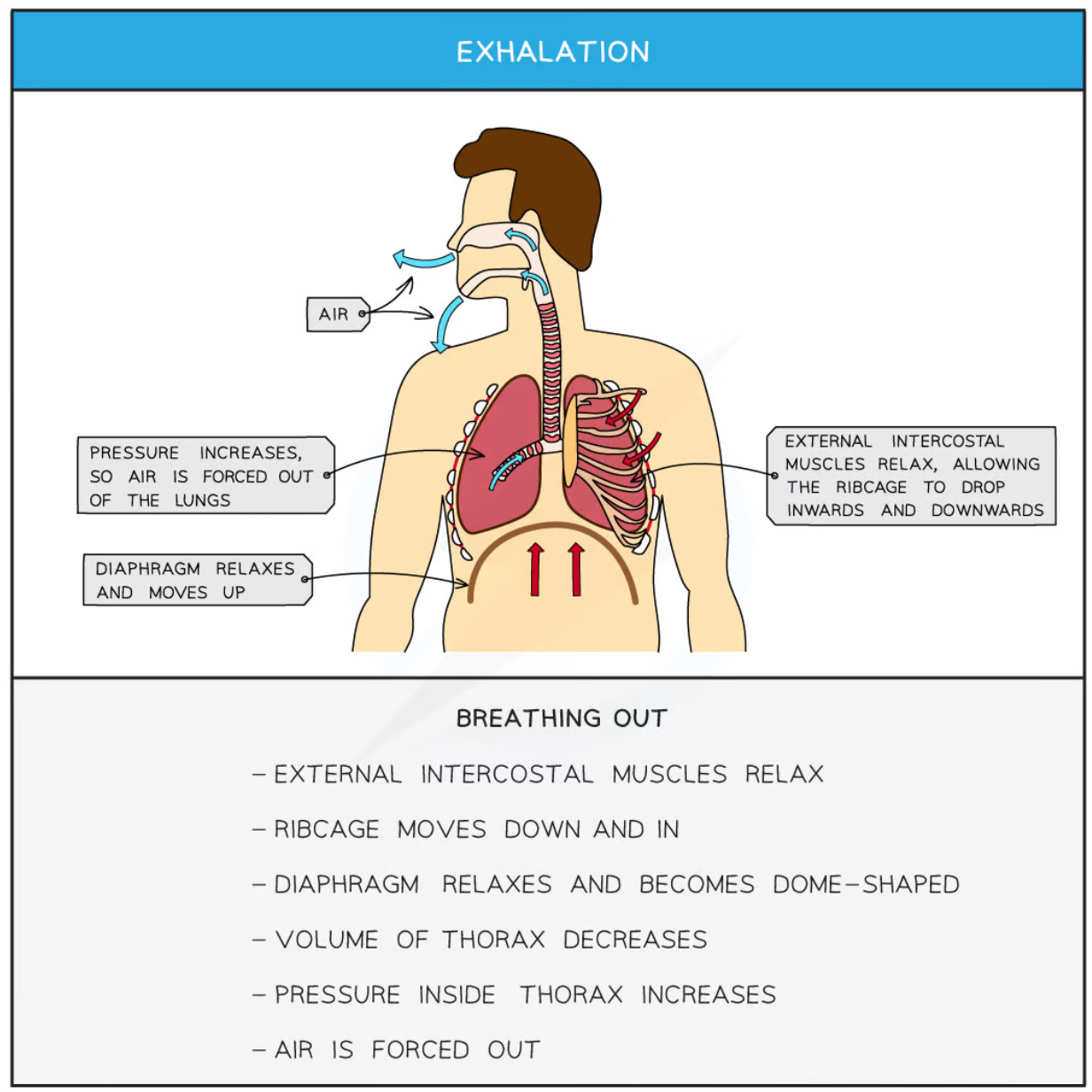
Describe the process of exhalation.
Describe the structure of fish gills.
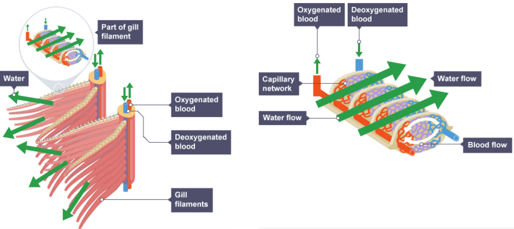
How are the gills adapted for gas exchange?
the large surface area of the gills
the large surface area of the blood capillaries in each gill filament
the short distance required for diffusion – the outer layer of the gill filaments and the capillary walls are just one cell thick
the efficient ventilation of the gills with water - there is a counter current flow of water and blood
Label the structures of the leaf, a plant organ.
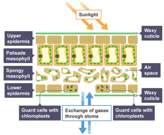
Explain the functions of different structures in a leaf.
waxy cuticle - transparent to let light through and waterproof to prevent water loss
upper epidermis - thin and transparent to allow light to enter palisade mesophyll layer underneath it
palisade mesophyll - column shaped cells tightly packed with chloroplasts to absorb more light, maximising photosynthesis
spongy mesophyll - contains internal air spaces that increases the surface area to volume ratio for the diffusion of gases, contains chloroplasts for photosynthesis, cells are rounder for larger surface area
lower epidermis - contains guard cells and stomata guard cell absorbs and loses water to open and close the stomata to gas diffusion, transparent to let light through
stomata - where gas exchange takes place: opens during the day, closes during the night. evaporation of water also takes place from here. in most plants, found in much greater concentration on the underside of the leaf to reduce water loss
vascular bundle - contains xylem and phloem to transport substances to and from the leaf
xylem - transports water into the leaf for mesophyll cells to use in photosynthesis and for transpiration from stomata
phloem - transports sucrose and amino acids around the plant the leaf
State the adaptations of a leaf.
- broad and flat leaf increases surface area for the diffusion of carbon dioxide and absorption of light for photosynthesis and decreases diffusion distance for gas exchange
- thin chlorophyll network of veins stomata allows carbon dioxide to diffuse to palisade mesophyll cells quickly absorbs light energy so that photosynthesis can take place allows the transport of water to the cells of the leaf and carbohydrates from the leaf for photosynthesis (water for photosynthesis, carbohydrates as a product of photosynthesis) allows carbon dioxide to diffuse into the leaf and oxygen to diffuse out
- epidermis is thin and transparent allows more light to reach the palisade cells thin cuticle made of wax to protect the leaf without blocking sunlight palisade cell layer at top of leaf maximises the absorption of light as it will hit chloroplasts in the cells directly
- spongy layer vascular bundles air spaces allow carbon dioxide to diffuse through the leaf, increasing the surface area thick cell walls of the tissue in the bundles help to support the stem and leaf
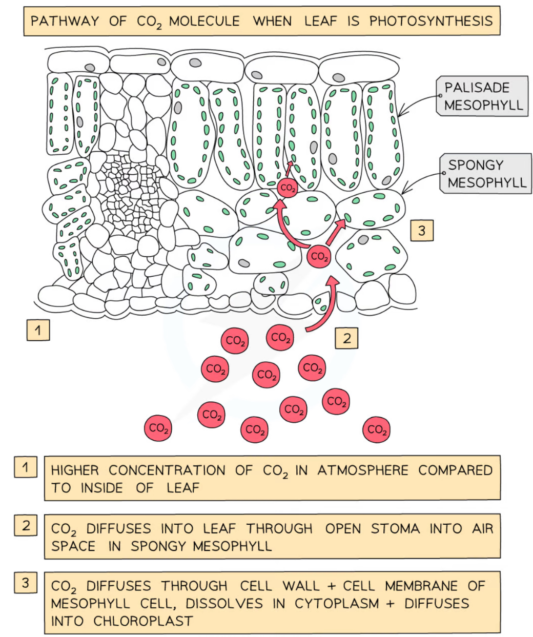
Explain the gas exchange occuring in the leaf.
How does the structure of meristem relate to its function?
Meristematic tissue has a number of defining features, including small cells, thin cell walls, large cell nuclei, absent or small vacuoles, and no intercellular spaces. The apical meristem (the growing tip) functions to trigger the growth of new cells in young seedlings at the tips of roots and shoots and forming buds.
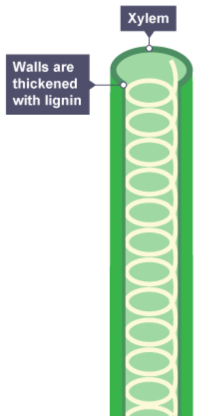
Explain the adaptations of xylem.
The xylem transports water and minerals from the roots up the plant stem and into the leaves.
In a mature flowering plant or tree, most of the cells that make up the xylem are specialised cells called vessels.
Lose their end walls so the xylem forms a continuous, hollow tube.
Become strengthened by a chemical called lignin. The cells are no longer alive. We call lignified cells wood. Lignin strengthens the plant to help it withstand the pressure of the water movement.
Explain the adaptations of phloem.
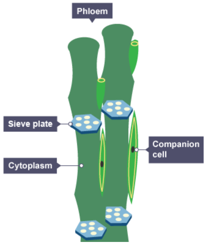
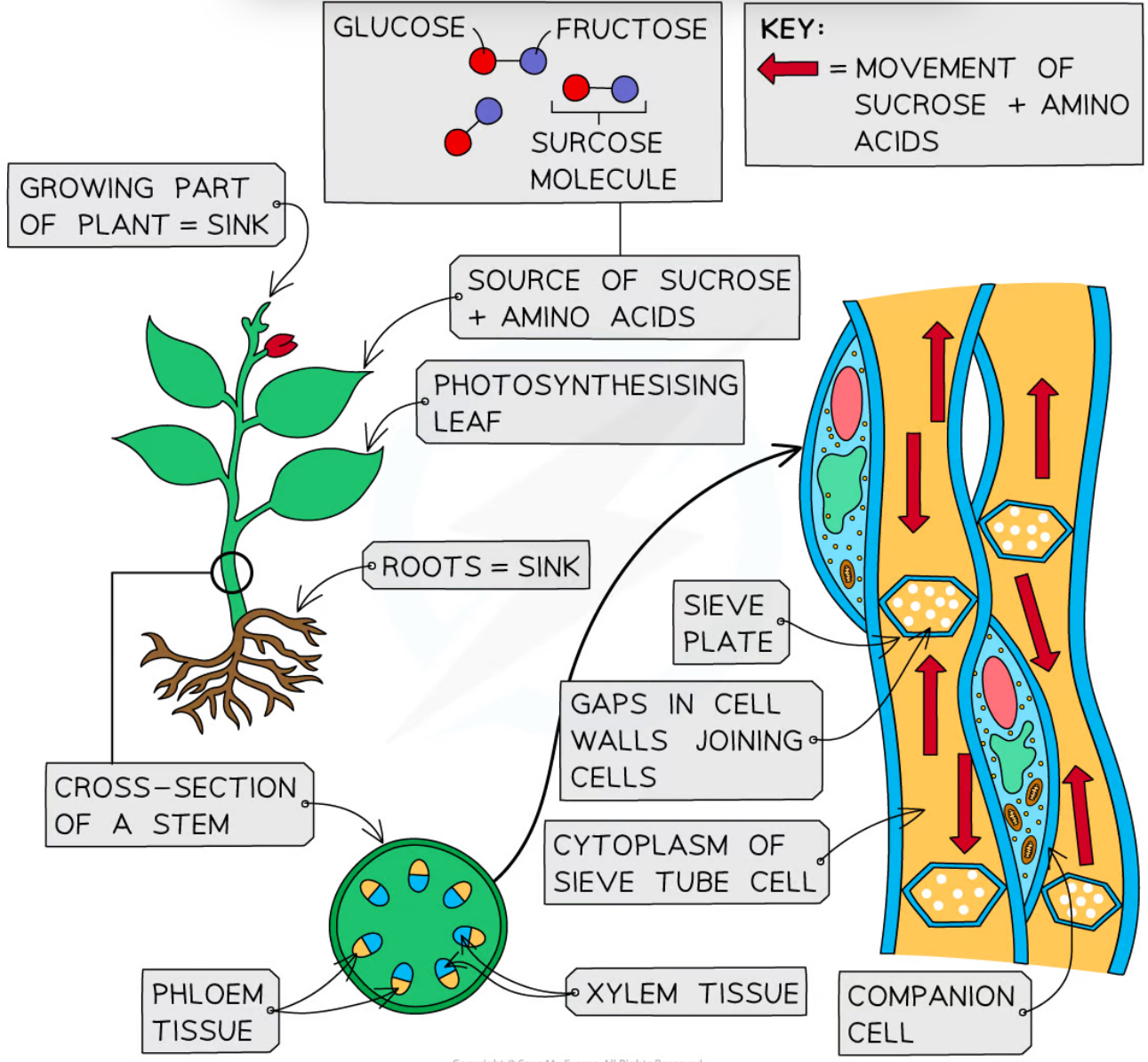
Sieve tubes - specialised for transport and have no nuclei. Each sieve tube has a perforated end so its cytoplasm connects one cell to the next.
Companion cells - transport of substances in the phloem requires energy. One or more companion cells attached to each sieve tube provide this energy. A sieve tube is completely dependent on its companion cell(s).
Compare the transport in the xylem and phloem.

Explain the pathway of water and ions through the plant.
Describe the process of transpiration.
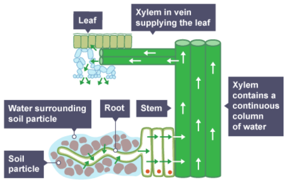
Transpiration is defined as the loss of water vapour from plant leaves by evaporation of water at the surfaces of the mesophyll cells followed by diffusion of water vapour through the stomata
Water travels up xylem from the roots into the leaves of the plant to replace the water that has been lost due to transpiration
Explain the effect of changing temperature on the rate of transpiration.
At higher temperatures, particles have more kinetic energy so transpiration occurs as a faster rate as water molecules evaporate from the mesophyll and diffuse away faster than at lower temperatures.
Explain the effect of changing humidity on the rate of transpiration.
Humidity is a measure of moisture (water vapour) in the air; when the air is saturated with water vapour the concentration gradient is weaker so less water is lost.
Explain the effect of changing air movement(e.g.?) on the rate of transpiration.
Good airflow (e.g. wind) removes water vapour from the air surrounding the leaf which sets up a concentration gradient between the leaf and the air, increasing water loss.
Explain the effect of changing light intensity on the rate of transpiration.
Increases the rate of photosynthesis; stomata open more so oxygen diffuses out of the leaf but water also diffuses out of the leaf. Guard cells are responsive to light intensity; when it is high they are turgid and the stomata open allowing water to be lost.
Describe a method to investigate water loss from plant leaves.
Various factors that affect water loss from the leaf can be investigated using this method, for instance:
air movement - direct a fan on the leaves
temperature
obstructing the stomata, eg with petroleum jelly
Method
Remove a number of leaves from a bush or tree.
Find the mass of each leaf.
Apply petroleum jelly on different sides of the leaf, e.g. neither(for control value), upper side, lower side, both sides.
Suspend each leaf from a piece of wire or string.
After a set period of time, re-measure the mass.
Describe a method to measure water uptake using potometers.
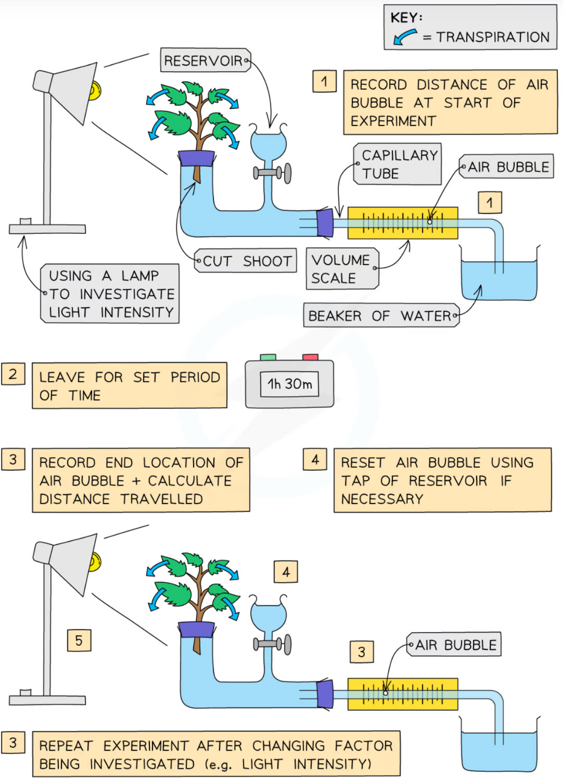
Cut a shoot underwater to prevent air entering the xylem and place in tube
Set up the apparatus as shown in the diagram and make sure it is airtight, using vaseline to seal any gaps
Dry the leaves of the shoot (wet leaves will affect the results)
Remove the capillary tube from the beaker of water to allow a single air bubble to form and place the tube back into the water
Set up the environmental factor you are investigating
Allow the plant to adapt to the new environment for 5 minutes
Record the starting location of the air bubble
Leave for a set period of time
Record the end location of the air bubble
Change the light intensity or wind speed or level of humidity or temperature (only one - whichever factor is being investigated)
Reset the bubble by opening the tap below the reservoir
Repeat the experiment
The further the bubble travels in the same time period, the faster transpiration is occurring and vice versa
Explain how this experiment can be adapted to investigate the effect of different factors on transpiration.
Airflow: Set up a fan or hairdryer
Humidity: Spray water in a plastic bag and wrap around the plant
Light intensity: Change the distance of a light source from the plant
Temperature: Temperature of room (cold room or warm room)
Describe the process of translocation.
The soluble products of photosynthesis is cell sap (sugars (mainly sucrose) and amino acids). This is transported around the plant in the phloem tubes which are made of living, elongated cells.
Phloem tissue transports dissolved sugars from the leaves to the rest of the plant for immediate use or storage.
The cells are joined end to end and contain pores in the end cell walls (called sieve plates) which allow easy flow of substances from one cell to the next.
The transport of sucrose and amino acids in phloem, from regions of production to regions of storage or use, is called translocation.
Transport in the phloem goes in many different directions depending on the stage of development of the plant or the time of year; however, dissolved food is always transported from source (where it’s made) to sink (where it’s stored or used):
During winter, when many plants have no leaves, the phloem tubes may transport dissolved sucrose and amino acids from the storage organs to other parts of the plant so that respiration can continue
During a growth period (eg. during the spring), the storage organs (eg roots) would be the source and the many growing areas of the plant would be the sinks
After the plant has grown (usually during the summer), the leaves are photosynthesizing and producing large quantities of sugars; so they become the source and the roots become the sinks – storing sucrose as starch until it is needed again
Define health.
Health is the state of physical and mental well-being.
What causes ill health?
Diseases, both communicable and non-communicable, are major causes of ill health. Other factors including diet, stress and life situations may have a profound effect on both physical and mental health.
How can different types of disease interact?
• Defects in the immune system mean that an individual is more likely to suffer from infectious diseases.
• Viruses living in cells can be the trigger for cancers.
• Immune reactions initially caused by a pathogen can trigger allergies such as skin rashes and asthma.
• Severe physical ill health can lead to depression and other mental illness.
discuss the human and financial cost of these non-communicable diseases to an individual, a local community, a nation or globally
What are risk factors?
Risk factors are linked to an increased rate of a disease; but exposure to a risk factor doesn’t guarantee that an individual will suffer a disease (a person who smokes regularly isn’t guaranteed to develop lung cancer but their risk compared to someone who doesn’t smoke is much, much higher)
Certain risk factors correlate with certain diseases (are related to them); but correlations are not always causations
Risk factors can be:
Aspects of a person’s lifestyle; such as the food they eat or whether or not they drink alcohol
Substances in the person’s body or environment; such as air pollution in a crowded city or asbestos in old buildings
Explain the effects of diet, smoking and exercise on cardiovascular disease.
Obesity leads to high blood pressure and the build-up of fatty deposits in the arteries, which lead to cardiovascular disease. It also increases the likelihood of developing diabetes, another risk factor cardiovascular disease.
Being obese - with deposits of lipids in the abdomen - increases blood pressure beyond normal levels and increases levels of blood lipids.
Smoking increases the risk of cardiovascular disease in several ways:
Smoking damages the lining of the arteries, including the coronaryarteries. The damage encourages the build-up of fatty material in the arteries. This can lead to a heart attack or a stroke.
Inhalation of carbon monoxide in cigarette smoke reduces the amount of oxygen that can be carried by the blood.
The nicotine in cigarette smoke increases the heart rate, putting strain on the heart.
Chemicals in cigarette smoke increase the likelihood of the blood clotting, resulting in a heart attack or stroke.
Explain obesity as a risk factor for Type 2 diabetes.
Body fat also affects the body's ability to use insulin.
Type 2 diabetes is where the body's cells lose their sensitivity to insulin - they no longer respond, or respond less effectively, to the insulin that's produced.
Obesity accounts for 80 to 85% of the risk of type 2 diabetes. Rising obesity is linked with 'western diet' - a diet that includes energy-rich 'fast foods' and an inactive lifestyle.
Explain the effect of alcohol on liver function.
Drinking excess alcohol can damage the liver, the organ responsible for processing and breaking down alcohol.
The liver can regenerate its cells, but long-term alcohol abuse causes serious damage:
the patient begins by feeling sick, experiences weight loss, loss of appetite, there is a yellowing of the eyes, confusion, drowsiness and vomiting blood
alcohol causes lipids to build up in the liver - fatty liver disease
alcohol damage leads to alcoholic hepatitis, which can lead to death
cirrhosis of the liver can develop – the liver becomes scarred and loses its ability to function
changes are now irreversible and the reduced ability to process alcohol can also lead to brain damage
Explain the effect of alcohol on brain function.
Alcohol affects the brain in several ways, it:
slows reaction time
causes difficulty walking
can impair memory
causes slurred speech
causes changes in sleep patterns and mood, including increased anxiety and depression
Longer term drinking of excess alcohol:
causes brain shrinkage
leads to memory problems
leads to psychiatric problems
may result in the patient requiring long-term care
Explain the effect of smoking on lung disease.
Smoking can result in many lung diseases.
A person may develop COPD – chronic obstructive pulmonary disease. This condition includes the diseases chronic bronchitis and emphysema. In COPD:
smoking damages the bronchioles and can eventually destroy many of the alveoli in the lungs
the airways become inflamed and mucus, which normally traps particles in the lungs, builds up
the patient becomes breathless, and finds it more and more difficult to obtain the oxygen required for respiration
The damage caused by COPD is permanent. The disease cannot be cured, and can result in death. It is essential that the person seeks medical help to try to prevent progression of the disease.
Explain the effect of smoking on lung cancer.
The carcinogens in cigarette smoke also cause lung cancer. Almost all cases of lung cancer are caused by smoking – smaller numbers of cases are linked with air pollution and ionising radiation from radon gas, a radioactiveelement found in the environment in some parts of the country.
The vast majority of cases of lung cancer lead to death.
Explain the effects of smoking on unborn babies.
For mothers who smoke during pregnancy:
smoking increases the risk of miscarriage
the babies and children are more likely to suffer from respiratory infections and an increased risk of asthma
the long-term physical growth and intellectual development of the baby/child is affected
there is an increased risk of birth defects
the birthweight of the baby is reduced
Explain the effects of alcohol on unborn babies.
Alcohol can lead to a variety of physical, developmental and behavioural effects on the fetus. The most serious is foetal alcohol syndrome – the fetus:
is smaller in size
has a smaller brain with fewer neurones
will have long-term learning and behavioural difficulties
has distinct facial features
Explain carcinogens, including ionising radiation, as risk factors in cancer.
There are genetic factors that increase the likelihood of developing some cancers.
Chemicals and other agents that can cause cancer are called carcinogens.
Carcinogens cause cancer by damaging DNA. Carcinogens cause mutations to occur. A single mutation will not cause cancer – several are required. For this reason, we are more likely to develop cancer as we get older.
Define cancer.
Cancer is the result of changes in cells that lead to uncontrolled growth and division
Cells grow then divide by mitosis only when we need new ones – when we’re growing or need to replace old or damaged cells.
When a cell becomes cancerous, it begins to grow and divide uncontrollably. New cells are produced – even if the body does not need them.
A group of cancerous cells produces a growth called a tumour.
Explain the two different types of tumours.
Benign tumours are slow growths of abnormal cells which are contained in one area, usually within a membrane. They do not invade other parts of the body.
Malignant tumour cells are cancers. They invade neighbouring tissues quickly and spread to different parts of the body in the blood where they form secondary tumours.
What are the risk factors for cancer?
Scientists have identified lifestyle risk factors for various types of cancer. There are also genetic risk factors for some cancers.