Intro to cross sectional, Ultrasound, Nuc Med
 Call Kai
 Learn
Learn Practice Test
 Spaced Repetition
 Match
Match Flashcards
 Knowt Play
1/54

There's no tags or description
Looks like no tags are added yet.
Name | Mastery | Learn | Test | Matching | Spaced | Call with Kai | Chat |
|---|
No analytics yet
Send a link to your students to track their progress
55 Terms
ultrasound
Uses high-frequency (3-20 mHz; megahertz) sound waves with a transducer generated at 20,000 vibrations per second (hertz/second)
Reflected soundwaves echoes are recorded and shown as an image in real time
Transducer transmits inaudible soundwaves into the body, as it echoes from the body’s fluid and tissues, transducer records the strength and character of the waves, then displayed
3-20mHz
ultrasound frequency
20,000
transducer vibrations
attenuation coefficient
How much of the sound wave will get absorbed
How much passes through
How much bounces back
piezoelectric crystal
Converts the electricity into sound waves- and then echoes into electricity
functions of the transducer
generates sound waves
detects reflected sound waves
types of transducers
curvilinear
linear
endocavity, transvaginal, transrectal
intraoperative probe
curvilinear
Low frequency 5mHz
Abdominal, obstetrics, pelvis
linear
High frequency 15mHz
Great detail
Superficial structures
Thyroid, scrotal, musculoskeletal, soft tissue masses
curvilinear
transducer type?

linear
transducer type?

endocavity
transvaginal
transrectal
8mHz
intraoperative probe
Very high frequency
Directly on organ or vessel
Liver transplants, vascular
endocavity, transvaginal, transrectal
transducer type?

intraoperative probe
transducer type?

sagittal (long)
transverse
imaging planes
echogenicity
describes the tissue in relation to another tissue, or as compared to its normal state
benefits of ultrasound
No radiation, or harmful effects to patient
Low cost
Live imaging
Virtually no pain to patient
Quick results
Quick test time
No (measurable) weight limit
Portable
doppler ultrasound
Evaluates blood flow of major arteries and veins in the extremities - arms/legs
hyperechoic
white on image hy
hypoechoic
dark on image
anechoic
no color on image
indications of doppler ultrasounds
Arteriosclerosis of the arms or legs
Blood clot (deep vein thrombosis)
Venous insufficiency
Evaluate injury to the arteries
Monitor of arterial reconstruction and bypass grafts
carotid ultrasound
Evaluates how well blood is flowing through the carotid arteries
indications of carotid ultrasound
Blood clotting (thrombosis)
Narrowing in the arteries (stenosis)
Blood flow; other causes of blockage in the carotid arteries (surgery related complications)
baby hip ultrasounds
Evaluation for Developmental Dysplasia of the Hip (DDH)
why babies get hip ultrasounds
Typically performed if baby is breech or Hx of DDH in family
cranial ultrasound (neonatal)
hydrocephalus (enlargement of the ventricles)
detect bleeding within the brain tissue or the ventricles
assess for damage to the white matter brain tissue surrounding the edges of the ventricles during difficult birthing process
evaluate for congenital abnormalities
locate the site of an infection or tumor.
physical relationship among structures
3-d concept
evaluation = soft tissue structures
what does cross sectional anatomy show
advantages of cross sectional anatomy
eliminates overlapping structures
helps to differentiate between adjacent structures
offers view of anatomy in a variety of planes
sagittal
1
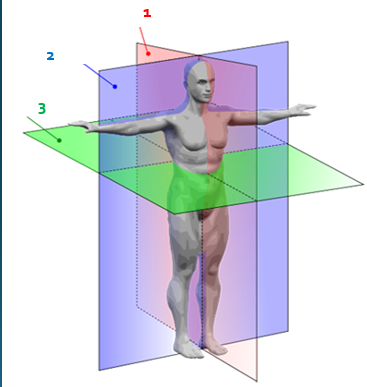
coronal
2

axial
3
mm to cm
slice thickness can vary from?
AP
how are images oriented?
CT
Generates a three-dimensional image of the body from a large series of two-dimensional X-ray images taken around a single axis of rotation
AP and lateral
what scout images are taken to designate where the slices will be?
iodine based and barium sulfate
what contrast is used in CT?
advantages of CT
Visualization of anatomy without superimposition of structures
Axial imaging
Computerized mathematical reconstruction of digital numbers
Ability to differentiate small differences in density of anatomic structures and abnormalities
2 tissues with similar densities distinguished
CT indications
Diagnose muscle and bone disorders
Detect internal injuries and internal bleeding
Pinpoint the location of a tumor, infection or blood clot
Guide procedures such as surgery, biopsy and radiation therapy
Detect and monitor diseases and conditions
MRI
Utilizes a powerful magnetic field to align the magnetization of some atoms in the body
Magnetic properties of hydrogen atoms
Human body = 80% hydrogen
1973
when was MRI discovered?
coil
what is placed around the area of interest in MRI to obtain the info of that specific area of anatomy
the magnet aligns the single protons in the hydrogen nuclei within the field
T1 weighted MRI images
Fat appears bright
Water appears dark
Best for anatomy
T2 weighted MRI images
Water appears bright (white)
Fat appears dark
Best for pathology
gadolinium
what contrast is used in MRI?
Less adverse reactions and eliminated through kidneys easier
why is gadolinium used in MRI?
advantages of MRI
No exposure to ionizing radiation
Superior contrast resolution
Superior tissue characterization
Multi-planar capabilities
Possibly safer intravenous contrast agents
Visualization of angiography without using contrast
MRI indications
A noninvasive way to examine organs, tissues and the skeletal system by producing high-resolution images that help diagnose a variety of problems
contraindication of CT
high dose
extraversion of veins
allergic reaction
claustrophobia
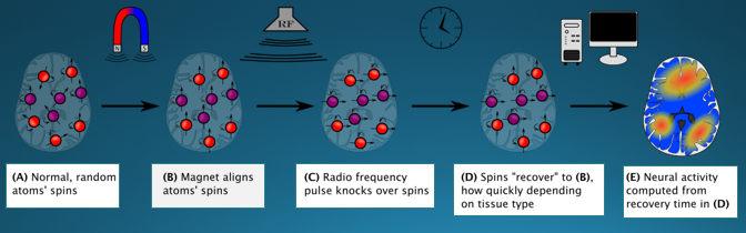
how MRI works
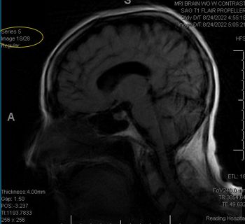
axial
what plane?

coronal
what plane?

sagittal
what plane?
contraindications of MRI
take longer
claustrophobia
contrast involved
magnet → can affect pacemakers/ defibrillators, metal pieces in body
narrow bore → larger patients may not fit