Exam 2 - new material
 Call Kai
 Learn
Learn Practice Test
 Spaced Repetition
 Match
Match Flashcards
 Knowt Play
1/141

There's no tags or description
Looks like no tags are added yet.
Name | Mastery | Learn | Test | Matching | Spaced | Call with Kai | Chat |
|---|
No analytics yet
Send a link to your students to track their progress
142 Terms
Causes of sacroiliitis that are positive HLA-B27
(1) Ankylosing Spondylitis (AS)
(2) Enteropathic
(3) Psoriatic
(4) Reactive Arthritis
Causes of sacroiliitis that are HLA-B27 negative
(1) HPT (calcium levels)
(2) Gout (uric acid levels)
(3) DJD
(4) Infection
(5) Trauma
(6) Pregnancy
(7) DISH
(8) OCI
Which two seronegative spondyloarthropathies primarily affect the axial skeleton? Which two affect the appendicular skeleton?
Axial = AS and enteropathic arthritis
Appendicular = psoriatic and reactive arthritis
PEAR (keep the first two together and then the second two together)
Which is less common: AS or Enteropathic Arthritis?
Enteropathic Arthritis
Enteropathic Arthropathies
Group of rheumatologic conditions that share a link to gastrointestinal pathology
Commonly associated with inflammatory bowel disease (IBD)
What infections are commonly linked to enteropathic arthritis?
Bacterial: shigella, salmonella, campylobacter, yersinia, clostridium difficile
Parasitic: Giardia lamblia, Ascaris lumbricoides, Cryptosporidium species
Enteropathic Arthritis: etiology
UNKNOWN
Inflammation in the GI tract may increase permeability, resulting in absorption of antigenic material, which may then localize to musculoskeletal tissues and elicit an inflammatory response
Alternate idea: autoimmune response through molecular mimicry
Additional clinical symptoms with enteropathic arthritis
— malaise
— anorexia
— weight loss
Arthropathy is self-limiting, but symptoms worsen with flare-ups of bowel disease
Enteropathic Arthritis: axial skeleton clinical presentation
Insidious onset of low back pain, especially in younger persons
— morning stiffness
— exacerbated by prolonged sitting or standing
— improved by moderate activity
— independent of GI symptoms
Is enteropathic arthritis more common with Crohn Disease or ulcerative colitis?
More common in Crohn Disease than ulcerative colitis
Enteropathic Arthritis: peripheral skeleton
Nondeforming and nonerosive
May precede intestinal involvement, but usually concomitant or subsequent to bowel disease
Common locations: heel (insertion of Achilles tendon and plantar fascia) and knee (tibial tuberosity and patella)
Enteropathic Arhtritis Management
Focused on the bowel disease with anti-inflammatory medications for arthropathy
Bowel resection may halt the peripheral arthropathy, but not axial arthropathy
Features of Enteropathic Arthritis
Looks the same as Ankylosing spondylitis! — same features!
Psoriatic and Reactive Arthritis Features
— soft tissue edema
— asymmetrical small joints (mostly DIP)
— peripheral erosions with periostitis ("mouse ears")
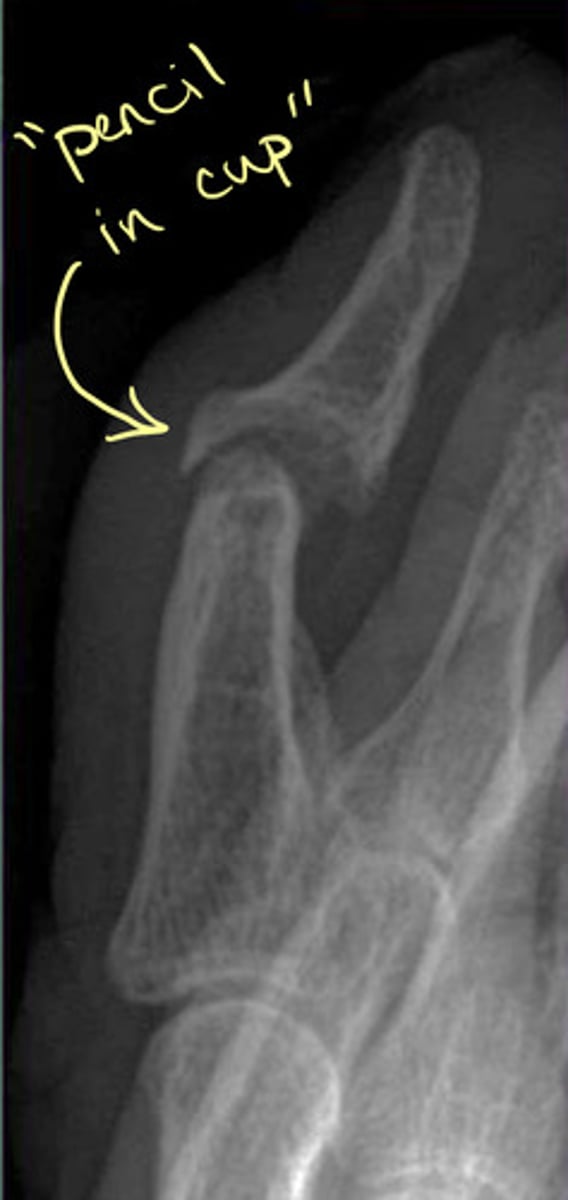
— central erosions ("pencil in cup")
— ankylosis and deformity
— normal bone density
— unilateral or bilateral AND Asymmetrical SI erosions and/or sclerosis
— unilateral or bilateral SI fusion
— syndesmophytes (asymmetrical, bulky, non-marginal, paravertebral)
Remember: primarily appendicular skeleton
Psoriatic Arthritis
Chronic inflammatory arthritis that develops in at least 5% of patients with dermatological psoriasis
Chronic disease of the joints and the entheses
Risk factors of Psoriatic Arthritis
— nail, inverse (immune-mediated disease that causes a rash in areas where skin rubs against itself), and scalp psoriasis
— family history
— severity of skin disease
Psoriasis precedes the onset of psoriatic arthritis in what percent of patients?
60-80%
Pathogenesis of psoriatic arthrits?
— genetics
— environmental factors
— immune-mediated inflammation
Prognosis of Psoriatic Arhtritis
As many as 40% develop erosive and deforming arthritis
Possible increased risk of: hypertension, obesity, hyperlipidemia, Type II diabetes, and cardivascular events
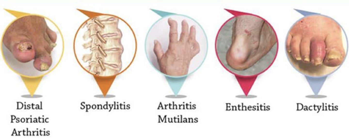
Types of Psoriatic Arthritis
(1) Distal Psoriatic Arthritis
(2) Spondylitis
(3) Arthritis Multilans
(4) Enthesis
(5) Dactylitis
Age of onset of psoriatic arthritis
30-50 years
Psoriatic arthritis mainly targets which joints?
Hands
Less common in feet
Does psoriatic arthritis commonly have a symmetrical distribution?
No >> asymmetrical
Proliferative synovitis with psoriatic arthritis results in what?
Pannus
Main features of Psoriatic Arthritis
— soft tissue edema
— asymmetrical small joints (mostly DIP)
— peripheral erosions with periostitis ("mouse ears")
— central erosions ("pencil in cup")
— ankylosis and deformity
— normal bone density*
— unilateral or bilateral and Asymetrical SI erosions and/or sclerosis
— unilateral or bilateral SI fusion
— syndesmophytes (asymmetrical, bulky, non-marginal, and paravertebral)
Extremity Findings of Psoriatic Arthritis: similar features as RA
— joint space narrowing
— soft tissue edema
— marginal erosions ("bare area") erosions
— ankylosis and deformity
Extremity Findings of Psoriatic Arthritis: distinctive features from RA
— mostly DIP (acro-osteolysis possible)
— normal bone density
— periostitis ("mouse ears")
— central erosions ("pencil in cup" appearance)
— less symmetrical within a joint and throughout the body (distribution may involve all joints of one digit - "ray pattern")
What is a key bone change that can be seen in Psoriatic arthritis but NOT in RA?
New bone formation
Occurs at the periarticular area owing to enthesitis
The surface of bone erosion and proliferation in psoriatic arthritis may appear "fluffy"
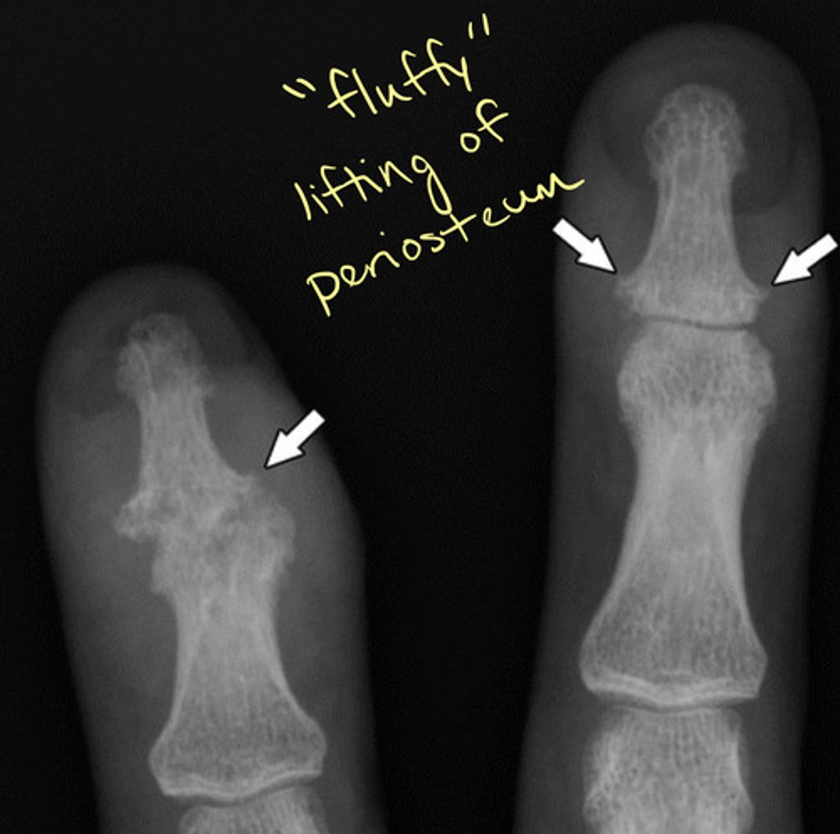
Psoriatic arthritis at the DIP joint of the hand
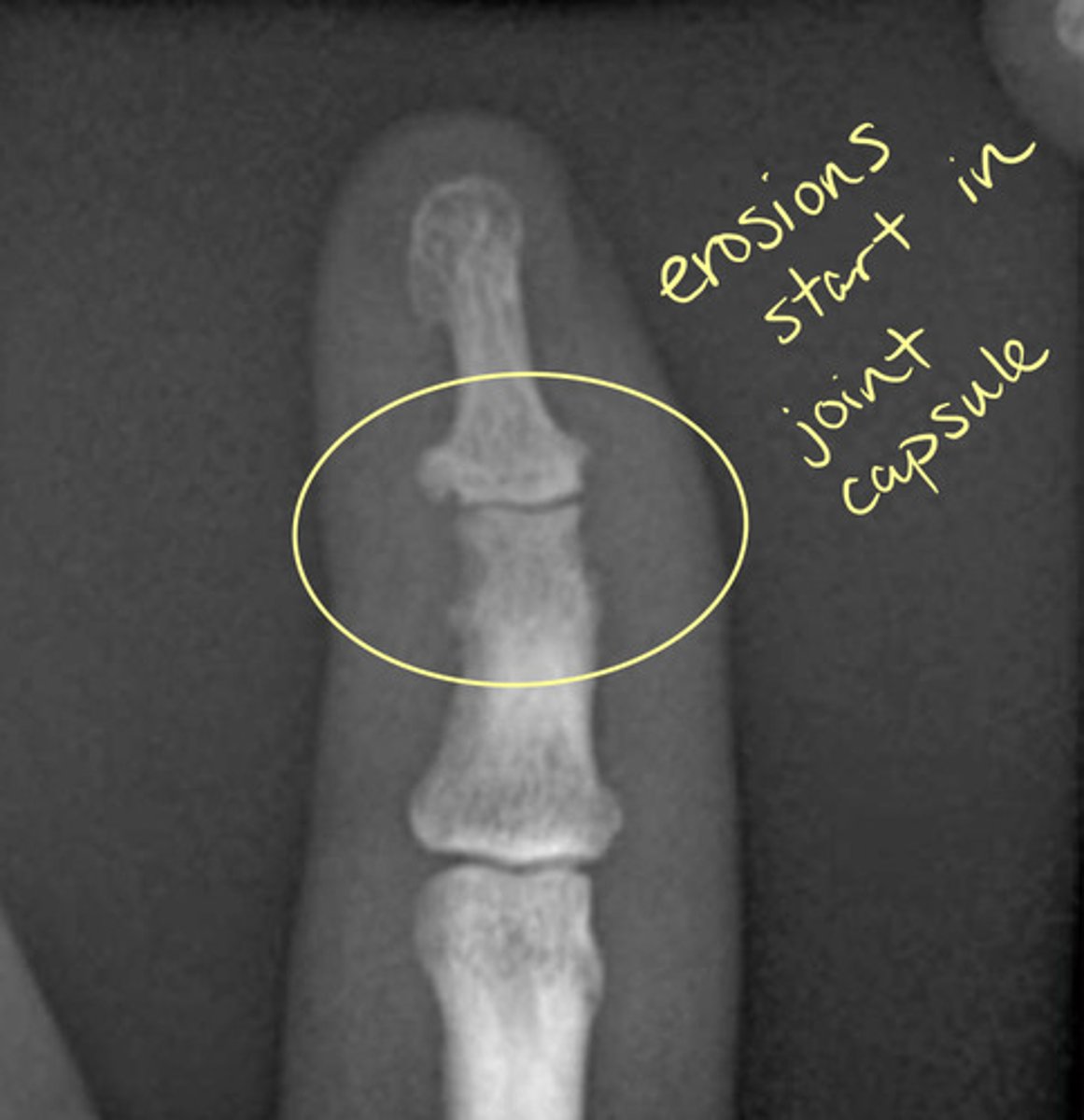
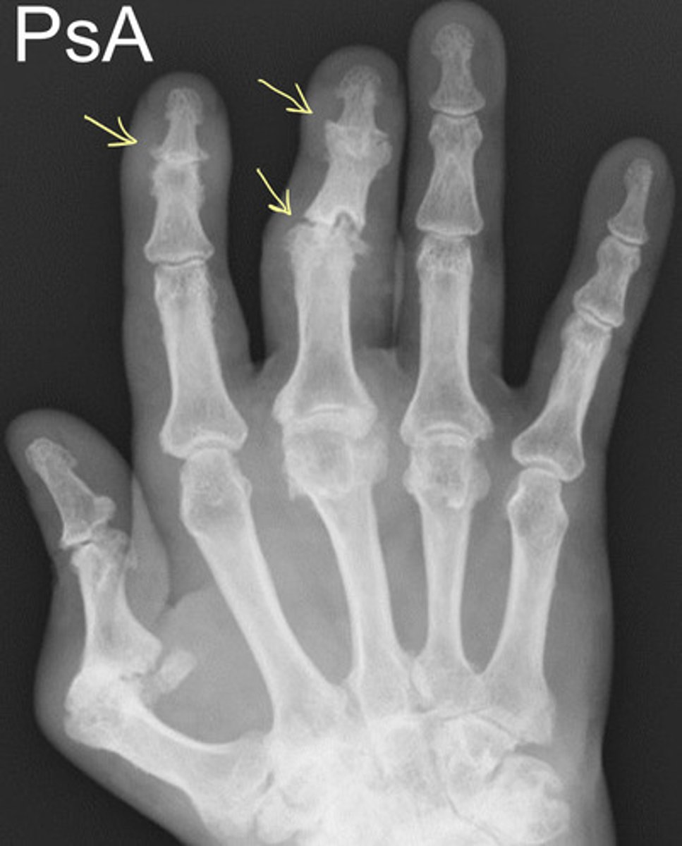
Psoriatic Arthritis in the hand with joint deformity
"Pencil in cup" deformity with Psoriatic Arthritis
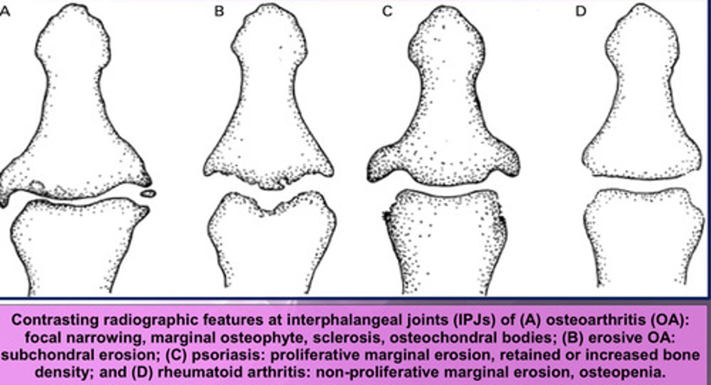
Drawing Comparison between different types of arthritis in the hand
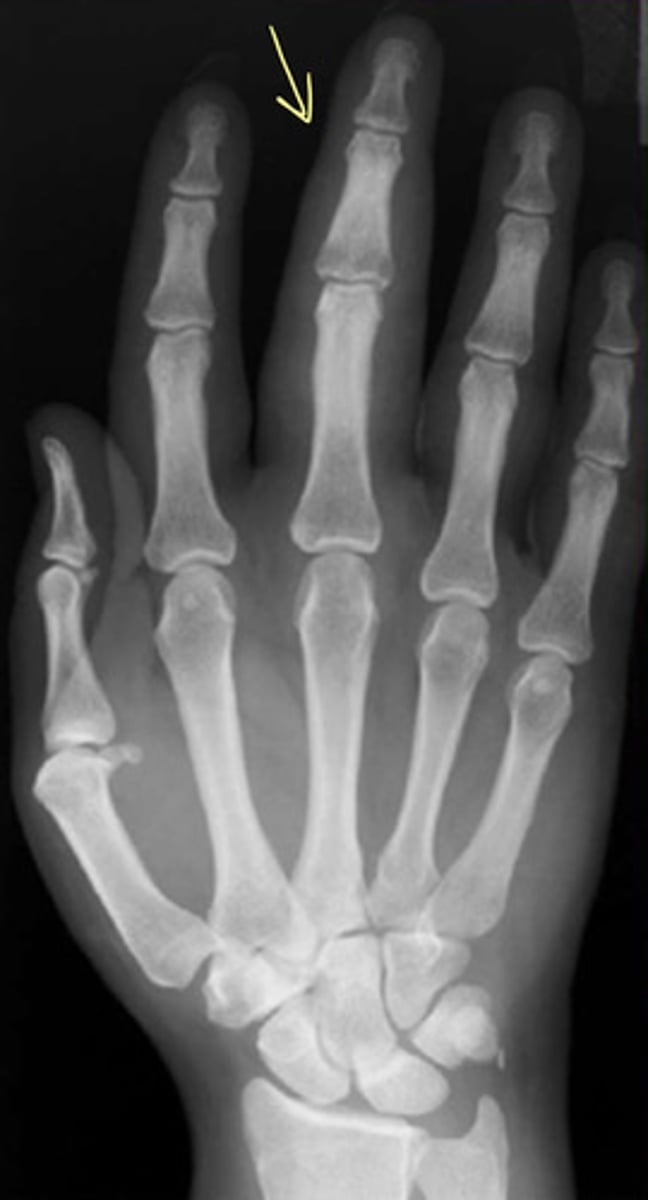
"Sausage Digit" from Psoriatic Arthritis
How is psoriatic arthritis in the spine similar to ankylosing spondylitis?
— erosions
— sclerosis
— possible ankylosis
— syndesmophytes (but different appearance)
How is psoriatic arthritis in the spine different from ankylosing spondylitits?
— unilateral or bilateral
— asymmetrical
— thick, bulky nonmarginal (don't come from just the endplate) syndesmophytes
Syndesmophytes in psoriatic arthritis of the spine
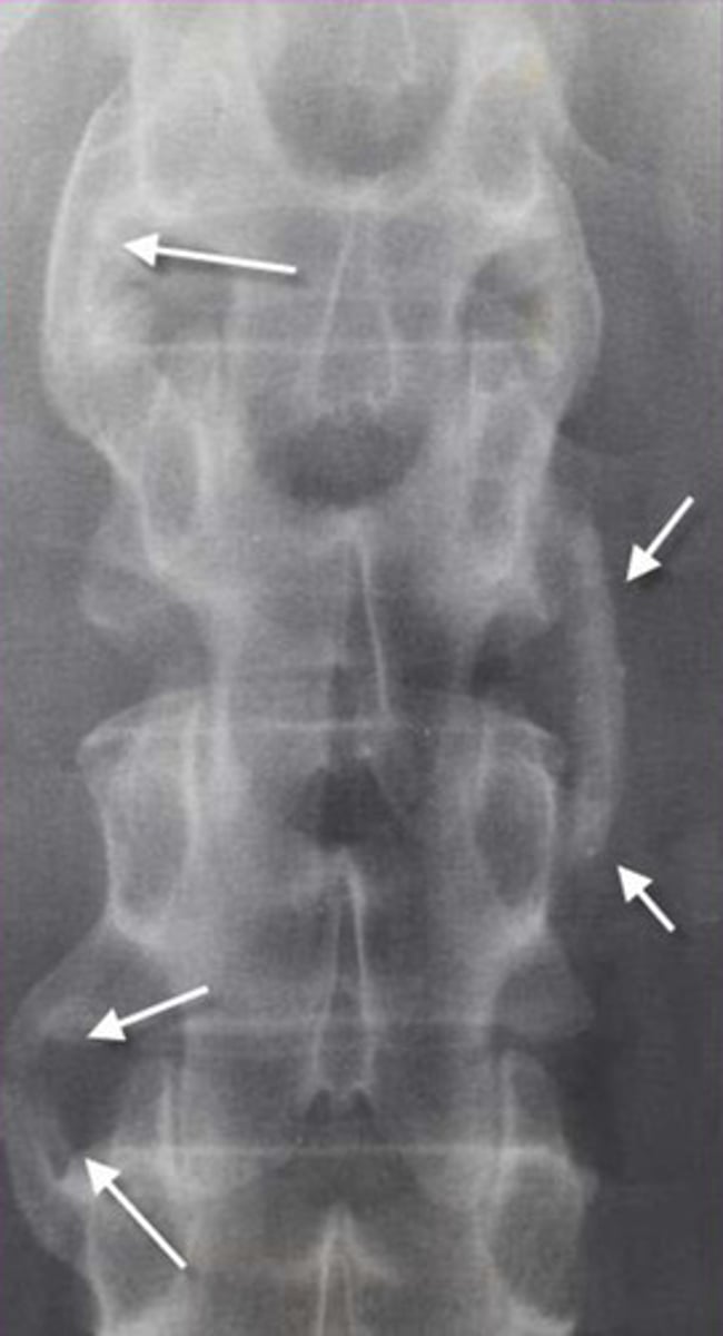
Psoriatic Arthritis on the SI Joints
Similar to AS, indistinct articulations
BUT...AP view identifies thick, nonmarginal syndesmophytes
For psoriatic arthritis, SI joint is usually not the first sign, and it is usually asymmetrical
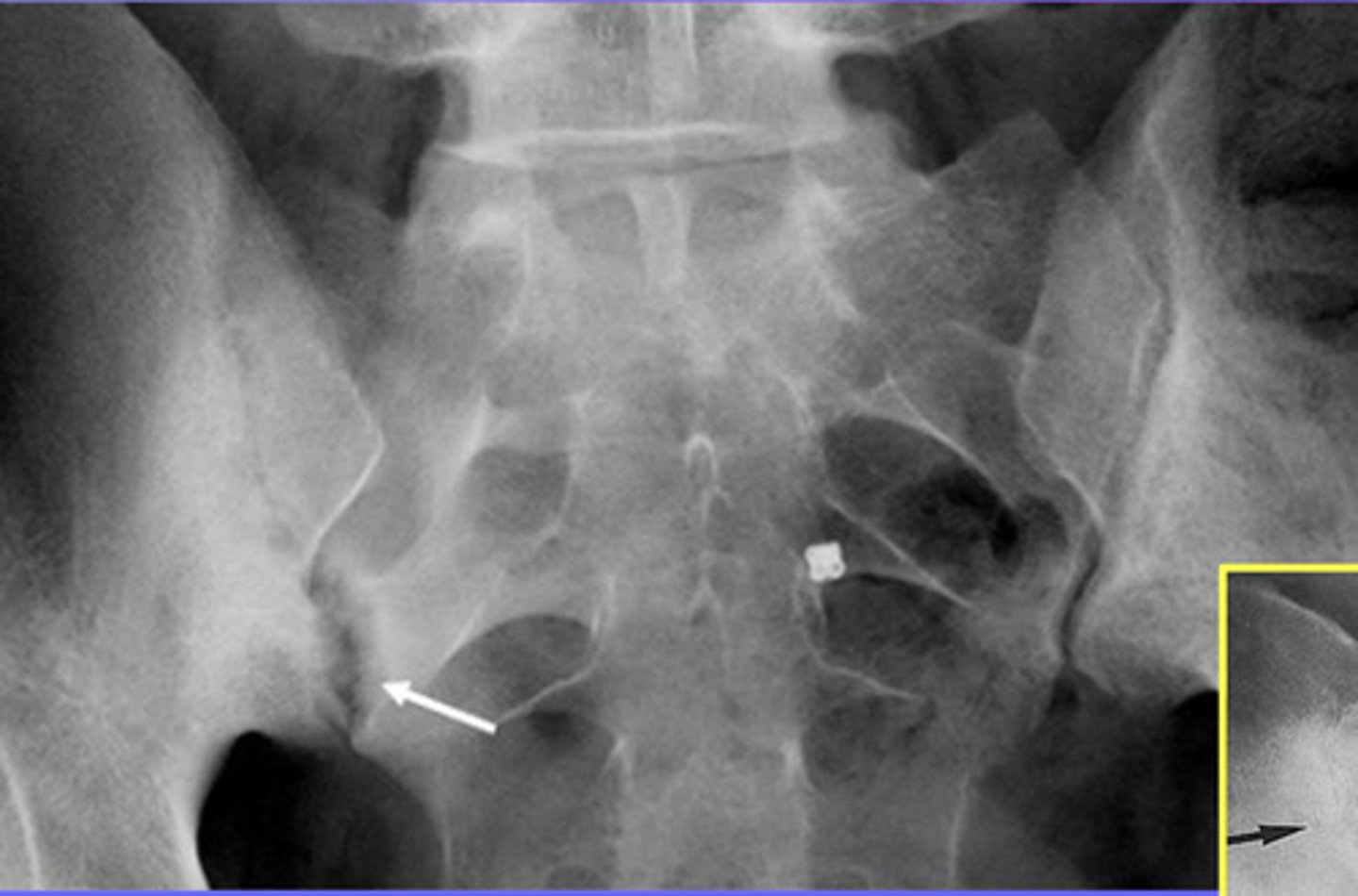
Reactive Arthritis Etiology
Autoimmune condition
Usually caused by GI or GU infections (most commonly Shigella, Salmonella, Campylobacter, Chlamydia trachomatis)
What is the classic triad of reactive arthritis?
(1) Noninfectious urethritis
(2) Arthritis
(3) Conjunctivitis
"Can't see, can't pee, and can't dance with me"
Other S/S of reactive arthritis?
— malaise, fever, fatigue, myalgia
— diarrhea, sometimes abdominal pain
— asymmetrical oligoarthritis (MC lower extremity)
— about 50% have LBP
Reactive Arthritis Onset
Usually develops 2-4 weeks after GU or GI infection (about 10% have no preceding symptomatic infection
Classic triad is seen in only about 30% BUT has a specificity of about 99%
Physical Examination Findings of Reactive Arthritis
— asymmetric oligoarthritis (predominately LE)
— dactylitis ("sausage digits")
— toe or heel pain and/or other enthesitis
— cervicitis or urethritis
— conjunctivitis or iritis
— genital ulcerations
— skin lesions
Common locations for reactive arthrtitis
Weight-bearing joints
— Asymmetrical distribution in the knees, hips, and ankles
— Shoulders, wrists, and elbows can be affected
— Small joints of fingers and toes usually in severe cases
Joints are tender, warm, edematous, and sometimes red
Imaging Findings of Reactive Arthritis
Enthesopathy
— achilles insertion is most common site
— ischial tuberosities, iliac crest, tibial tuberosities, and ribs
Sacroilitis also can occur
Reactive Arthritis Management
No curative treatment exists
— Treatment aimed at relieving symptoms
— Includes corticosteroids, NSAIDs, antibiotics, and disease-modifying antirheumatic drugs (DMARDs)
About 2/3 are self-limiting BUT approximately 30% develop chronic symptoms
Reactive Arthritis Complications
— recurrent arthritis
— chronic arthritis or sacroiliitis
— ankylosing spondyliyis
Reactive Arthritis: similarities of Psoriatic Arthritis at the extremity
— joint space narrowing
— soft tissue edema
— normal bone density
— periostitis ("mouse ears")
— less symmetrical within a joint and throughout the body
Reactive Arthritis: distinctions from psoriatic arthritis at the extremity
— Reactive arthritis is more common in the lower extremity vs psoriasis is more common in the upper extremity
— Lack of nail pitting and psoriatic scaling
— Central erosions ("pencil in cup") are less common
— ankylosis and deformity are less common
Reactive Arthritis: SI similarities to psoriatic arthritis
— thick, bulky nonmarginal syndesmophytes
— unilateral or bilateral
— asymmetrical
Reactive Arthritis: SI distinctions from psoriatic arthritis
Nearly identical!
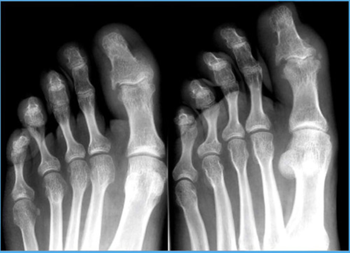
Reactive Arthritis of the foot
Normal bone density, but shaggy periosteum
What are the seropositive inflammatory joint diseases?
— Rheumatoid arthritis
— Systemic Lupus Erythematosis (SLE)
— Jaccoud Arthropathy
— Scleroderma
RA diagnosis
Diagnosis is clinical, serological, and radiological
— requires the presence of several criteria
— signs and symptoms usually begin as insidious and may be proceeded by emotional or physical stress
What are the most common articular complaints of RA?
— pain
— tenderness
— swelling
— stiffness especially in the morning ("jelling phenomenon")
Most common symptoms begin in the PIP and MCP and proceed proximally
What are some other possible clinical abnormalities with RA?
— Raynaud
— osteopenia (joint capsule)
— cervical spine subluxations/dislocations
— vasculitis with skin ulcerations
— ocular complications
— lacrimal gland atrophy leading to dry eyes
What is the checklist for Rheumatoid Arthritis Diagnosis? How many do you need for diagnosis?
(1) Morning stiffness for at least 6 weeks
(2) Pain on joint motion for at least 6 weeks
(3) Swelling of at least 1 joint for at least 6 weeks
(4) Swelling in at least 1 other joint for more than 6 weeks
(5) Bilateral symmetrical joint swelling
(6) Subcutaneous nodules
(7) Radiographic changes
(8) Laboratory
Need 4 or more to qualify for RA!
RA prognosis
Most patients will experience periods of remission and exacerbation with gradual deformity and disability
Minority of patients will experience complete remission
Poor prognosis is associated with subcutaneous nodules, high titer levels of RH factor, sustained disease for one year or more, onset before 30, and extra-articular manifestations
What is the common distribution of RA at the hands/wrist?
— MCP
— PIP
— ulnar aspect of the wrist
Rheumatoid Arthritis (RA)
Generalized multi-system connective tissue autoimmune disease involving synovial tissue and resulting in polyarticular joint inflammation
Antibodies directed against Fc fragments of IgG = Rheumatoid Factors (RFs)
Usually composed of IgM and are used as a marker for rheumatoid arthritis and other autoimmune conditions
Rheumatoid Factor (RF) is positive in approximately how many of those diagnosed with RA?
75%
Note: 5% of the general population is RF positive without RA diagnosis
Higher RF titers indicate what?
Poorer prognosis
Tends to have a more severe disease
What is the most common inflammatory arthritis? Who does this usually affect?
RA
Age 20-60 = F > M (3:1)
Age >60 = F=M
RA targets what?
Synovial tissue
Particularly of the hands, feet, and cervical spine
Other systems: heart, lungs, small blood vessels, nervous system, eyes, and reticuloendothelial system
RA is marked by what?
Inflammatory hyperplastic synovitis = synovial tissue proliferation = pannus
Pannus = late, inactive and irreversible manifestation of RA
How does pannus affect the bones?
Proliferating pannus erodes and narrows the cartilage as the pannus releases chondrolytic collagenase enzymes and interferes with nutrition (to the region/joint)
Proliferating synovium results in marginal erosions in the bare area = window for pannus to intrude into subchondral region, which produces cysts
Eventually the entire joint is filled with proliferating panus and undergoes progressive fibrosis and eventually ankylosis
What is arthritis mutilans?
Severe joint deformity/destruction
What is a Baker's cyst?
Enlargement of the gastrocnemius bursa
What is Felty's Syndrome?
Leukopenia, splenomegaly, and RA together
What are Haygarth's Nodes?
Soft tissue swelling at the MCP
What is the jelling phenomenon?
Stiff joints after inactivity
What is a rheumatoid nodule?
Accumulation of inflammatory cells with necrotic area and fibrosis seen on extensor surfaces
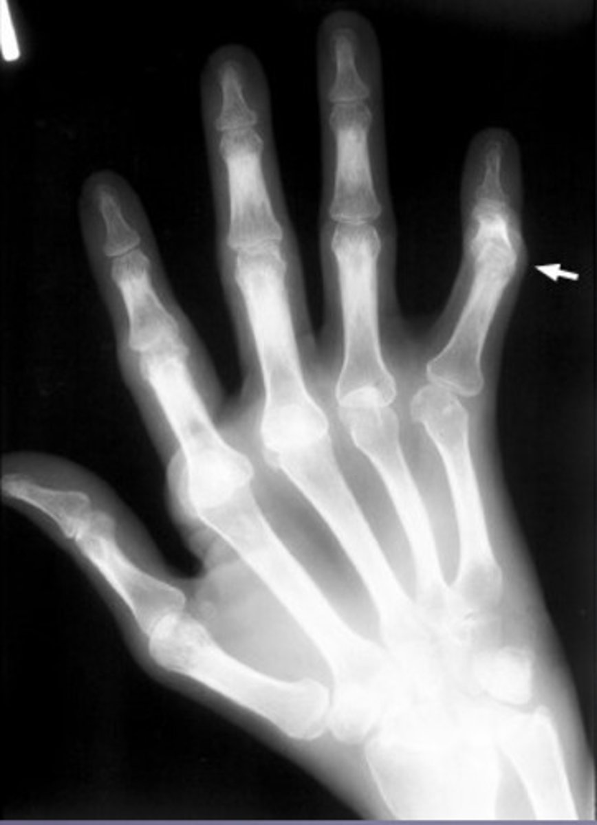
What is Boutonniere deformity?
PIP flexion and DIP extension
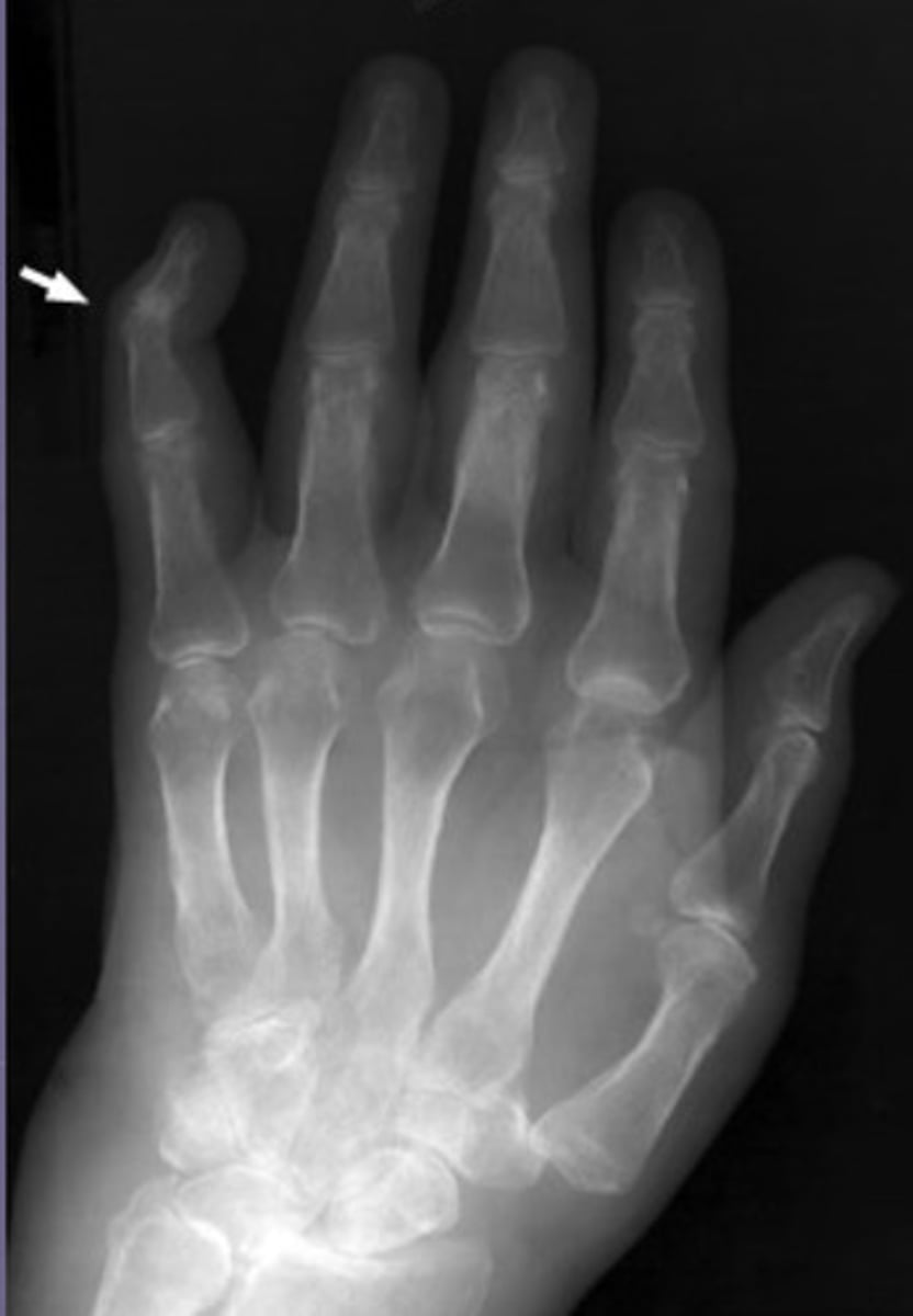
Swan neck deformity
PIP extension and DIP flexion
Radiographic features of RA
(1) Marginal erosion ("rate bite erosions") = localized loss of intraarticular cortex adjacent to the capsular insertion due to pannus erosion at the anatomical bare area
(2) Dot-dash appearance = intermittent absence of the articular cortex (due to erosions)
(3) Periarticular soft tissue swelling
(4) Juxta-articular osteopenia (lateral generalized osteopenia)
(5) Uniform narrowed joint spaces
(6) Erosions (beginning in bare areas)
(7) Subchondral Geodes
(8) Joint deformity
(9) possible ankylosis
Bare Area with RA
Unprotected area that is usually the first site of erosions
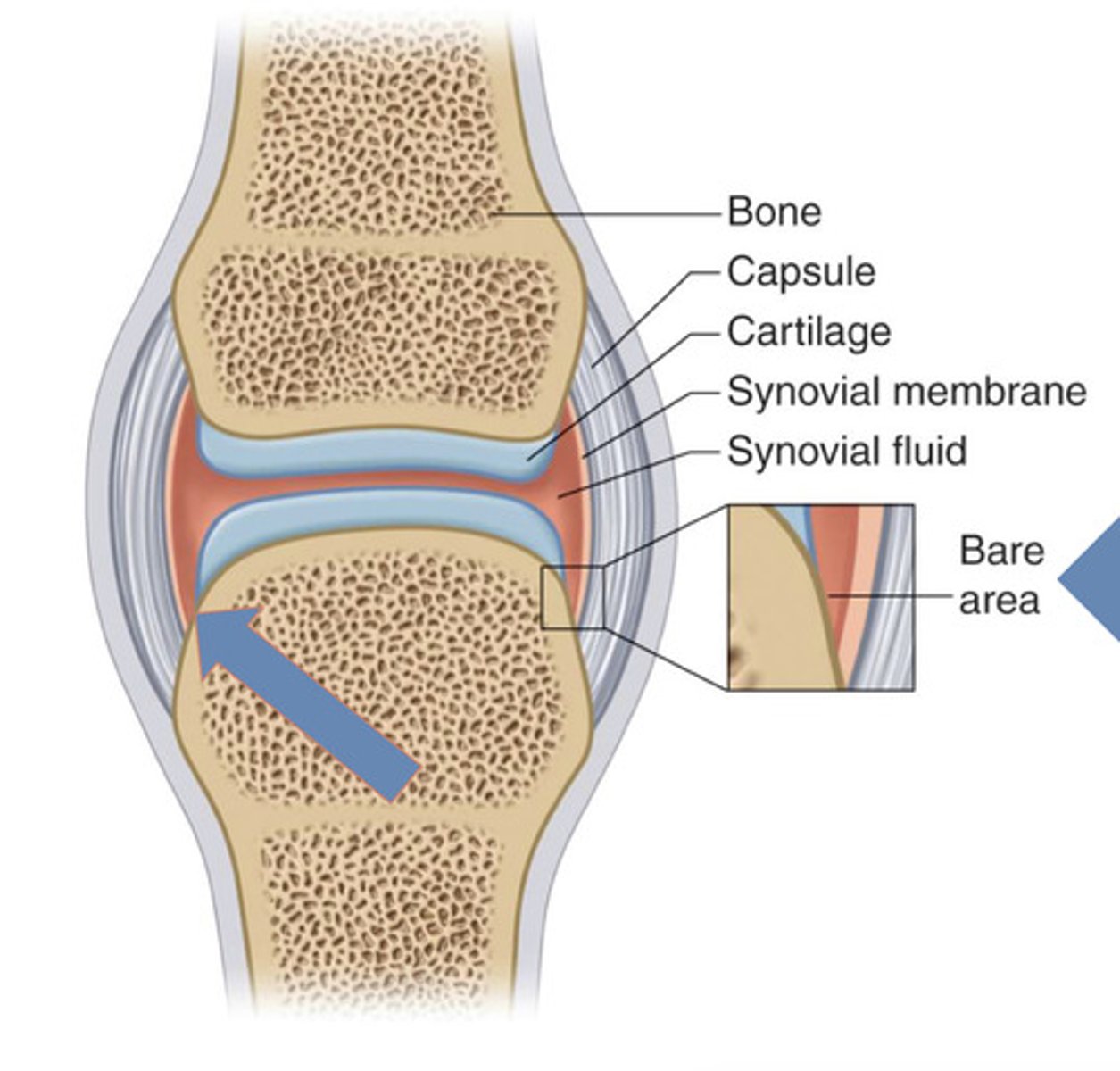
"Rat Bite" Erosions from RA
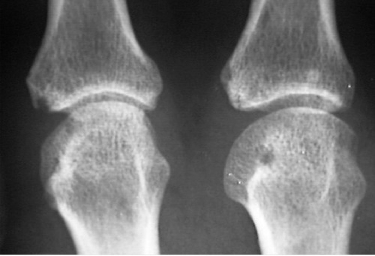
Periarticular Osteoporosis from RA
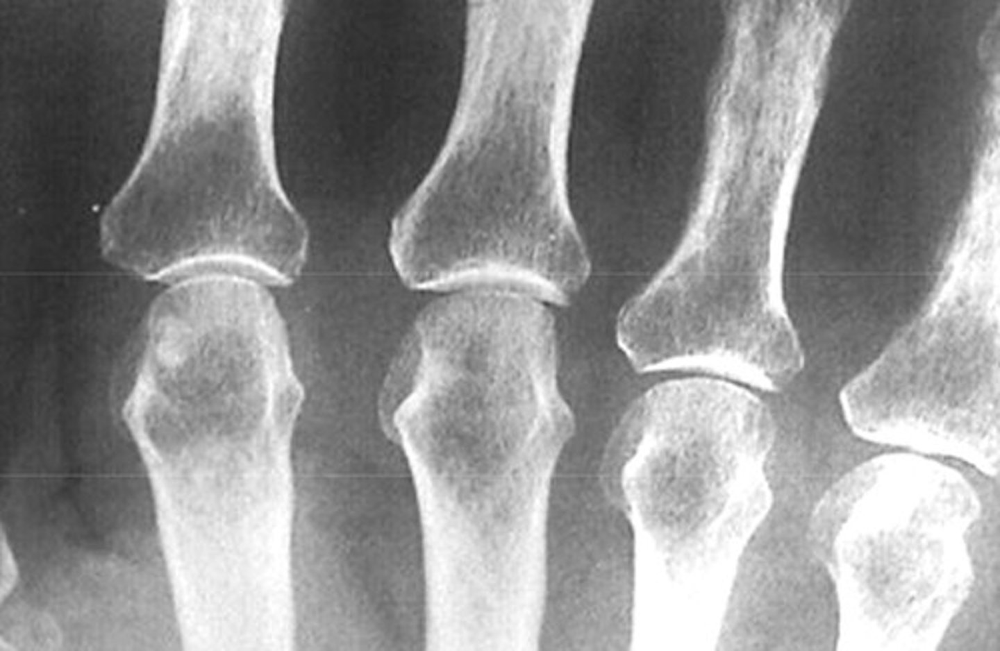
RA Soft tissue nodes
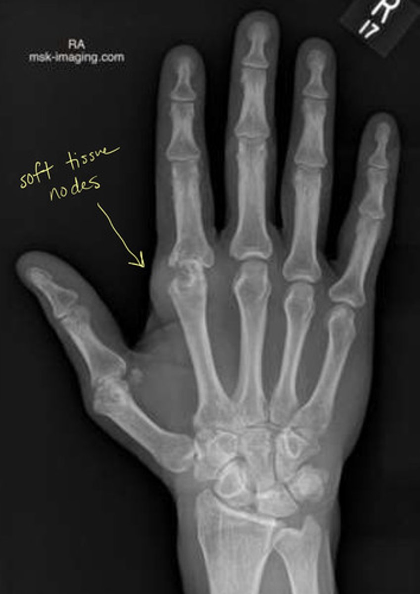
What area should you check with RA?
WRIST! Loves to attack the carpals and may see ankylosis of the carpals
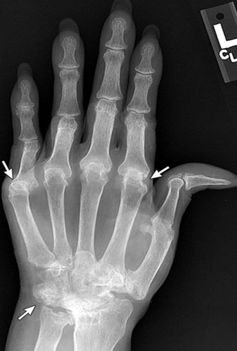
What can happen to long bones with RA?
Erosions can cause narrowing of the long bones
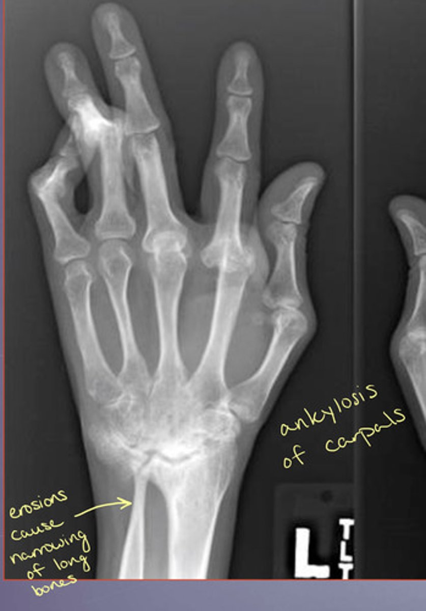
Boutonniere Deformity on X-ray
Swan Neck Deformity on X-ray
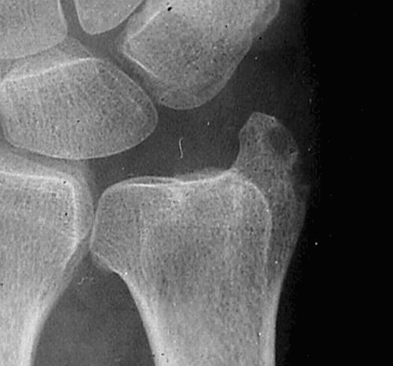
What is a common early location of RA in the wrist?
Ulnar styloid process erosions
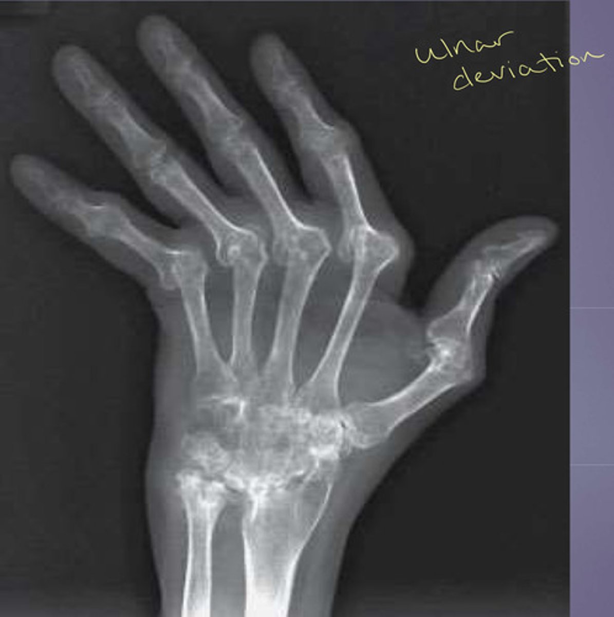
RA causes deviation of the fingers in which direction?
Ulnar deviation
RA at the hip can cause what?
Axial migration or acetabular protrusion
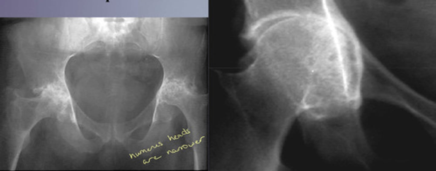
Arthritis mutilans can be caused by what?
Psoriatic or rheumatoid arthritis
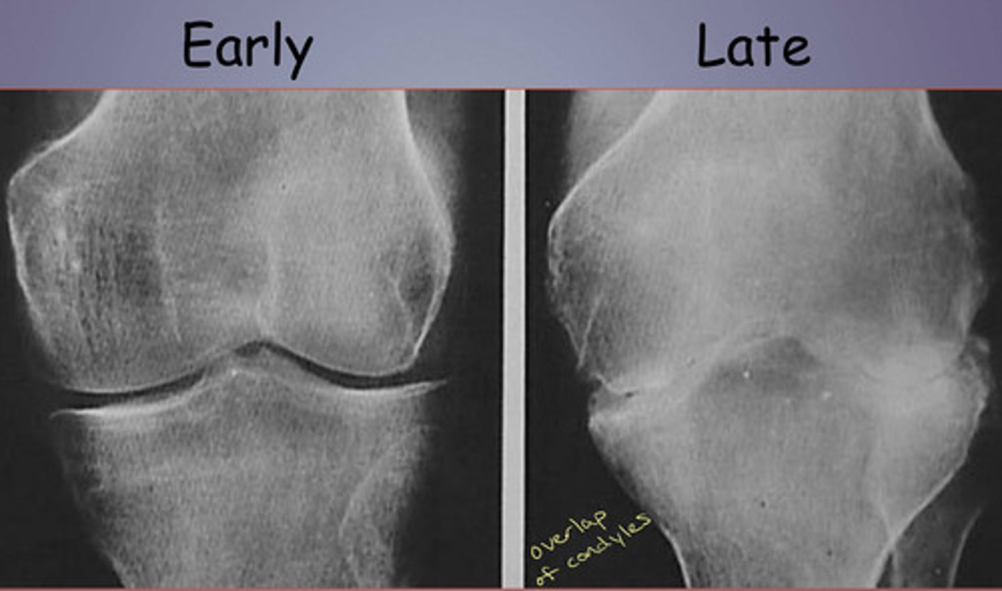
RA at the knees
Symmetric loss of joint space (vs osteoarthrosis = more at medial side)
Erosions and Baker Cyst
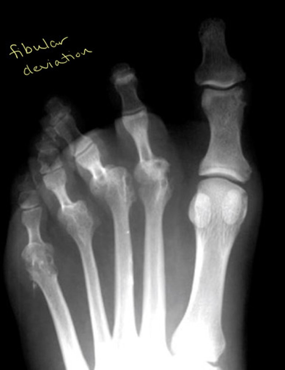
RA at the feet causes what kind of deviation?
Fibular deviation
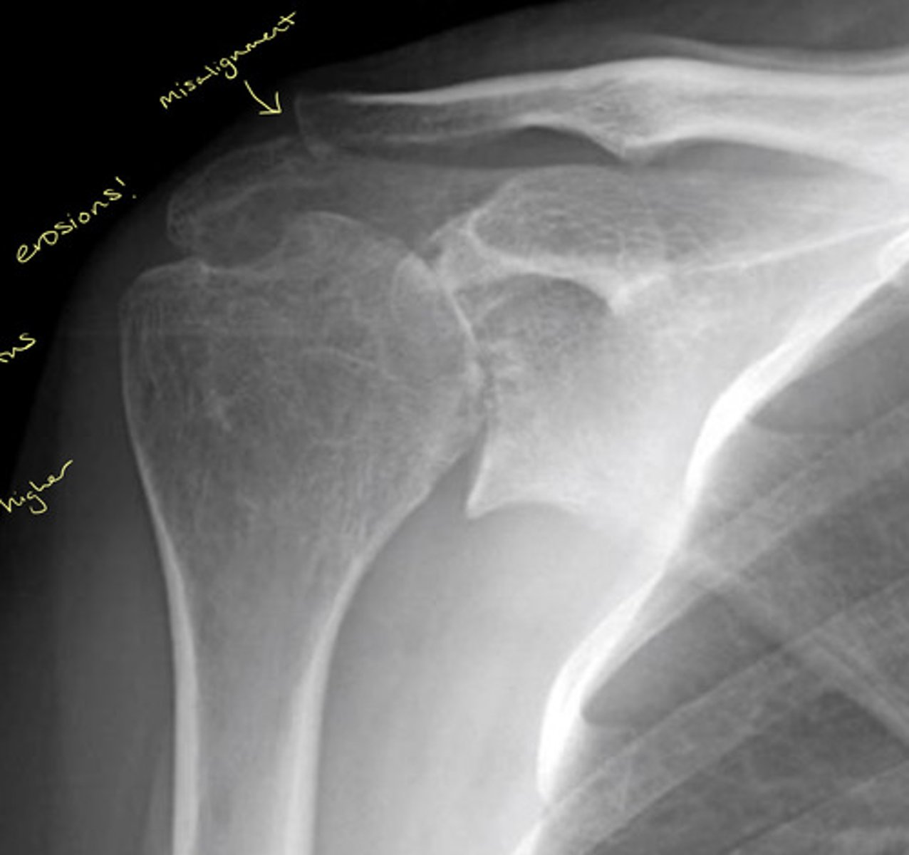
What joint in the shoulder is common for RA
AC joint
NOTE: take other side image to ensure not from trauma (RA is systemic so most likely bilateral changes)
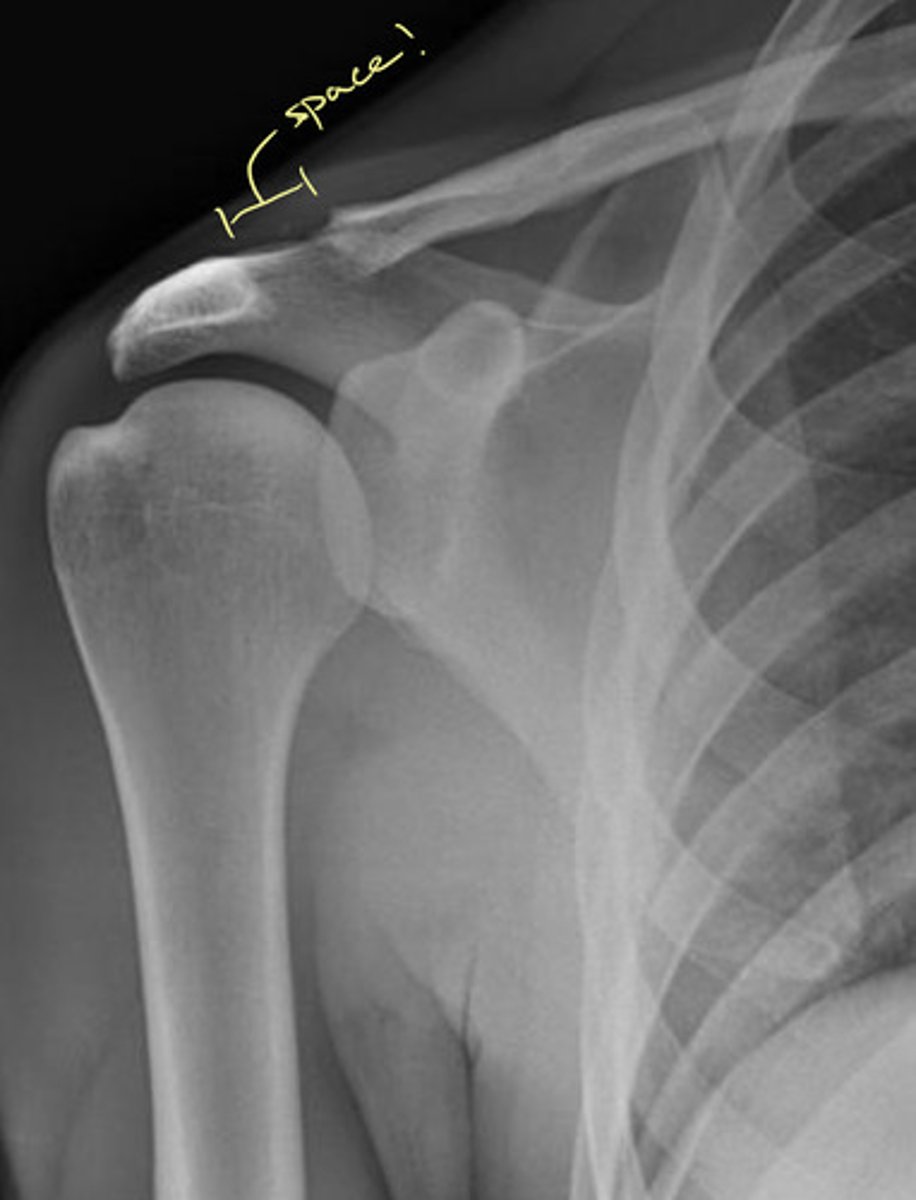
RA at the shoulder image
Erosions, misalignment, and the humerus head is higher (most likely due to supraspinatus tendon erosion)
RA of the cervical spine impacts how many RA patients?
About 50% within 10 years of diagnosis
Where does RA mainly attack in the cervical spine?
Upper cervical
Review: anterior and posterior ADI measurements
Anterior ADI = 3mm or less
Posterior ADI = 14 mm or greater
What can happen to axis with RA?
Odontoid erosions could lead to "whittled dens"
Atlanto-axial impaction is the most common cause of upper cervical neurological symptoms in patients with RA
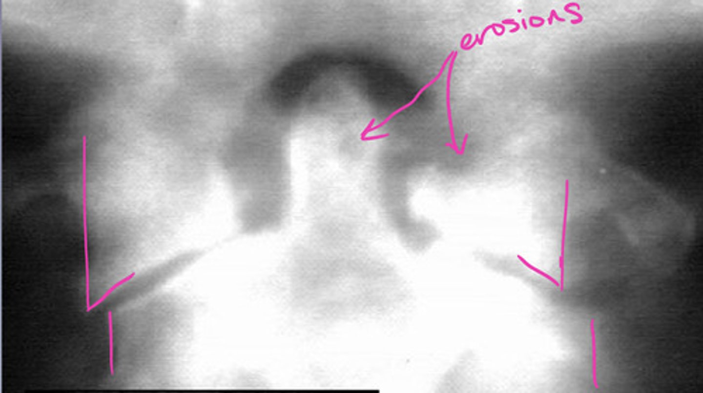
RA presentation in the cervical spine
— apophyseal joint disease
— decreased disc height
— spinous process erosions
— malalignment/subluxation
— osteopenia
Odontoid Erosions with RA can occur where?
(1) Between the dens and anterior arch
(2) Between the dens and transverse ligament
(3) Tip of the dens
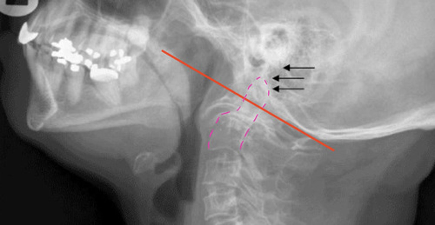
Cranial Settling from RA
Occ-C1 and/or C1-C2 facet erosions
Risk of basilar invagination and potential neurological compromise
Juvenile Chronic Arthritis (Aka JRA or JIA)
RA onset younger than 16 years of age
Can be seropositive (like adult type RA) or seronegative (MC)
Seropositive = more aggressive and poorer prognosis (but only about 10% of cases)
Polyarticular Form of JRA
Most common variety
Females > males
Bilateral and symmetrical involvement with systemic signs (mild fever, lymphadenopathy, and rash)
Radiological signs of JRA
Similar to adult RA:
— soft tissue swelling
— osteopenia
— loss of joint space
— articular erosions
— malaignments/subluxations
Characteristic to children:
— growth disruptions (potential growth plate closure)
— periostitis (lifting periosteum)
— ankylosis