The Patient Exam 1
 Call Kai
 Learn
Learn Practice Test
 Spaced Repetition
 Match
Match Flashcards
 Knowt Play
1/22

There's no tags or description
Looks like no tags are added yet.
Name | Mastery | Learn | Test | Matching | Spaced | Call with Kai | Chat |
|---|
No analytics yet
Send a link to your students to track their progress
23 Terms
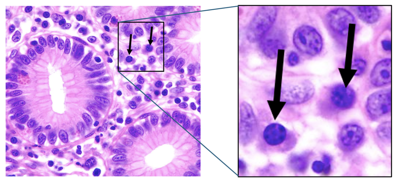
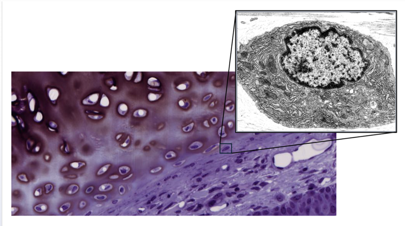
Plasma Cell (specialized lymphocyte)
Produces antibodies and has eccentric nuclei with "clock-face" chromatin
Lymphocyte photo → makes antibodies + condensed chromatin
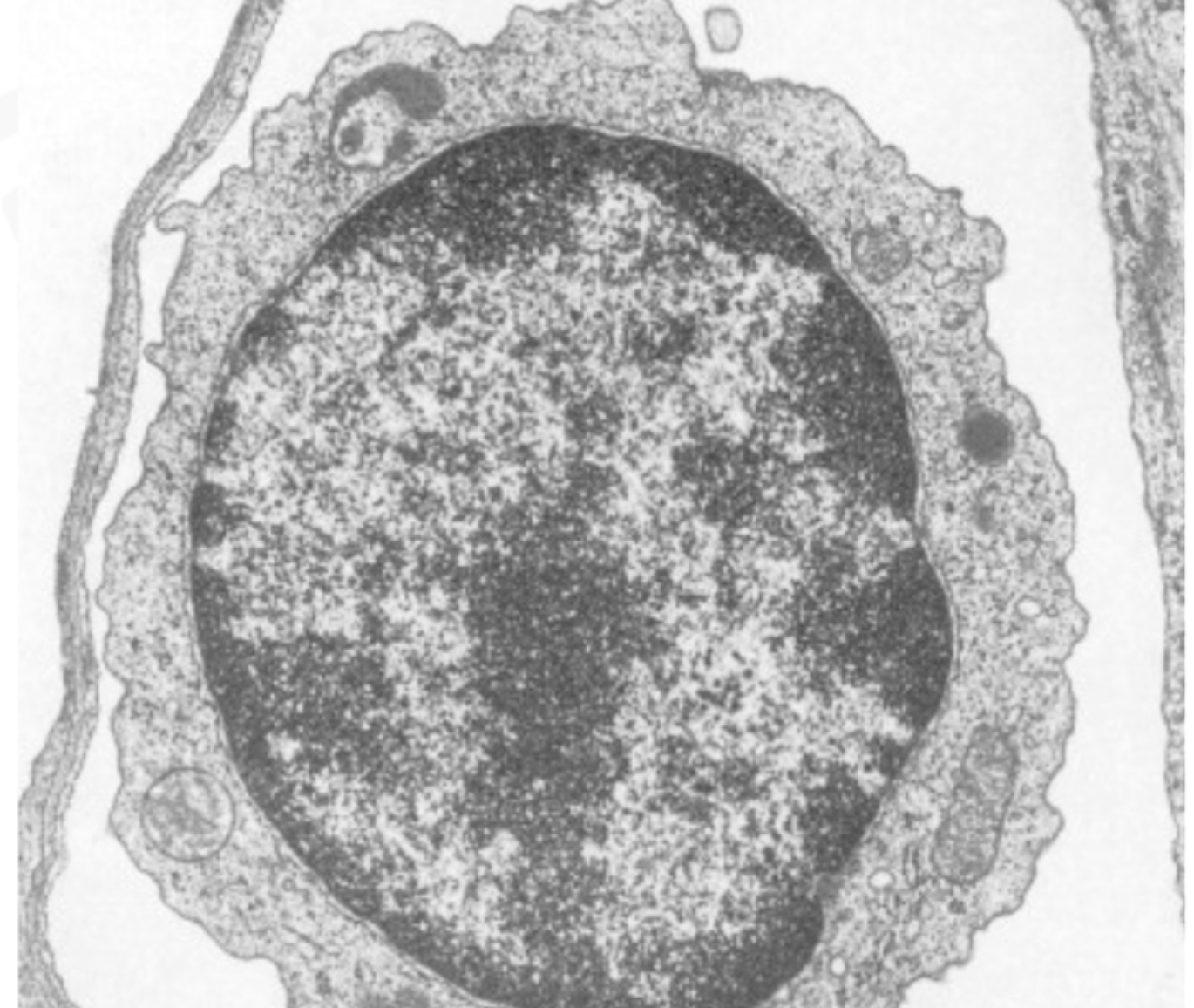
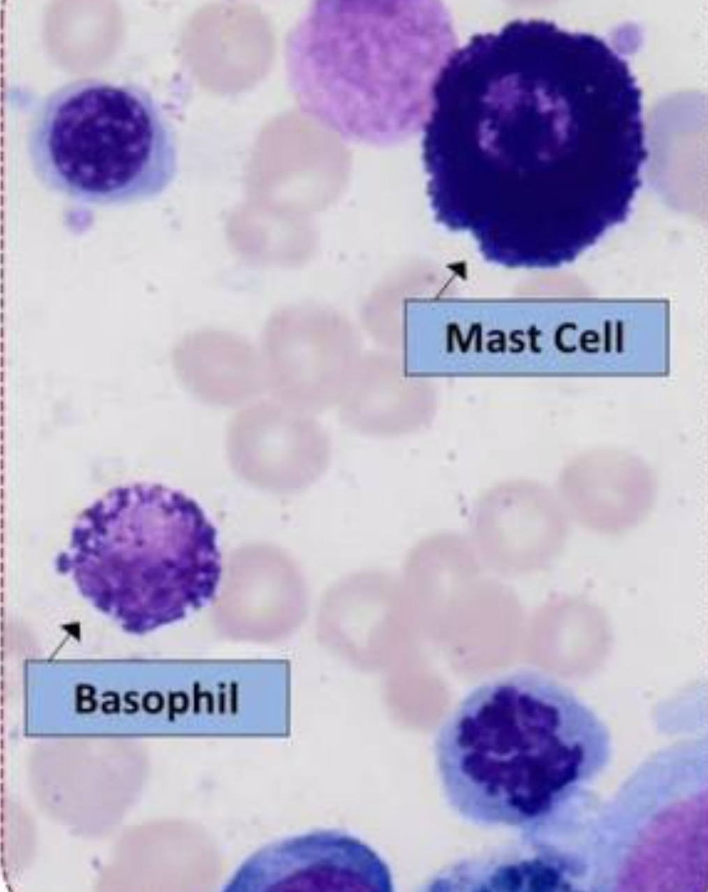
Mast Cells
Contain granules rich in histamine and heparin (+ release to start inflammatory response)
Macrophages
Phagocytic cells involved in tissue defense
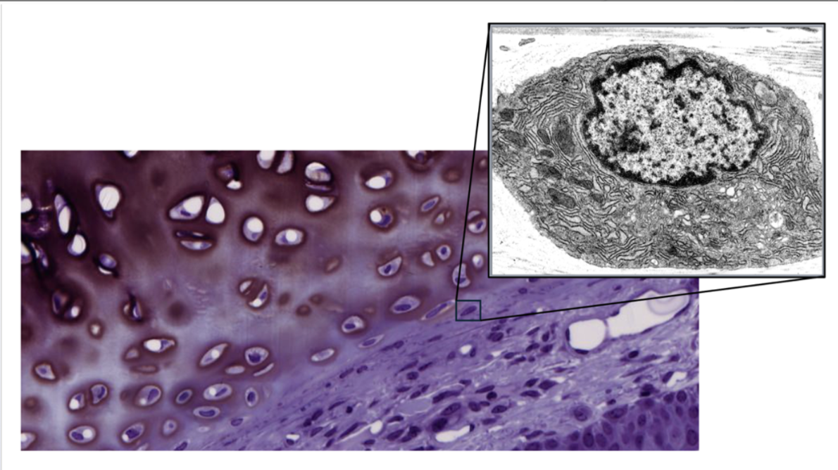
Fibroblasts
Synthesis of extracellular matrix components, including collagen and ground substance
specific alpha chains (α1 and α2)
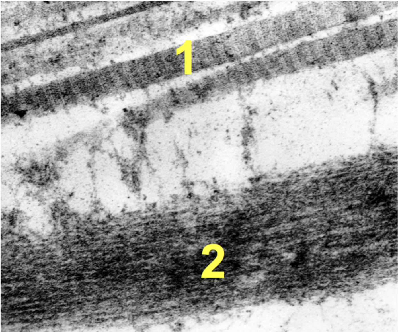
What are 1 and 2?
1= collagen fibers; 2 = elastic fibers
Collagen molecules; these fibers provide tensile strength (maximum stretch a fiber can withstand) and resist stretching forces.
Elastic fibers, often seen branching, consist of an electron-dense amorphous elastin core surrounded by fibrillin-containing microfibrils and function to allow tissues to stretch (up to 150%) and recoil.
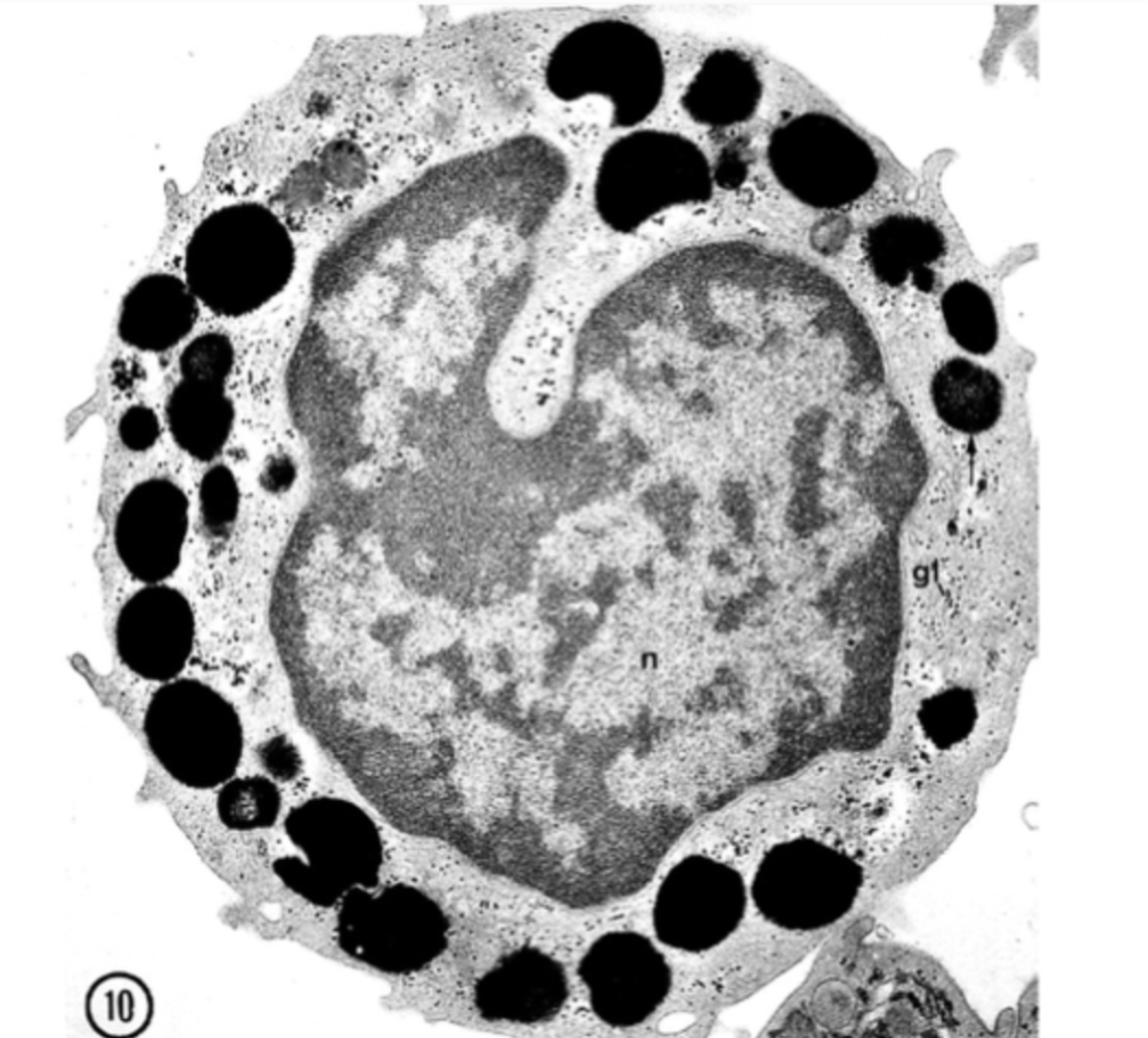
Basophil
Produce histamine during allergic reaction
Large black areas are storage of histamine and heparin
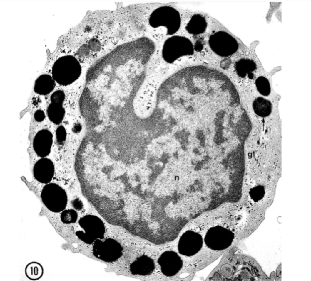
Dendritic Cell
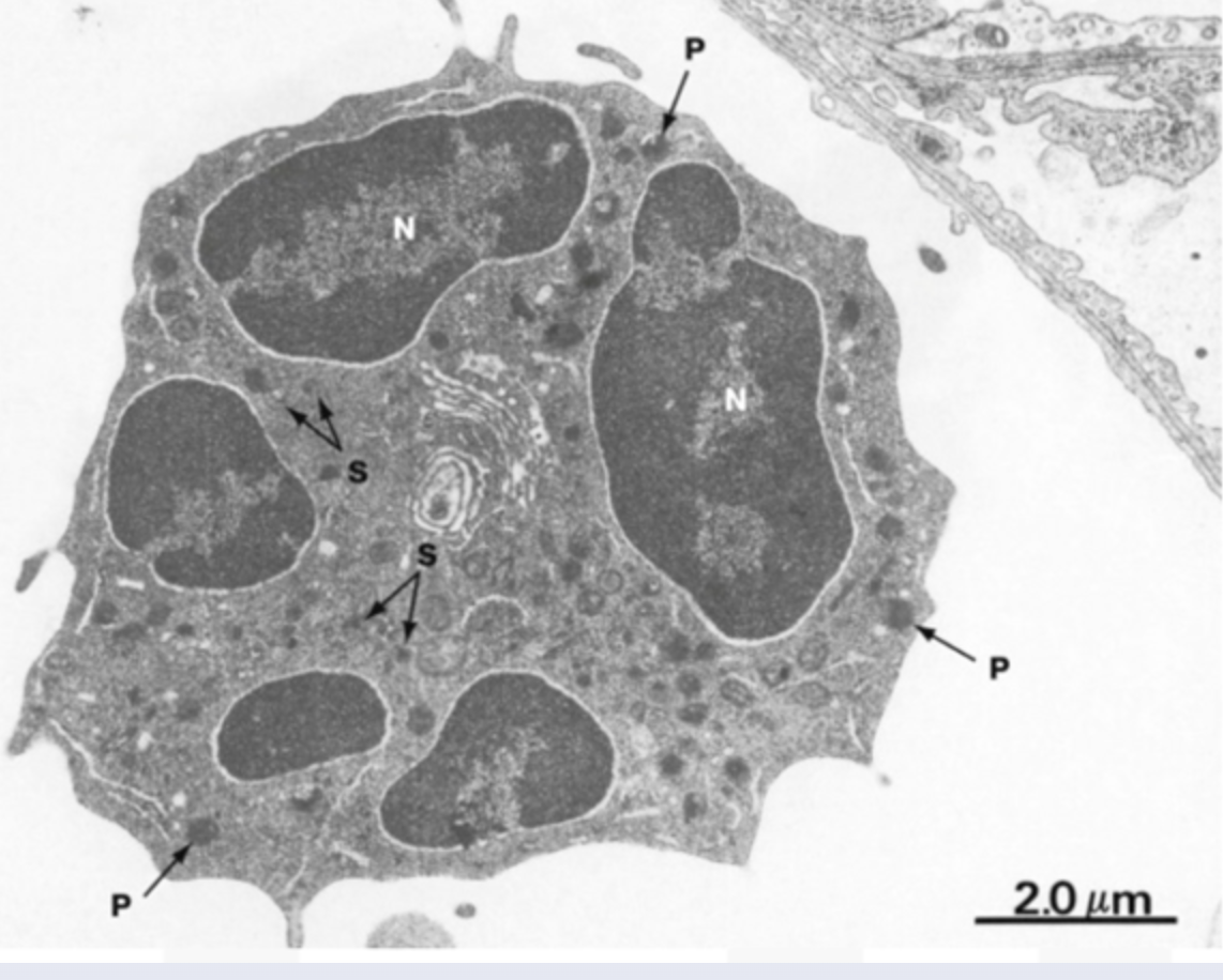
Neutrophils
Phagocytosis and destruction of microorganisms
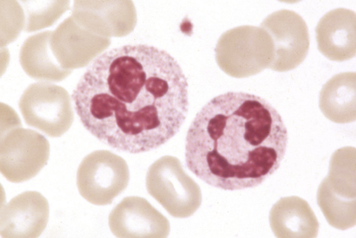
Osteogenesis Imperfecta
History of recurrent fractures after minor trauma and evidence of discolored teeth. OI is a genetic bone disorder that results from defects in collagen synthesis, most commonly in the COL1A1 or COL1A2 genes. Most patients with OI have an autosomal-dominant mutation leading to decreased production of normal type I collagen. Type I collagen is found in bone, skin, tendons, dentin, and the corneas. In addition to skeletal fractures, patients can develop blue sclerae, secondary to translucent connective tissue covering the choroidal veins, and opalescent (discolored) teeth due to lack of dentin (dentinogenesis imperfecta).
Type IV Collagen
basement membrane of epithelium
alpha chains (α1 and α2)
Type III Collagen
Reticular fibers in lymphoid organs and healing tissue.
Type 1 Collagen
bone, tendon, and dermis.
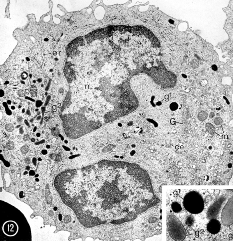
Monocyte
No specific granules, indented nucleus
Migrate into tissues and differentiate into macrophages or dendritic cells, which phagocytose material and present processed antigens to T cells
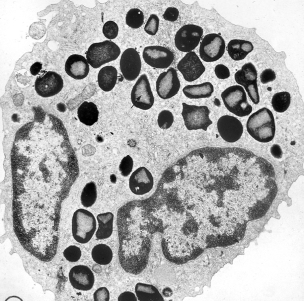
Eosinophil
dual-colored specific granules
bi-lobed nucleus
Inflammation of Common Flexor Origin
Medial epicondylitis
Golfer’s elbow from overactivity of the flexor muscles
Inflammation of common extensor origin
Lateral epicondylitis
“Tennis elbow” from overactivity/use of these muscles
What can be palpated in floor of the anatomical snuff box?
Radial artery and scaphoid bone
Thenar compartment
Opponents Pollicis, abductor pollicis brevis, flexor pollicis brevis
Recurrent branch of the median nerve
Lumbricals
#1 and #2 are median nerve innervated
#3 and #4 are ulnar nerve innervated
Flex the MCPs and help extend the PIPJs and DIPJs of fingers 2-5
What is injured in midshaft humeral fractures?
The radial nerve
B → brachoradialis
E → extensors
S → supinators
T → triceps
Ulnar nerve passes posterior to the medial epicondyle in the _____
Also passes through a groove ____ on the anterior aspect of the wrist/hand
cubital tunnel
canal of Guyon (on anterior aspect of the wrist, common site of ulnar nerve compression)
Adductor pollicis is innervated by the deep branch of the ulnar nerve
Median nerve then passes beneath the _______ and enters the hand
flexor retinaculum (through the carpal tunnel)
Symptoms include paresthesia, hypothesis, or anesthesia of lateral 3 ½ digits along the palmar surface. Weakness with opposition of thumb and motor loss of thenar muscles
Carpal Tunnel Syndrome