Bones of the Axial Skeleton
 Call Kai
 Learn
Learn Practice Test
 Spaced Repetition
 Match
Match Flashcards
 Knowt Play
1/114

There's no tags or description
Looks like no tags are added yet.
Name | Mastery | Learn | Test | Matching | Spaced | Call with Kai | Chat |
|---|
No analytics yet
Send a link to your students to track their progress
115 Terms
The Skeletal System
Bones, joints, and other associated tissues
Epiphysis
Ends of long bones
Diaphysis
Middle shaft of long bones
Spongy bone
Interior of epiphysis & lining the medullary cavity
Trabeculae
Network of bony struts in spongy bone
Red bone marrow
In the empty space of spongy bone
Axial Skeleton
Trunk
Appendicular Skeleton
Limbs
Long bone
Bone is longer than it is wide (Humerus)
Short bone
Bone is about as long as it is wide (Trapezium - carpal bone)
Flat bone
Bone is broad, flat, and thin (Sternum)
Irregular bone
Bone’s shape does not fit into other classes (Vertebra)
Sesamoid bone
Round, flat bone found within tendon (patella)
Inorganic Matrix
Makes bones hard - designed to resist compressive forces
Hydroxyapatite crystals
Composed of calcium & phosphorus salts (inorganic matrix)
Organic matrix
Contribute to bone’s ability to resist twisting forces and tension
Osteogenic cells
Stem cells of bones (first stage of bone cells)
Osteoblast
Immature cells that secrete the bony matrix → bone deposition
Osteocytes
Mature cells of bone, maintain bone tissue
Osteoclasts
Break down bone by secreting H+ and enzymes → bone resorption
Osteon
units of compact bone (tree trunks)
Central canal
Center of osteon → contains blood vessels and nerves
Lamellae
Rings around central canal
Lacunae
Holes where osteocytes sit
Canaliculi
Channels connecting lacunae (branches)
Interstitial lamellae
Rings around individual osteons
Circumferential lamellae
Rings around entire diameter of bones
Periosteum
Covers outer surface of bone → attachment site for tendons and ligaments; route for blood vessels to enter the bone
Perforating fibers
Connect periosteum to bone
Endosteum
Covers the interior of bone surfaces
Medullary cavity
Empty space in bone → can be filled with yellow bone marrow
Epiphyseal line
Remnants of the growth plate (lines between spongy bone near the epiphyses)
Growth plate
Area of hyaline cartilage from which longitudinal bone growth occurs
Joint
Where two or more bones come together
Synarthrosis
Joints allow no movement
Amphiarthrosis
Joints allow very limited movement
Diarthrosis
Freely movable joint
Fibrous joints
Two bones are separated by dense regular connective tissue
Suture
Fibrous → found between the bones of the skull
Syndesmosis
Fibrous → Articulating bones joined by long piece of dense regular connective tissue ex. tibia & fibula
Gomphosis
Fibrous - Occurs between the teeth and the maxilla or mandible
Cartilaginous joints
Two bones are joined by cartilage
Synchondrosis
Cartilaginous → bones are connected by hyaline cartilage (ex. ribs to sternum; costochondral)
Symphysis
Cartilaginous → bones are connected by fibrocartliage (ex. intervertebral bodies)
Synovial joints
Two bones are separated by a synovial cavity, lined by a synovial membrane and filled with synovial fluid
Planar
Synovial → nonaxial movement (movement does NOT occur in relation to axis)
Pivot
Synovial → uniaxial movement (movement occurs around one axis) (ex. Atlantoaxial)
Hinge
Synovial → uniaxial movement (ex. temporomandibular)
Saddle
Synovial → biaxial movement (movement occurs around two axes)
Condylar
Synovial → biaxial movement (ex. Atlanto-occipital)
Ball & Socket
Synovial → triaxial movement (movement around 3 axes)
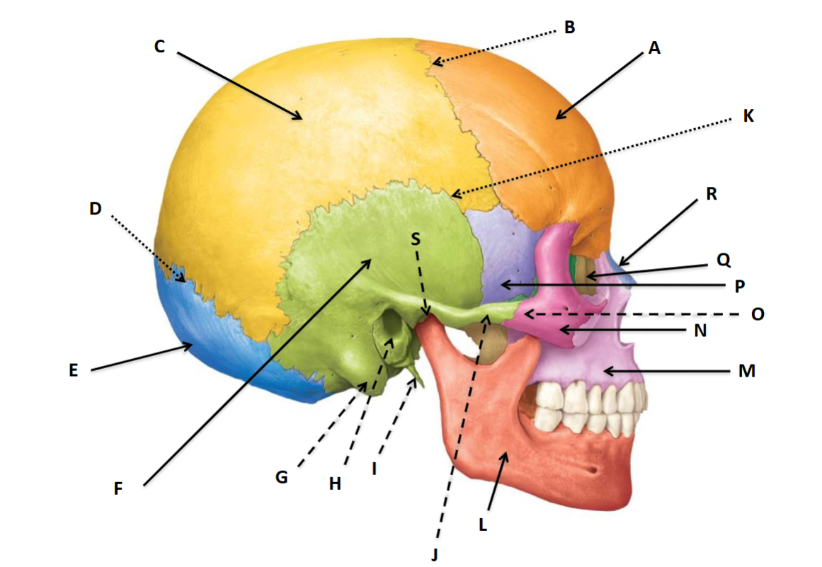
Cranial Skull
Houses and protects the brain

A
Frontal Bone

B
Coronal Suture

C
Parietal Bone

D
Lambdoid Suture

E
Occipital Bone

F
Temporal Bone
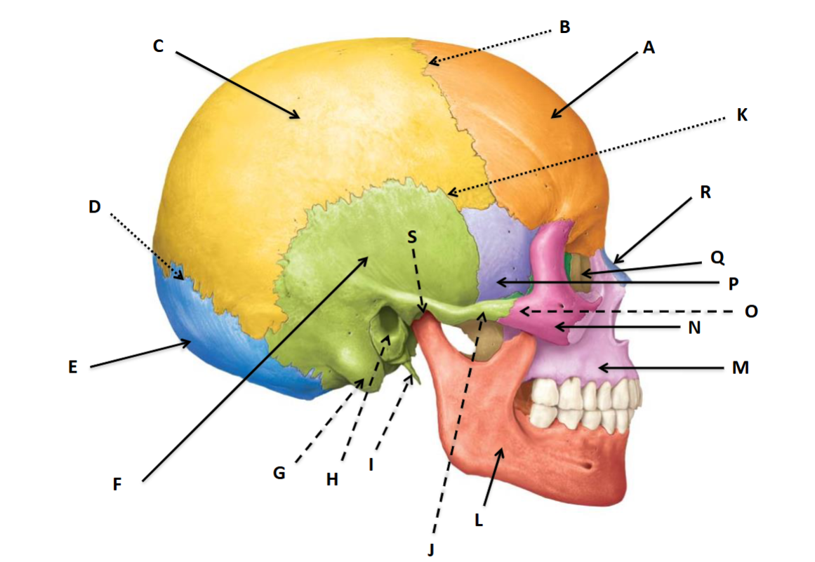
G
Mastoid Process of the temporal bone
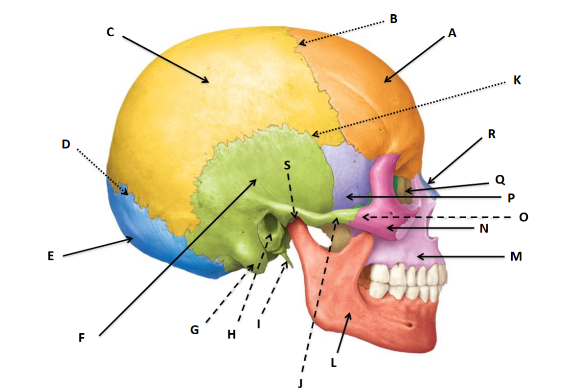
H
External acoustic meatus
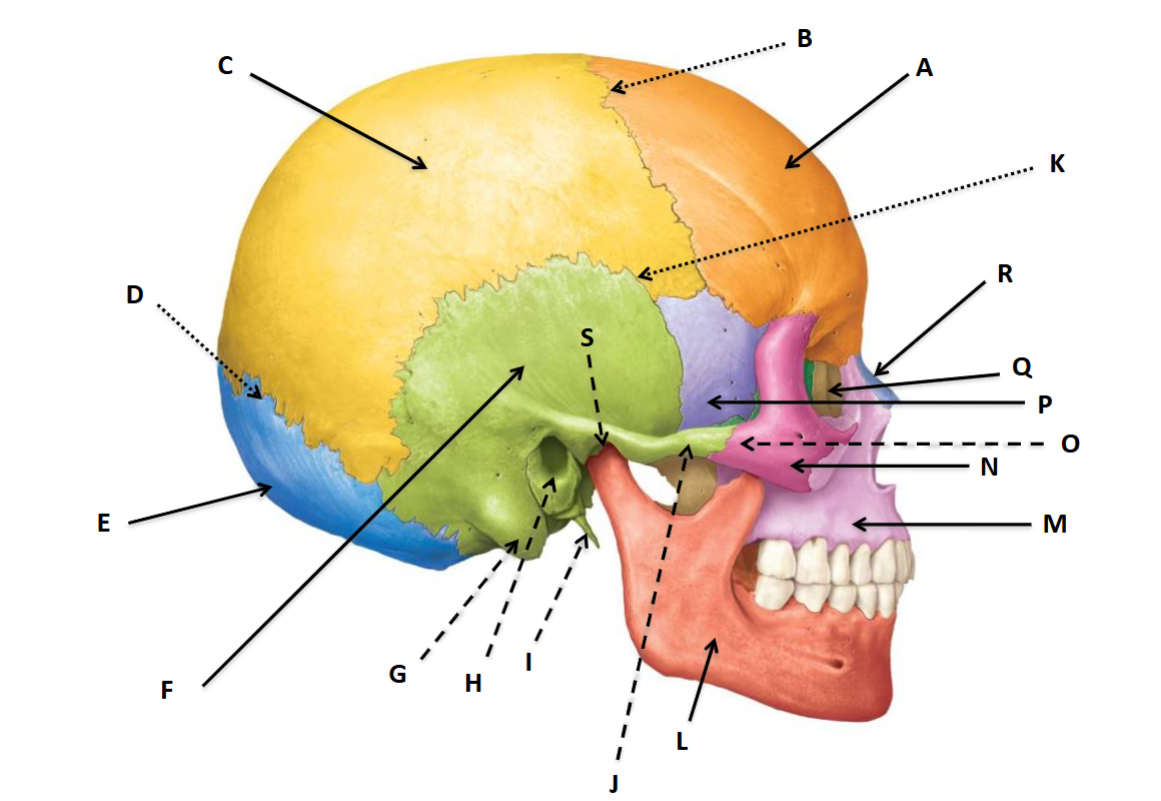
I
Styloid process of the temporal bone
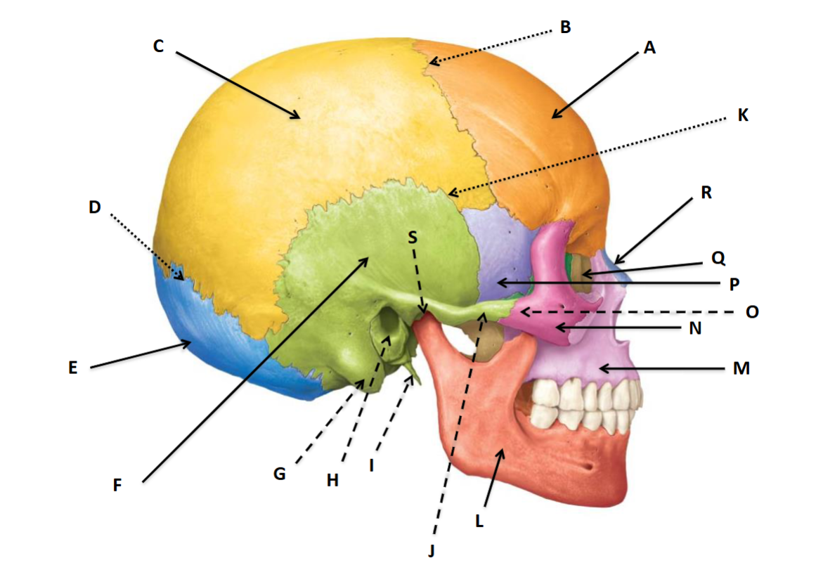
J
Zygomatic Process of the temporal bone

K
Squamous suture

L
Mandible
M
Maxilla

N
Zygomatic bone
O
Temporal process of the zygomatic bone

P
Sphenoid bone

Q
Lacrimal bone

R
Nasal bone
S
Zygomatic arch of the temporal bone

A
Frontal

B
Nasal bone

C
Zygomatic bone

D
Superior nasal concha

E
Inferior nasal concha

F
Maxilla
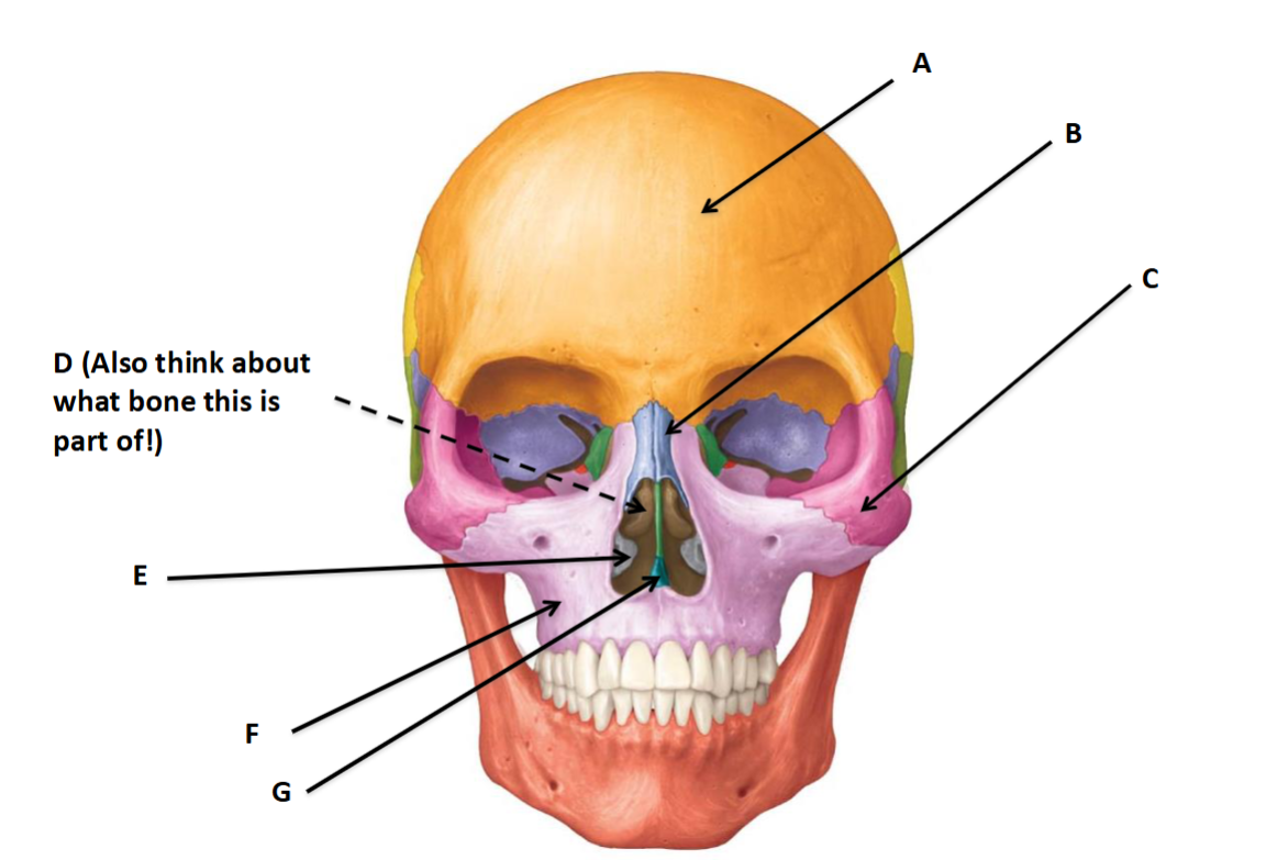
G
Vomer
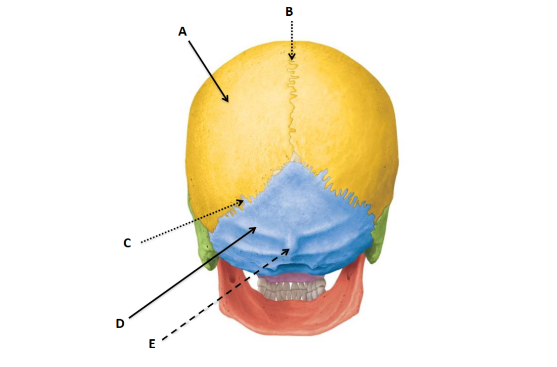
A
Parietal bone

B
Sagittal suture
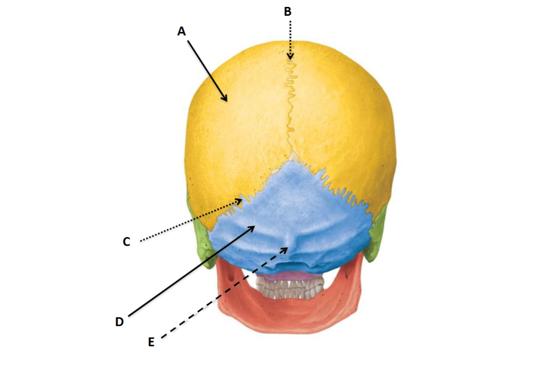
C
Lambdoid suture
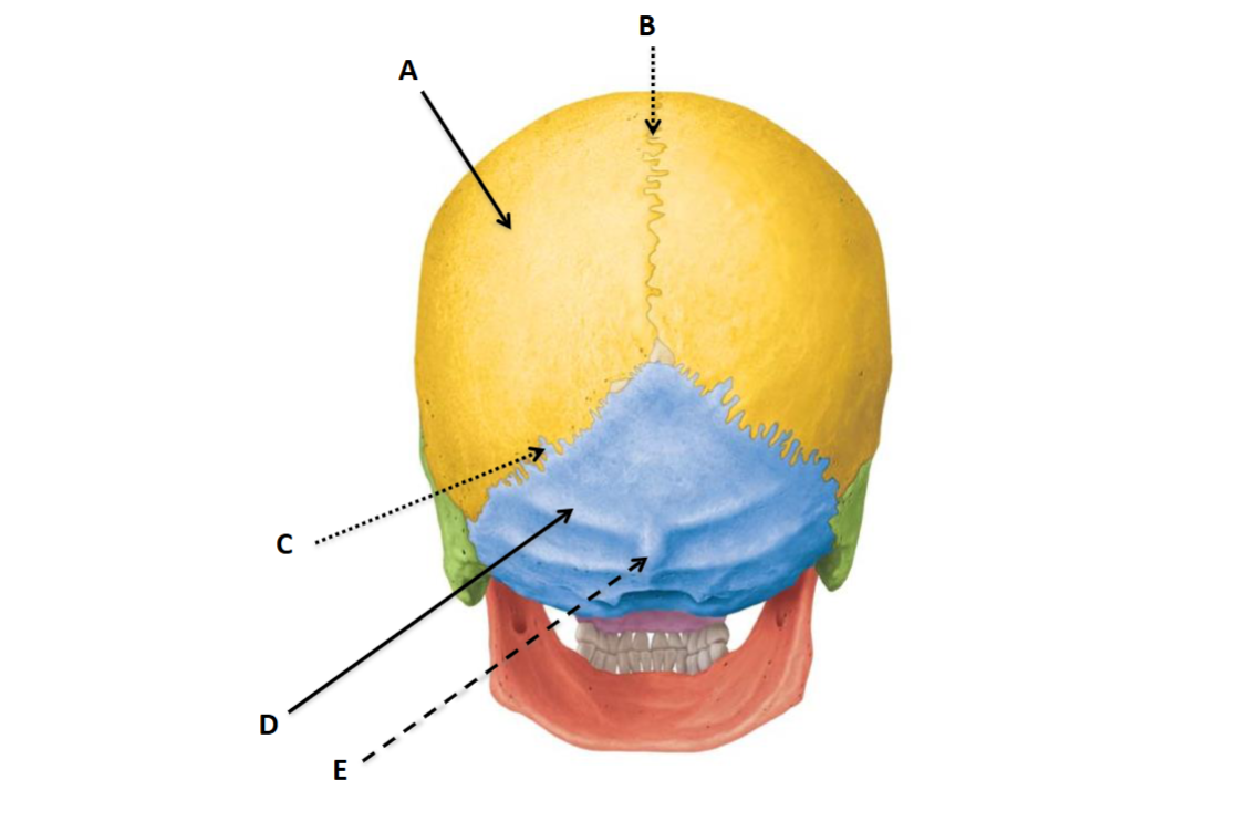
D
Occipital bone

E
External occipital protuberance

A
Occipital bone

B
Magnum foramen

C
Occipital condyle (connects to C1)

D
Palatine bone

E
Maxilla

F
Sphenoid bone
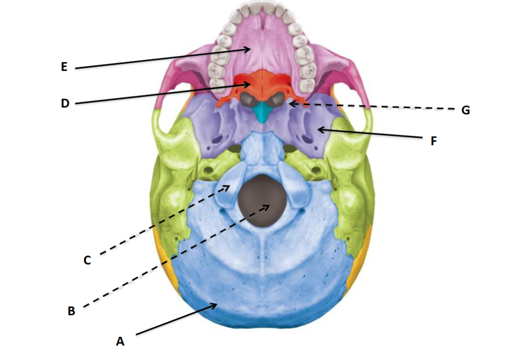
G
Pterygoid process

A
Sella Turcica

B
Lesser wing

C
Greater wing

A
Coronoid process of the mandible
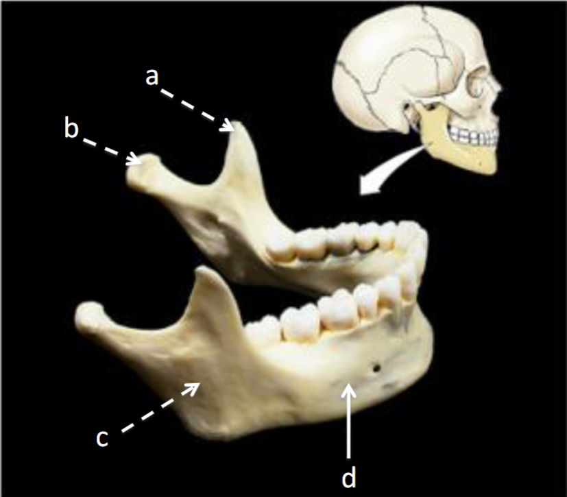
B
Mandibular condyle

C
Ramus of the mandible

D
Mandible
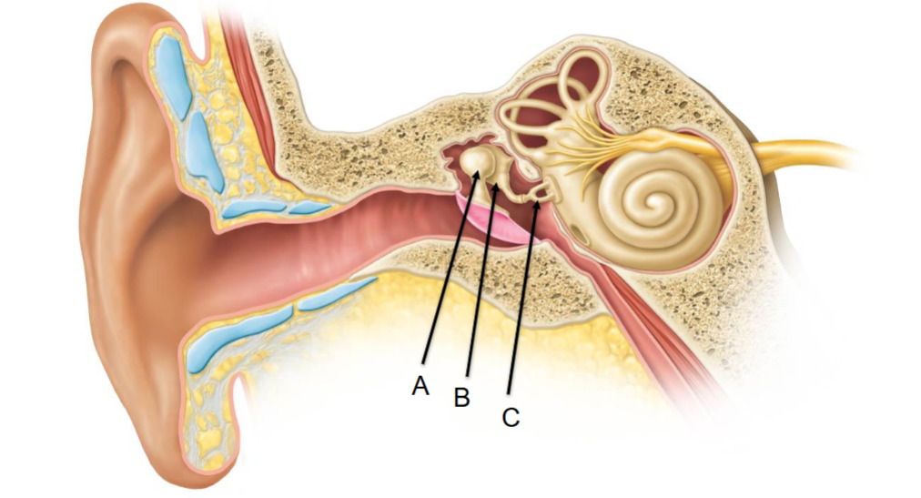
A
Malleus

B
Incus
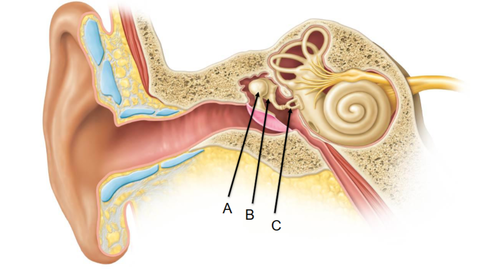
C
Stapes