ABD: CTL Anatomy 30% - Liver, GB, and Pancreas
 Call Kai
 Learn
Learn Practice Test
 Spaced Repetition
 Match
Match Flashcards
 Knowt Play
1/140

There's no tags or description
Looks like no tags are added yet.
Name | Mastery | Learn | Test | Matching | Spaced | Call with Kai | Chat |
|---|
No analytics yet
Send a link to your students to track their progress
141 Terms

Which of the following structures is labeled #5?
a. anterior right lobe
b. lateral left lobe
c. medial left lobe
d. posterior right lobe
b
The liver is divided into superior and inferior segments by the:
a. course of the fissures through the tissue
b. branching of the hepatic veins
c. branching of the portal veins
d. course of the biliary vessels through the tissue
c
Which of the following correctly describes the ducts of Luschka?
a. associated with porcelain GB and GB carcinoma
b. only visible on obese patients
c. located within the 3rd segment of the duodenum
d. bile thickening occurs within them
d

What structure/vessel is indicated by #6?
a. splenic vein
b. superior mesenteric artery
c. splenic artery
d. superior mesenteric vein
b
Which of the following correctly describes the location of the right hepatic vein?
a. courses inferiorly through the falciform ligament
b. courses superiorly through the main lobar fissure
c. courses superiorly through the falciform ligament
d. courses superiorly through the right intersegmental fissure
d
All the following laboratory tests are used to evaluate the liver function, except:
a. prothrombin time
b. aspartate aminotransferase
c. bilirubin
d. sodium bicarbonate
d
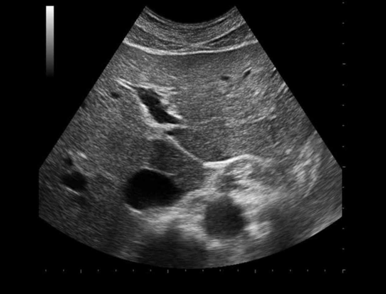
Use your mouse to place your cursor over the ligamentum venosum and click to mark the structure.

Which statement best describes the location of the ligamentum venosum?
a. it forms the posterior border of the right lobe
b. it lies between the right and left lobes of the liver
c. on ultrasound, it appears as a thick, triangular, echogenic foci in the left lobe of the liver
d. it forms the anterior border of the caudate lobe
d

What structure/vessel is indicated by #2?
a. head of pancreas
b. stomach
c. duodenum
d. left lobe of liver
a
Branches of which of the following vessels supply the pancreas with blood?
a. superior mesenteric artery and right gastric artery
b. proper hepatic artery and superior mesenteric artery
c. gastroduodenal artery and proper hepatic artery
d. superior mesenteric artery and gastroduodenal artery
d

What structure/vessel is indicated by #7?
a. medial left lobe
b. caudate lobe
c. ligamentum teres
d. ligamentum venosum
e. lateral left lobe
d
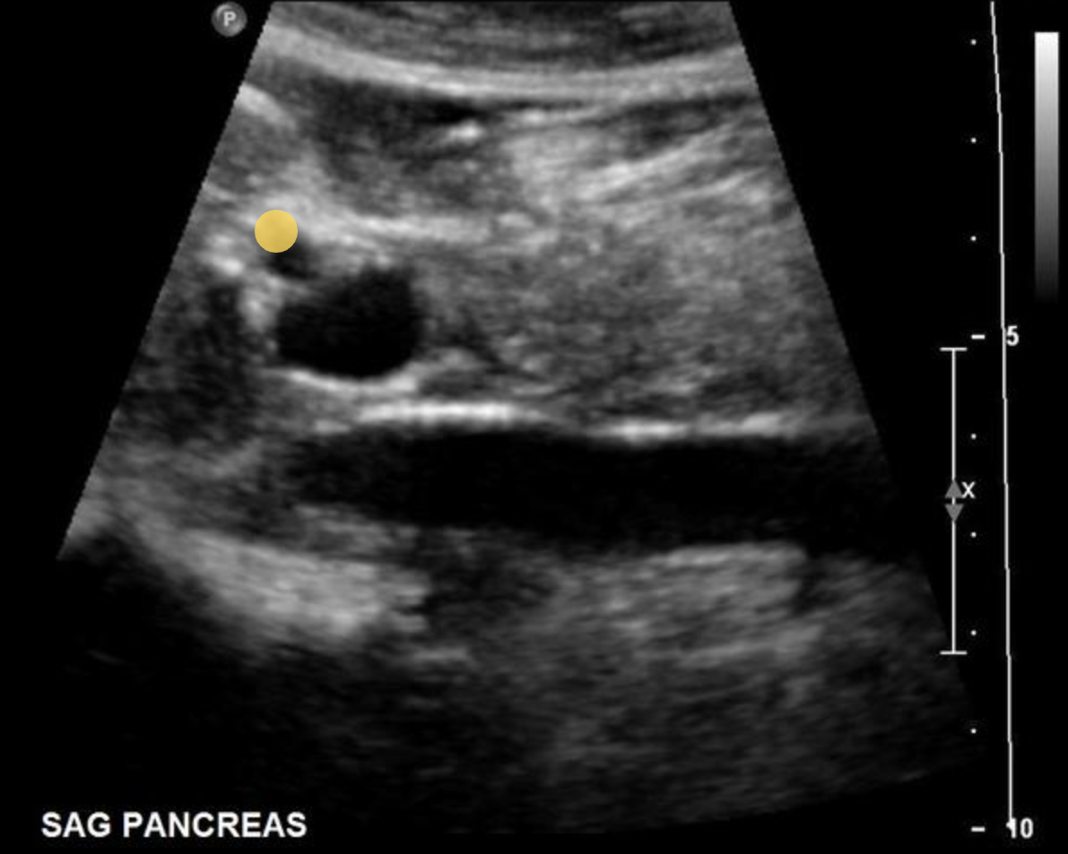
Use your mouse to place your cursor over the hepatic artery and click to mark the vessel.
In a normal adult patient, the intrahepatic common hepatic duct lumen measures less than or equal to _______.
a. 2 cm
b. 6 mm
c. 2 mm
d. 4 mm
d
Which of the following hormones is responsible for causing the gallbladder to contract?
a. insulin
b. lipase
c. trypsin
d. cholecystokinin
d

What causes the appearance of this normal variant of the uncinate process?
a. higher concentration of fat in the area of interest
b. polycythemia vera
c. reduced concentration of fat in the area of interest
d. hyperbilirubinemia
c
The cystic artery originates at the _______ and the cystic vein empties into the ________.
a. right hepatic artery, right hepatic vein
b. common hepatic artery, main portal vein
c. left hepatic artery, left portal vein
d. right hepatic artery, right portal vein
d
All of the following are intrasegmental vessels of the liver, except?
a. portal veins
b. hepatic arteries
c. bile ducts
d. hepatic veins
d
The pancreatic duct should normally be less than _______ in diameter in children and young adults.
a. 0.5 mm
b. 1 mm
c. 2 mm
d. 4 mm
c

Which of the following structures is labeled #2?
a. superior mesenteric artery
b. hepatic artery
c. gastroduodenal artery
d. common bile duct
c

Which of the following statements is true regarding the image displayed?
a. the gallbladder has a normal variant called a phrygian cap near the neck
b. the gallbladder has a normal variant called a phrygian cap near the fundal area
c. the gallbladder has a septation within the fundus
d. the gallbladder has a normal variant called Hartmann pouch near the fundal area
b
The hepatoduodenal ligament contains which of the following structures?
a. the umbilical arteries and vein
b. main portal vein, proper hepatic artery, and common bile duct
c. right portal vein, right hepatic artery, and cystic duct
d. the emissary veins
b
The normal gallbladder is usually less than ________ in a transverse diameter.
a. 8 cm
b. 5 cm
c. 3 cm
d. 3 mm
b
The right lobe of the liver is divided into _______ segments, while the left lobe is divided into ______ segments.
a. caudate and anterior, quadrate and posterior
b. anterior and posterior, medial and lateral
c. medial and lateral, anterior and posterior
d. caudate and posterior, quadrate and anterior
b
Which liver vessel courses within the main lobar fissure?
a. middle hepatic vein
b. main portal vein
c. common hepatic artery
d. right hepatic vein
a
A small rounded prominence on the anteroinferior aspect of the normal caudate lobe is called:
a. elongated left lobe
b. Reidel lobe
c. distal papillary process
d. quadrate lobe
c
A normal portal vein will:
a. increase in diameter by placing patient in an upright position
b. decrease in diameter after eating
c. increase in diameter by more than 20% with deep inspiration
d. increase in diameter after exercise
c
Which of the following correctly describes the pediatric pancreas?
a. cystic fibrosis leads to production of mucus and the gland is reduced in echogenicity
b. pancreatic echogenicity increases with age
c. the ducts are dilated at >2 mm at birth and decrease in size throughout the first year
d. it normally decreases in echogenicity as the patient reaches adulthood
b
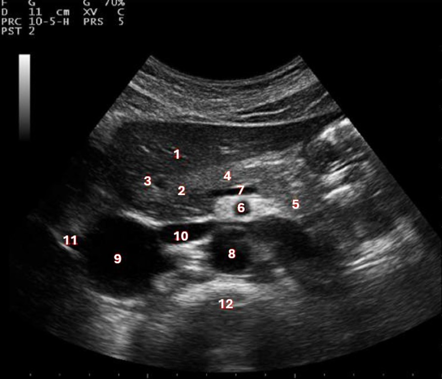
What structure/vessel is indicated by #5?
a. tail of pancreas
b. head of pancreas
c. left lobe of liver
d. body of pancreas
a

What structure/vessel is indicated by #3?
a. lateral branch of LPV
b. main portal vein
c. medial branch of LPV
d. left portal vein
a

What structure is indicated by the green arrow?
a. main portal vein
b. CBD
c. common hepatic artery
d. porta hepatis
a
An ERCP is commonly performed to evaluate:
a. the Ampulla of Vater
b. the left renal vein
c. the right renal vein
d. pyloric stenosis
a
What blood vessel separates the right and left lobes of the liver?
a. right portal vein
b. common hepatic artery
c. left hepatic vein
d. middle hepatic vein
d
The pancreatic tail is anterior to all of the following, except:
a. left adrenal gland
b. splenic hilum
c. upper pole of the left kidney
d. lesser omental sac
d
Which lobe of the liver receives blood from the left and right portal veins?
a. caudate lobe
b. anterior right lobe
c. posterior right lobe
d. medial left lobe
a
Normal hepatic venous flow will demonstrate:
a. two large retrograde diastolic and systolic waves followed by a small retrograde component that corresponds with the atrial contraction
b. two large antegrade diastolic and systolic waves followed by a small antegrade component that corresponds with the atrial contraction
c. two large antegrade diastolic and systolic waves followed by a small retrograde component that corresponds with the atrial contraction
d. two large retrograde diastolic and systolic waves followed by a small antegrade component that corresponds with the atrial contraction
c
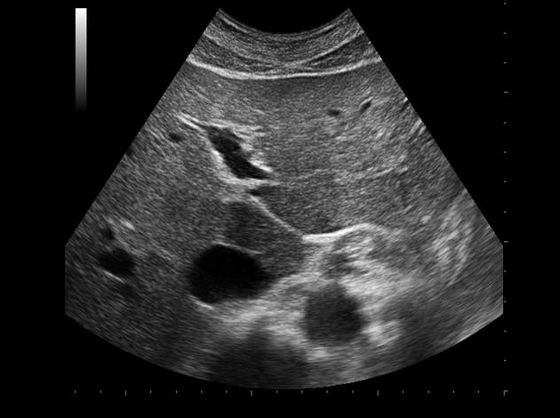
Use your mouse to place your cursor over the center of the lateral left lobe of the liver and click to mark the structure.

__________ is produced by the exocrine function of the pancreas and ________ is produced by the endocrine function of the pancreas.
a. amylase, lipase
b. sodium bicarbonate, insulin
c. insulin, tryplin
d. sodium bicarbonate, amylase
b
Trypsin breaks down _______, amylase breaks down ______, and lipase breaks down _______ during digestion.
a. protein, carbohydrates, fat
b. fat, carbohydrates, protein
c. carbohydrates, protein, fat
d. sugar, carbohydrates, fat
a
Which of the following produces insulin?
a. acini glands of the pancreas
b. Islets or Langerhans
c. Kupffer cells in the liver
d. pituitary gland
b

What lobe of the liver is indicated by #2?
a. anterior right lobe
b. caudate lobe
c. posterior right lobe
d. medial left lobe
a
The patient presents with a history of choledocholithiasis. How will you assess the common bile duct for a presence of a stone?
a. demonstrate a longitudinal view of the main portal vein and look for the CBD anterior to it
b. demonstrate a transverse view of the pancreatic head and look for the CBD entering the anterior aspect
c. demonstrate a long axis gallbladder image and look for the CBD superior to it
d. locate the main portal vein and look for the CBD posterior to it
a
The formation of a Hartmann pouch usually occurs in what portion of the gallbladder?
a. neck
b. fundus
c. body
d. cystic duct
a
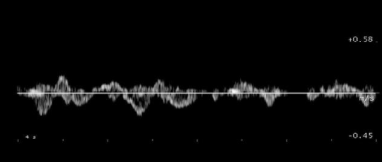
Which of the following statements is true regarding the Doppler tracing displayed?
a. it could represent a doppler tracing of normal flow in the right hepatic vein
b. it could represent a doppler tracing of normal flow in the main portal vein
c. it could represent a doppler tracing of normal flow in the portal or hepatic venous system
d. it could represent a doppler tracing of normal flow in the proximal aorta
a
Which of the following blood vessels delivers the majority of oxygenated blood that enters the liver?
a. common hepatic artery
b. hepatic veins
c. main portal vein
d. proper hepatic artery
c
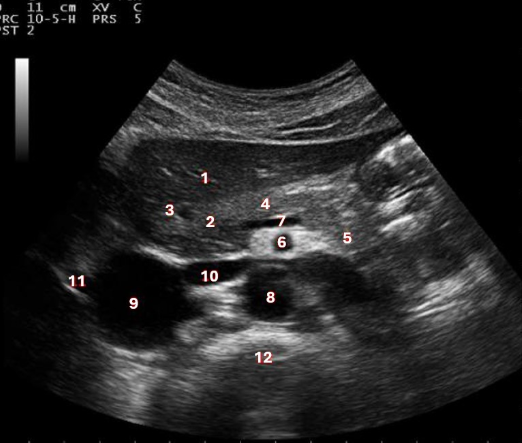
What structure/vessel is indicated by #7?
a. splenic artery
b. superior mesenteric vein
c. splenic vein
d. superior mesenteric artery
c
What causes the hepatic vein flow to have a triphasic waveform?
a. left atrial contraction and relaxation
b. right atrial contraction and relaxation
c. portal HTN
d. hepatic congestion
b
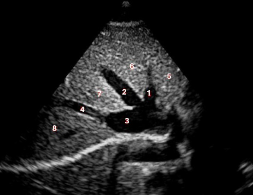
Which of the following structures is labelled #3?
a. IVC
b. left hepatic vein
c. middle hepatic vein
d. left portal vein
a
The bare area of the liver is in direct contact with:
a. right kidney
b. right portal vein
c. IVC
d. diaphragm
d
The first branch of the common hepatic artery is the:
a. duodenal artery
b. pancreatic artery
c. gastroduodenal artery
d. cystic artery
c
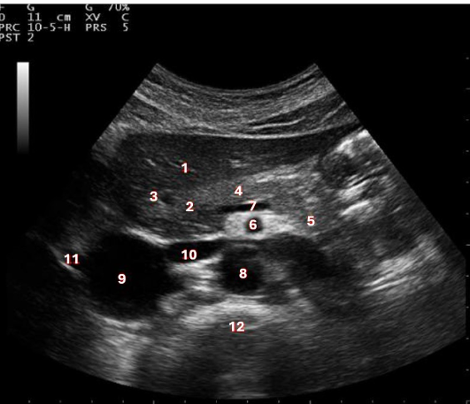
What structure/vessel is indicated by #4?
a. body of pancreas
b. head of pancreas
c. left lobe of liver
d. tail of pancreas
a
The main lobar fissure:
a. separates anterior right lobe and median left lobe
b. separates the anterior and posterior right lobes
c. separates the medial and lateral left lobes
d. separates the IVC and medial left lobe
a
Hepatomegaly in a 5 y/o is diagnosed using which of the following criteria?
a. multiply the patient’s age in years by the number 2 to obtain the maximum normal liver length for the patient’s age
b. divide the patient’s height in inches by the number 10 to obtain the maximum normal liver length for the patient’s age
c. divide the patient’s age in years by the number 2 to obtain the maximum normal liver length for the patient’s age
d. normal liver should not extend more than 1 cm below the costal margin
d
The ________ can be identified anterolateral to the pancreas tail.
a. hepatic flexure of the colon
b. stomach
c. left kidney
d. superior mesenteric artery
b
All the following are true regarding Couinaud liver segmentation except:
a. each of the 8 segments has its own branch of the portal vein, hepatic vein, and bile duct
b. right and left lobes are divided by the branches of the main hepatic vein
c. divides liver into 8 functional segments
d. allows for liver resection/surgery without negatively affecting other segments
b

What structure/vessel is indicated by #12?
a. superior mesenteric artery
b. aorta
c. IVC
d. spine
d
The ligamentum venosum travels between:
a. the main portal vein and the IVC
b. the right portal vein and the IVC
c. the left portal vein and the IVC
d. caudate lobe and the right lobe
c
In the normal liver, which of the following correctly describes the changes in flow in the hepatic artery after eating?
a. increased end diastolic flow velocities
b. no change occurs in the hepatic artery after eating
c. decreased peak and end diastolic flow velocities
d. increased resistive index (RI)
d

Which waveform represents a normal inferior vena cava waveform?
A

What structure is indicated by the purple circle?
a. main portal vein
b. Caroli disease
c. choledochal cyst
d. portal triad
d

Which of the following structures is labeled #8?
a. lateral left lobe
b. medial left lobe
c. anterior right lobe
d. posterior right lobe
d

What structure is labelled #8?
a. caudate lobe
b. medial left lobe
c. ligamentum teres
d. lateral left lobe
e. ligamentum venosum
b
If a liver mass is located between the middle hepatic vein and the right portal vein, in what lobe of the liver is the mass located?
a. posterior right lobe
b. caudate lobe
c. anterior right lobe
d. medial left lobe
c
The abdominal organ that produces the majority of alkaline phosphatase is?
a. gallbladder
b. liver
c. pancreas
d. spleen
b

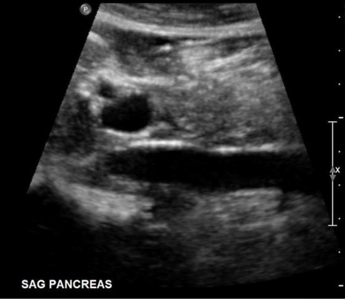
What structure is marked with X on the image?
a. superior mesenteric vein
b. splenic vein
c. common bile duct
d. main pancreatic duct
d
Which of the following statements correctly describes imaging of the dome of the liver?
a. the portal confluence should be used to locate the dome of the liver
b. deep inspiration is required to evaluate this portion of the liver
c. intercostal scanning is the preferred scanning approach to evaluate the dome of the liver
d. because the dome is bordered superiorly by the diaphragm, lobe artifact commonly inhibits complete evaluation of this portion of the liver
b

Use your mouse to place your cursor over the left medial superior segment of the liver and click to mark the structure.

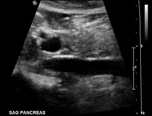
Use your mouse to place your cursor over the main portal vein and click to mark the vessel.
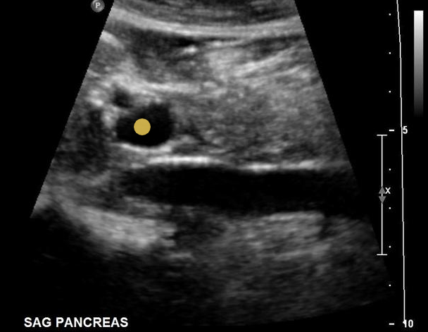

Which of the following structures is labelled #7?
a. anterior right lobe
b. lateral left lobe
c. medial left lobe
d. posterior right lobe
a

Use your mouse to place your cursor over the superior mesenteric vein and click to mark the vessel. If the vessel is not demonstrated on the image, mark the purple box that says “not demonstrated on this image.”
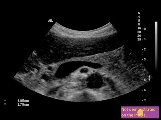
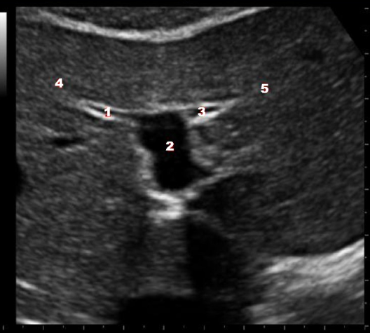
What structure/vessel is indicated by #4?
a. lateral left lobe
b. caudate lobe
c. medial left lobe
d. right lobe
c
The caudate lobe occupies much of the _______ surface of the liver.
a. posterior superior
b. anterior inferior
c. posterior inferior
d. anterior superior
a
In the porta hepatis, what structure is anterior to the portal vein and lateral to the proper hepatic artery?
a. duct of Wirsung
b. cystic duct
c. common hepatic artery
d. common hepatic duct
d

The purple arrow in the images represent which of the following structures?
a. medial left lobe
b. ligamentum teres
c. caudate lobe
d. ligamentum venosum
d
The liver’s exocrine function includes producing:
a. urea
b. heparin
c. bile
d. hemoglobin
c

Which of the numbered vessels is a part of the portal system?
a. 1
b. 2
c. 3
d. 1 and 3
b

Which of the following structures is labeled #4?
a. middle hepatic vein
b. IVC
c. right hepatic vein
d. right portal vein
c
The Rex-Cantle line is an imaginary line that extends from the ______ in the liver.
a. right to left portal vein
b. GB neck to right portal vein
c. middle hepatic vein to the main portal vein
d. GB fossa to IVC
d
Which of the following can be used to differentiate Reidel lobe from hepatomegaly?
a. the left lobe is normal in size with Reidel lobe but enlarged with hepatomegaly
b. the ligamentum venosum is not visible in cases of Reidel lobe but well seen in hepatomegaly
c. the right lobe is normal in size in Reidel lobe but enlarged in hepatomegaly
d. the ligamentum teres is not visible in cases of Reidel lobe but well seen in hepatomegaly
a
In a patient with complete situs inversus, the liver will be:
a. absent
b. located in the right lower quadrant
c. divided into 3 separate lobes
d. located in the left upper quadrant
d

What structure is indicated by the blue arrow?
a. common hepatic duct
b. main portal vein
c. common hepatic artery
d. common bile duct
a
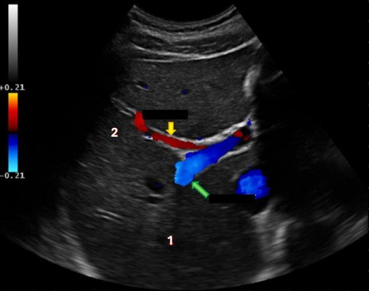
What liver vessel is indicated by the yellow arrow?
a. anterior right portal vein
b. medial left portal vein
c. posterior right portal vein
d. lateral left portal vein
a
The pancreas is found in what retroperitoneal space?
a. anterior pararenal
b. retrofascial
c. perirenal
d. posterior pararenal
a
The patient presents for an abdominal sonogram due to a history of hepatic congestion. What structure(s) should be closely evaluated for related findings?
a. portal system
b. renal arteries
c. celiac axis
d. left renal vein
a
All of the following are true regarding the spiral valves of Heister, except:
a. may simulate stone
b. may simulate shadow
c. located in the common bile duct
d. located in the cystic duct
c
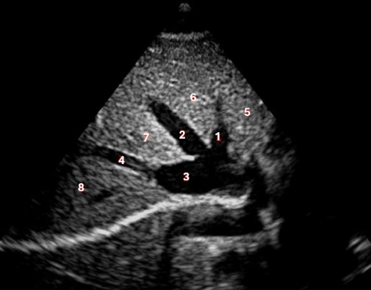
Which of the following structures is labelled #1?
a. left hepatic vein
b. right hepatic vein
c. IVC
d. left portal vein
a
The right and left hepatic ducts come together to form the common hepatic duct:
a. at the level of the Ampulla of Vater
b. at the pancreas head
c. inside the liver, near the porta hepatis
d. deep within the liver parenchyma
c
Sonographically, the ________ appears to connect the GB neck and the junction of the right and left portal veins.
a. intersegmental fissure
b. main lobar fissure
c. ligamentum teres
d. ligamentum venosum
b

What structure is indicated by the yellow arrow?
a. ligamentum venosum
b. falciform ligament
c. ligamentum teres
d. main lobar fissure
d
Which of the following ligaments separates the medial and lateral left lobes of the liver?
a. broad ligament
b. ligamentum venosum
c. ligamentum teres
d. coronary ligament
c
Which of the following terms can be used to describe a normal Doppler tracing from the hepatic veins?
a. continuous
b. non-pulsatile
c. triphasic
d. monophasic
c
The common bile duct is formed by:
a. varies per patient
b. common hepatic duct + cystic duct
c. cystic duct + duct of Wirsung
d. right and left hepatic ducts + cystic duct
b

Which of the following structures is labelled #2?
a. IVC
b. left hepatic vein
c. middle hepatic vein
d. left portal vein
c
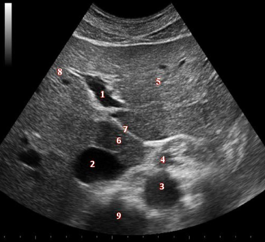
What structure/vessel is indicated by #9?
a. IVC
b. aorta
c. SMA
d. air within intestines
e. spine
e
What lab value is associated with the development of jaundice?
a. leukocytes
b. blood urea nitrogen
c. hematocrit
d. bilirubin
d

What structure/vessel is indicated by #5?
a. caudate lobe
b. right lobe
c. medial left lobe
d. lateral left lobe
d

What structure/vessel is indicated by #2?
a. main portal vein
b. lateral branch of LPV
c. medial branch of LPV
d. left portal vein
d
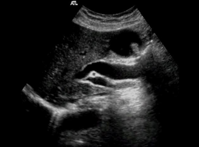
Use your mouse to place your cursor over the main portal vein and click to mark the vessel.

_______ merge to form the ampulla of Vater just prior to the duct entering the second portion of the duodenum.
a. the CHD and cystic duct
b. the CBD and duct of Wirsung
c. the CBD and cystic duct
d. the CBD and CHD
b

The purple arrows point to which of the following liver structures?
a. ligamentum venosum
b. falciform ligament
c. ligamentum teres
d. main lobar fissure
c
In the porta hepatis, what structure is anterior to the portal vein and medial to the common hepatic duct?
a. proper hepatic artery
b. cystic duct
c. common hepatic artery
d. Duct of Wirsung
a