Organelles and Movement of Materials Through the Cell
1/68
There's no tags or description
Looks like no tags are added yet.
Name | Mastery | Learn | Test | Matching | Spaced |
|---|
No study sessions yet.
69 Terms
Lysosome
Digests obsolete organelles
Nucleolus
Synthesizes ribosomal RNA
smooth endoplasmic reticulum (SER)
detoxifies many types of drugs and foreign toxins
the organelle primarily responsible for detoxifying many types of drugs and foreign toxins. This function is especially prominent in liver cells (hepatocytes), which contain a large amount of smooth ER.
Golgi apparatus
Glycosylates proteins destined to be part of the cell’s glycocalyx
Rough ER
Synthesizes peptide proteins destined to be released from the
cell into the bloodstream
Smooth ER
Storage depot of calcium used in skeletal muscle contraction
Chaperone proteins
Facilitates proper folding of proteins
Golgi Apparatus
Fails to stain with H&E and Romanovsky stains, resulting in a
negative image (i.e. pale area in the cytoplasm)
Proteasome
Breaks down improperly folded proteins
Lysosome
Breaks down phagocytosed (ingested) infectious agents
Mitochondria
Organelle, other than the nucleus, that has its own DNA
Mitochondria
Production of ATP through fatty acid oxidation
True or false: organelles are metabolically active
True
examples
nucleus
mitochondria
smooth & rough ER
golgi apparatus
lysosomes
cytoskeletal elements
What types of inclusions are metabolically inactive?
fat
glycogen
melanin
hemosiderin
lipofuscin
Membrane Bound Organelles
•Lipid membranes separate different compartments of the cell, many containing mutually incompatible biochemical reactions.
What organelles have a double membrane?
nucleus
mitochondria
What organelles have a single membrane?
•Rough endoplasmic reticulum
•Smooth endoplasmic reticulum
•Transport vesicles
•Lysosomes
•Endosomes
•Peroxisome
•Golgi apparatus
Which organelles can be seen with visible light microscopy?
nucleus
golgi (negative image)
nucleolus
***most organelles require electron microscopy to be seen
True or false: some organelles influence the color of the cytoplasm
True
•Nucleus, ribosomes (nucleic ACIDS): Basophilic (blue)
•Mitochondria, Smooth ER: Eosinophilic (pink).
*proteins are usually eosinophilic (pink)
What color does the nuclei appear to be with an H&E stain?
Basophilic (blue)
negatively charged nucleic acids bind basic (usually blue/purple) dyes
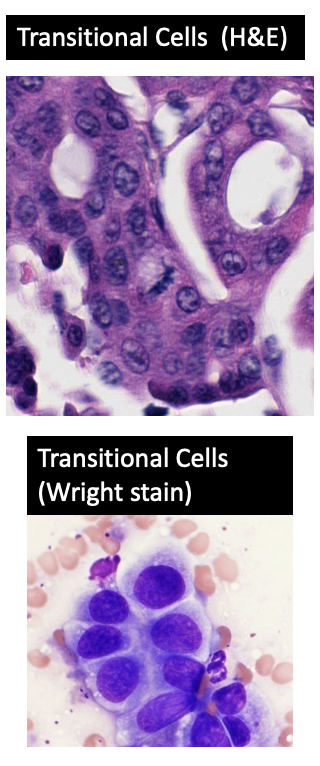
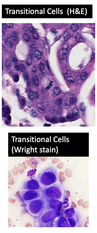
What color does the nuclei appear to be with a Romanovsky stain (e.g. Wright Stain, DifQuik)?
Purple
Cytoplasm is lightly basophilic or eosinophilic
True or false: histologic appearance of the nucleus can vary
True
•Usually round to oval
•Can be lobular or cigar shaped
— Nuclei vary in shape within cells, but will also be affected by plane of section – nuclei that are cigar shaped will appear round in a histologic section if cut in cross section.
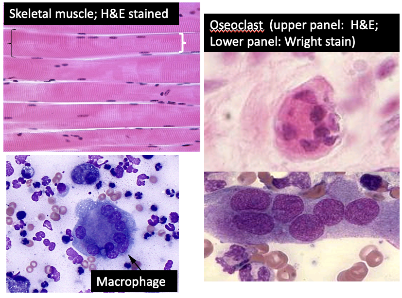
What types of cells are multinucleated?
osteoclasts
skeletal muscle
inflammatory macrophages
Heterochromatin
most highly condensed form of interphase chromatin in which genes cannot be expressed
Used to silence genes
Some DNA is permanently in heterochromatin (e.g. second X chromosome)
examples…
—> it is used to permanently inactivate one of the two X chromosomes in each female cell.
—> It can also silence genes that encode or regulatory proteins important in early embryonic development but unnecessary in an adult cell.
Euchromatin
uncoiled chromatin with active DNA
Most cell types express about 20 to 30% of their genes.
Active
“____” cells tend to have more euchromatin than quiescent cells
Transcribing a lot of DNA
Making a lot of proteins
Undergoing mitosis
less
More heterchromatin = a _____ (less/more) active cell
highly condensed chromatin
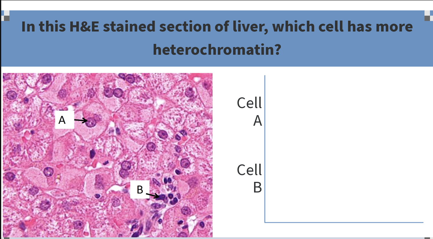
Cell B
B has a darker staining nucleus. This is probably a lymphocyte - the nucleus is much darker (i.e. more heterochromatin/more condensed chromatin) and it looks like there are some blood vessel cells in this area (endothelial cells). A is the nucleus of a hepatocyte (liver cell). These cells are very actively involved in protein synthesis — and therefore they need to continuously transcribe their DNA
terminally differentiated
Circulating neutrophils, eosinophils, and basophils are “____ _____”
•NOT capable of transcribing their DNA
•NOT capable of mitosis
***Most circulating WBCs have inactive nuclei (exception: monocytes & lymphocytes)
Which WBCs are capable of producing new proteins and capable of mitosis?
Monocytes and lymphocytes
True or false: Some cells can increase the amount of euchromatin when needed
True
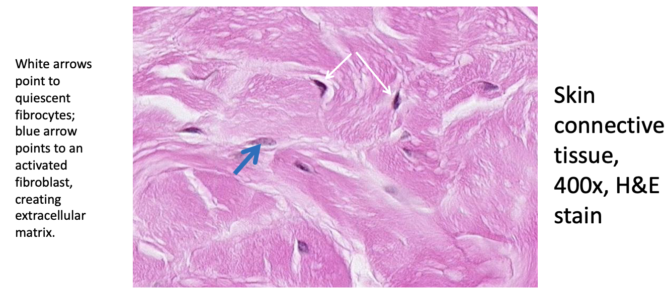
Most of these connective tissue cells (fibrocytes, black arrows) are quiescent, not actively producing connective tissue proteins and their nuclei contain a high proportion of heterochromatin. If there is a wound, these cells can “wake up” and start producing the extracellular proteins that strengthen the tissue. When this happens the chromatin unravels to increase the amount of euchromatin (fibroblasts, blue arrow).
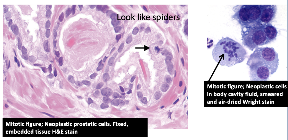
mitosis
During ____, chromatin is looped and further condensed into a chromosome
Chromatin is not only tightly condensed but is free - no longer contained within a nuclear envelope
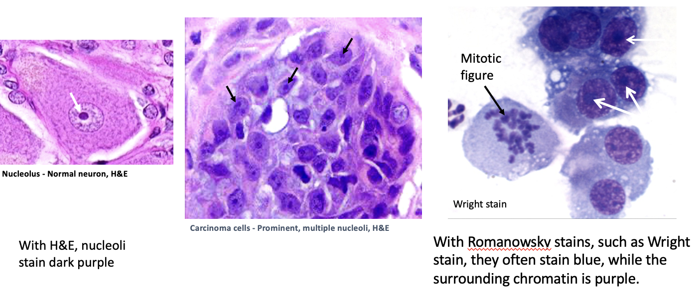
nucleolus
responsible for synthesis of ribosomes
more prominent in cells actively involved in protein synthesis
contains
rRNA
DNA w/ genes responsible for:
- production & processing of rRNA
- production of tRNA
Ribosome subunits exported from nucleus and assemble in cytoplasm
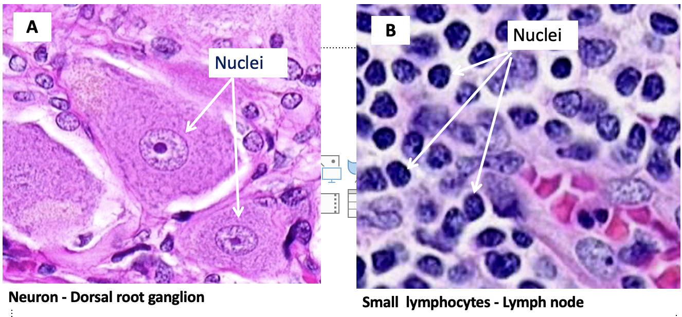
Based on the nuclear features, which cell type is the most likely to be transcribing a lot of its DNA?
Cell type A appears more active
explanation…
Cell A:
diffuse, fine granular chromatin (euchromatin; loosely condensed chromatin)
prominent nucleolus
lots of uncoiled DNA
Active transcription = busy cell
Cell B:
clumped, course granular chromatin
mostly coiled DNA
Can’t see a nucleolus
low level transcription = inactive cell
necrosis
cell suicide
Cells die by 2 distinct mechanisms called ___ and ____
Necrosis
Cell murder
→ initiated by external factors such as hypothermia, excessive heat, low pH, microbial pathogens and ionizing radiation.
—> Cells that die by this process undergo swelling and membrane rupture. Necrosis always results in a secondary inflammatory response.
mechanical injury
hypoxia
infectious agents
toxins
Apoptosis
Cell suicide
→ involves internal programming to eliminate cells no longer needed or cells with irreparable internal damage.
cells no longer needed
irreparably damaged cells
extrinsic & intrinsic signals
TNFa(alpha)
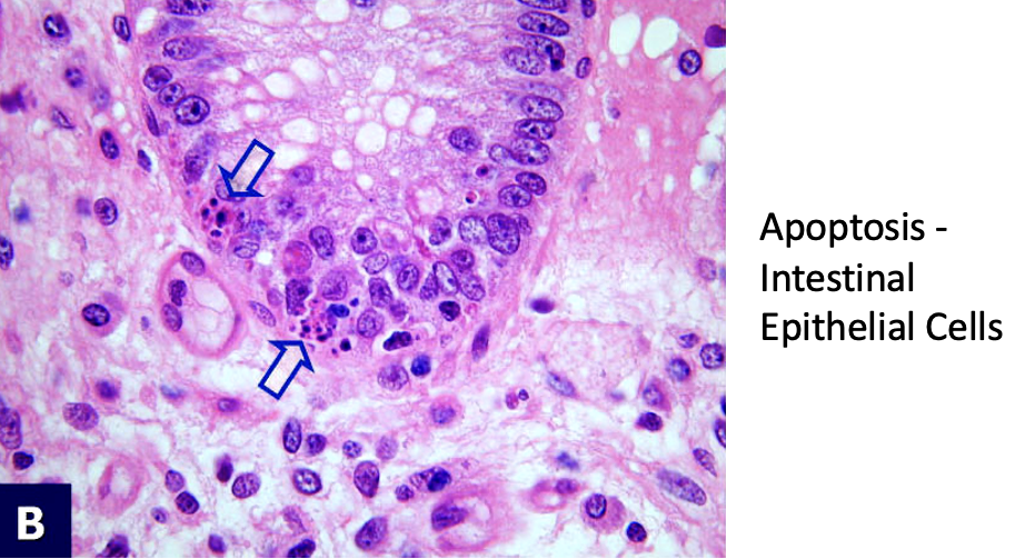
Nuclear appearance in Cell Death (Necrosis vs Apoptosis)
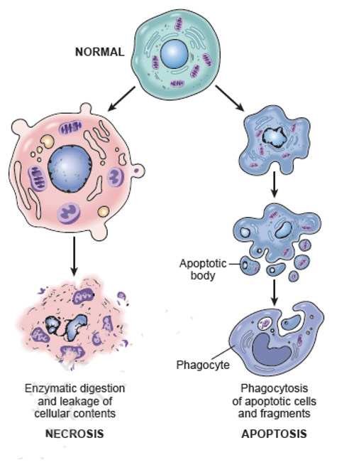
Necrosis:
•Cells swell
•Nucleus swells
•Membrane dissolves
•Many cells involved
•Cells burst & contents trigger an inflammatory reaction
Apoptosis
•Cells shrink & cytoskeleton collapses
•Nuclear chromatin condenses (pyknosis & karyorrhexis)
•Nuclear envelope disassembles
•Cell forms blebs into fragments (apoptotic bodies)
•Macrophages “eat” them
—> microscopic appearance: fragments of dark nucleus, few dead cells, little to no inflammation; Because the cells are being eaten (phagocytized) and digested quickly, there usually few dead cells to be seen in a given area, even when large numbers of cells have died by apoptosis.
Which organelles is responsible for protein synthesis?
Ribosomes
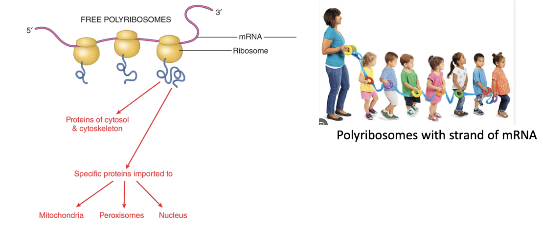
proteins that will remain in cytoplasm are synthesized by free ribosomes that float in cytoplasm
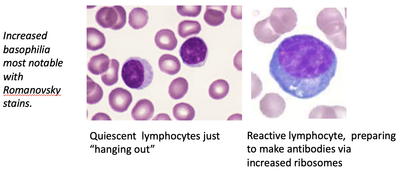
True or false: cells making lots of protein have large numbers of ribosomes which cause cytoplasm to appear basophilic (blue-purple)
True
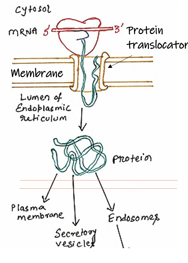
If a protein being made by the ribosome is destined for export it lands on the ER and passes out by Vesicle Transport.
If a protein intends to stay within the cell, ribosomes merely float around within the cell (polysome – chain of ribosomes on mRNA strand) and don't attach to the ER.
Ribosomes are stained more vividly by a Romanowsky stain than with H&E.

Protein folding is usually facilitated by ___
Chaperone proteins (aka heat shock proteins)
Network of molecular chaperones promote efficient protein folding
Several modes of action
These increase in response to heat and attempt to refold denatured proteins
Aberrantly folded proteins can cause aggregation → possible cellular damage.
What happens to proteins that fail to fold properly?
They are ubiquinated and then destroyed by a proteasome (an ATP-dependent protease)
The proteolytic machinery of the cell and the chaperones compete with one another

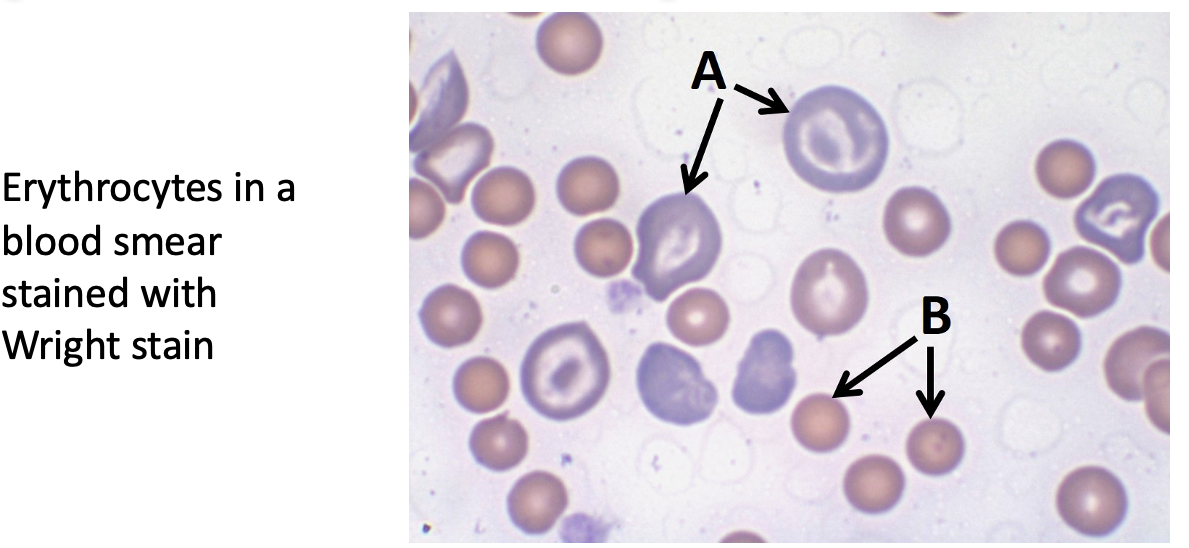
Immature RBC contain ribosomes, while mature RBC do not. Which of the indicated cells are immature RBC?
A is the immature RBC
cells making lots of protein have large numbers of ribosomes which cause cytoplasm to appear basophilic (blue-purple)
What organelle can ribosomes be bound to?
rER
Ribosomes bound to rER creates proteins for export to other organelles, to the plasma membrane, or out of body
Each type of organelle has characteristic membrane & lumen proteins, phospholipids and other specialized molecules related to its function
Ribosomes dock transiently on the rER
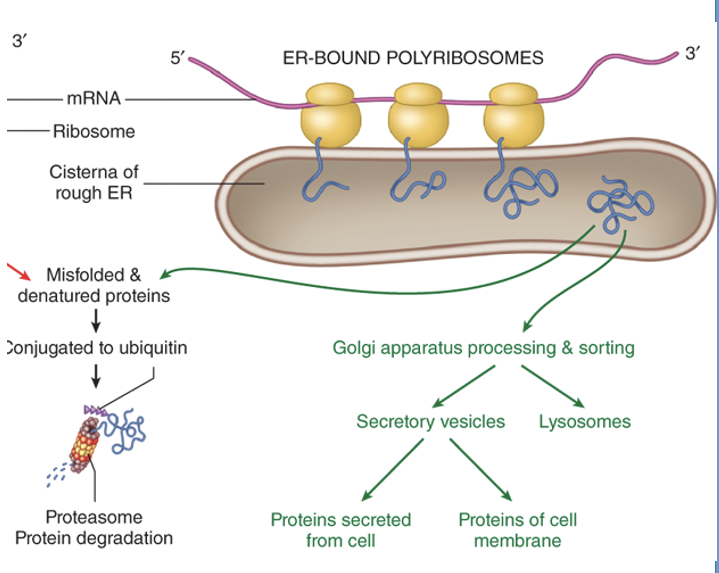
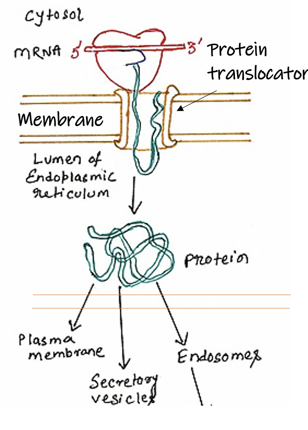
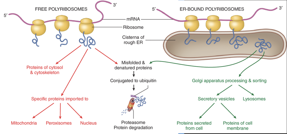
Free ribosomes vs ER-bound ribosomes
The main difference is their location and protein destination:
free ribosomes in the cytoplasm make proteins for use within the cell
while ribosomes bound to the rough endoplasmic reticulum (RER) synthesize proteins destined for export, secretion, or incorporation into cellular membranes, lysosomes, or other organelles. Ribsomes are released after synthesizing protein
***Structurally, they are identical, and a ribosome can switch between free and bound states depending on the protein it is synthesizing
What are the types of proteins produced in the rER?
Water soluble proteins (injected into lumen by ribosome)
proteins are modified and folded
packed into vesicles which are ultimately transported to Golgi (and beyond)
Transmembrane proteins (become embedded in rER membrane)
destined for plasma membrane
OR
destined for membrane of endomembrane organelle
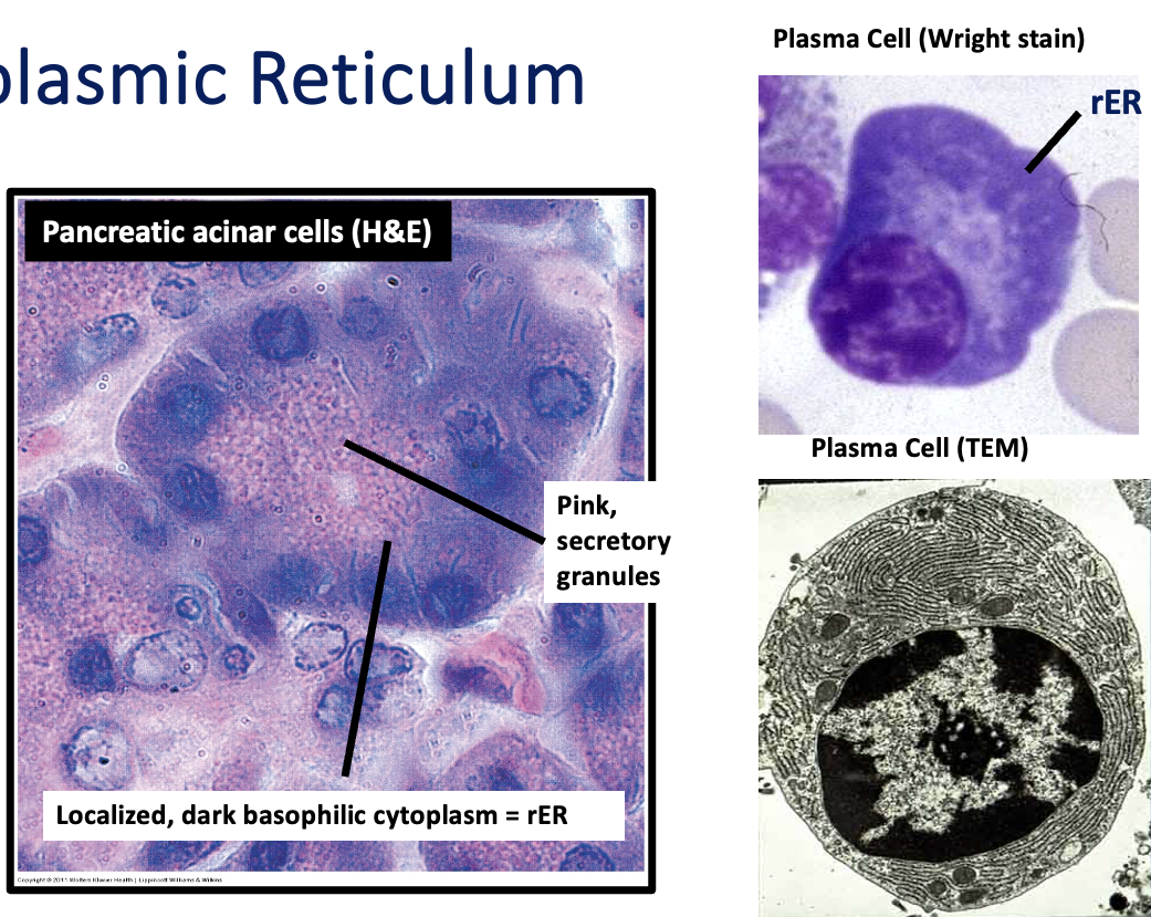
rER is well developed in ____cells
Secretory
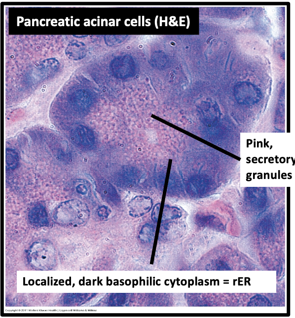
Embedded ribosomes (RNA) cause what color tinge to cytoplasm
blue (especially w/ Romanovsky stains)
Romanovsky stains, such as Wright stain, include a basic dye (Methylene Blue) which binds to nucleic acids and their phosphate groups. These stains are very sensitive indicators of ribosomes and rER and almost all cell types will show some degree of cytoplasmic basophilia (i.e. some blue coloration) unless they are free of ribosomes and rER (e.g. mature erythrocytes).
Relatively quiescent cells will have pale blue cytoplasm, reflecting the low numbers of these organelles present. A really deep blue color reflects a large number of ribosomes, either free or associated with rER – indicating active protein synthesis.
Hematoxylin is not a true basic dye, so in sections stained with H&E, the basophilia associated with ribosomes and rER is not as pronounced.
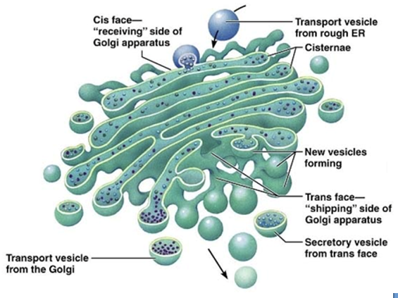
Golgi Apparatus
Organelle responsible for post-translational modification, sorting, and packaging
Stack of flattened saccules , near nucleus, without ribosomes
Usually curved – shaped dependent on cytoskeletal element and Golgi matrix proteins
Proteins are modified as they move through the saccules becoming glycosylated (sugars added), phosphorylated (phosphates added), sulfated, etc.
Golgi apparatus
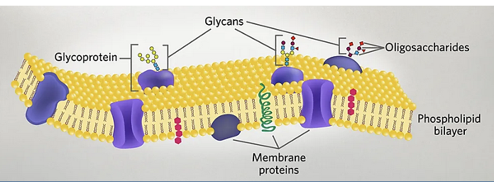
Organelle that modifies proteins and generates the heterogeneous oligosaccharide strucutre seen in mature proteins
List the role of glycoproteins for protein function
folding and stability
interactions w/ other molecules
cell signaling
cell-cell communication
cell protection, etc. (protection against proteosomes)
part of glycocalyx on outer membrane
carbohydrate rich layer surrounding the cell membrane of many cells
→ aids in protection
→ permeability layer
→ aid in cell-cell communication/signaling
What are some of the destinations that the golgi sends vesicles to?
secretory vesicles (e.g. collagen, hormones) (*material in them will be expelled*)
components for plasma membrane (e.g. receptors)
components of intracellular organelles (e.g. lysosomes)
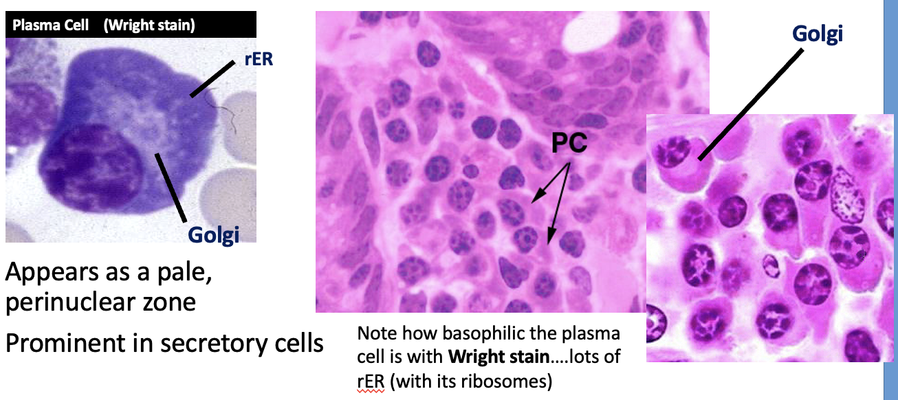
True or false: golgi apparatus does not stain well with H&E or Romanovsky stains
True
appears as a pale, perinuclear zone
prominent in secretory cells
As a result, in secretory cells, you can sometimes see a negative image where the Golgi apparatus sits next to the nucleus.
Romanovsky(wright stain) = mostly membrane → methylene blue won’t stain
Vesicular transport
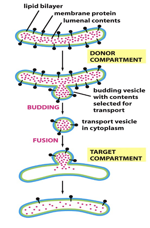
_____ _____ moves materials between organelles & the plasma membrane
Process of Vesicular transport
Vesicles bud off from one organelle and fuse with another (or the plasma membrane) contributing
Proteins from rER (modified in Golgi)
Lipids from sER
Things in lumen remain in lumen or pass into the extracellular space
Vesicles move from place to place by motor proteins that pull them along the cytoskeletal proteins
Vesicular transport
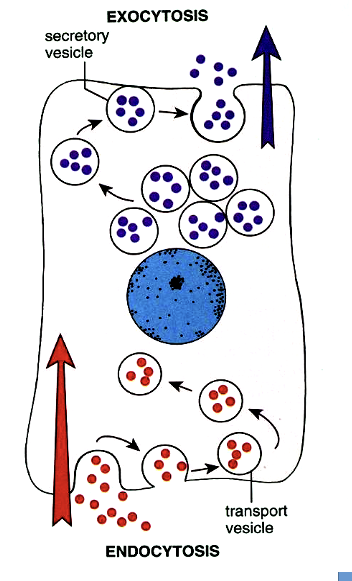
____ _____ allows movement of materials in & out of cell
Exocytosis
outward secretory pathway
Delivers new proteins, carbohydrates, and lipids to plasma membrane or the extracellular space
Endocytosis
inward secretory pathway
Plasma membrane components delivered to endosomes
Components can be recycled or delivered to lysosomes for degradation
Used to import various nutrients or ingest organisms or debris
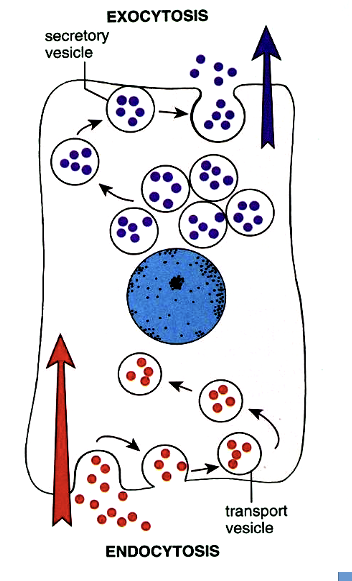
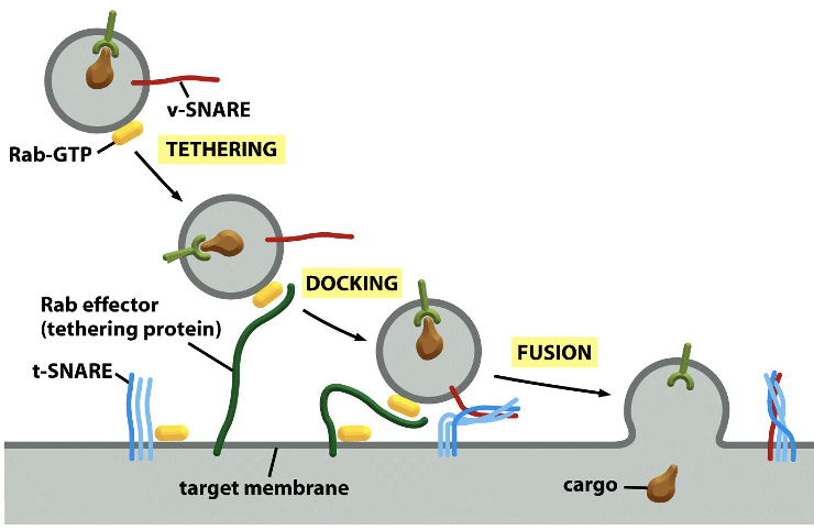
Vesicular transport is facilitated by which proteins? Also describe process.
SNARE proteins
Vesicle docks with the appropriate organelle (e.g. lysosome) through molecular markers on vesicle membrane and SNARE proteins
SNARE on vesicle (red line) interacts with SNARE on target (blue squiggles)
Holds vesicle in place
•SNARE proteins also catalyze vesicle fusion with membrane of target organelle or plasma membrane
How does the Clostridium botulinum neurotoxin cause botulism?
Acts on key SNARE proteins
Synaptic vesicles containing neurotransmittors are released from neurons and stimulate muscles to contract.
Neurotoxin cleaves one of the SNARE proteins (exocytosis no longer works)
prevents fusion of vesicle
prevents neurotransmitter from stimulating muscle
Result is muscle paralysis
as toxin spreads, it can block nerves controlling the respiratory tract and heart, leading to death
How does exocytosis occur?
Through fusion of vesicle’s membrane with the cell’s membrane