Anatomy-General Intro & Review of Systems
 Call Kai
 Learn
Learn Practice Test
 Spaced Repetition
 Match
Match Flashcards
 Knowt Play
1/159

There's no tags or description
Looks like no tags are added yet.
Name | Mastery | Learn | Test | Matching | Spaced | Call with Kai | Chat |
|---|
No analytics yet
Send a link to your students to track their progress
160 Terms
Hollow area of organs, vessels, and airways
Lumen
The inner region or core of an organ
Medullary
The outer region or layer of an organ/body part
Cortex (cortical)
T/F: basal refers to the bottom part of an organ
F, the base is not always the bottom (eg. heart)
What are the 2 dorsal cavities?
Cranial and spinal
What are the 2 ventral cavities?
thoracic and abdominopelvic
What makes up the thoracic cavity?
Mediastinum and left/right pleural cavities
What creates a thin, watery fluid that lubricates organs as they rub against one another?
serous membranes
T/F: serous membranes are one continuous sheet
true (balloon example)
____________ membranes line the wall of the body cavity
Parietal
_____________ membranes cover the surface of the organ
Visceral
The mediastinum contains these 12 things
Heart, trachea, left and right bronchi, esophagus, thymus, aortic arch, thoracic aorta, vena cava, thoracic duct, lymph nodes, nerves
The heart is covered in a double-layered ______________
pericardium
Pericarditis is inflammation of the ____________ ____________ often attributed to infection
serous membrane
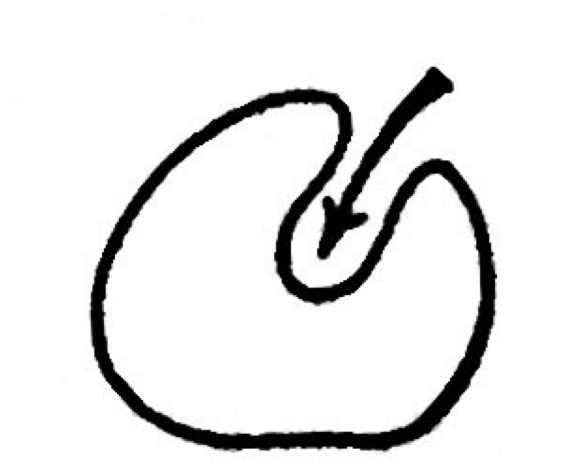
Invagination
Depression

Evagination
Protrusion
What movements can you perform in the sagittal plane?
flexion and extension
What movements can you perform in the coronal plane?
abduction, adduction
What movements can you perform in the transverse plane?
Rotation
Fingers spread apart is _____________
abduction
Fingers all together is ____________
adduction
Supination is rotating __/__
out/up
Pronation is rotating ___/___
in/down
Moving your toes/foot to the ceiling is called?
dorsiflexion
Pointing your toes/foot down to the ground is called?
plantarflexion
Moving the sole of your foot medially is called?
inversion
Moving the sole of your foot laterally is called?
Eversion
Which has thinner walls-veins or arteries?
veins
Which has larger lumens-veins or arteries?
veins
The innermost layer of blood vessel tissue is called?
tunica intima
The tunica intima is lined with ____ layer of ____________ cells
one, endothelial
The middle layer of blood vessel tissue is called?
tunica media
Tunica media contains _________ __________ cells
smooth muscle
The outermost layer of blood vessel tissue is called?
tunica adventitia
Tunica adventitia is made of?
fibroblasts, collagen, elastin
Vasa vasorum
a blood vessel for the blood vessel
The basic flow of blood in blood vessels is?
artery, arterioles, capillary, venule, vein
Capillaries only possess which blood vessel layer?
tunica intima
When arteries and veins create an imbalance of fluid (lymph) in the tissue, ______________ vessels will take the excess fluid out, filter it through _______ ________ and give it to the _______ which will then recirculate it properly
lymphatic, lymph nodes, veins
How many liters of blood is pumped every minute by an adult?
5-6
The right atrium receives?
de-oxygenated blood
The left atrium receives?
oxygenated blood
The right ventricle receives?
de-oxygenated blood
The left ventricle receives?
oxygenated blood
What is the order of circulation through the heart?
right atrium, tricuspid valve, right ventricle, pulmonary valve, left atrium, bicuspid (mitral) valve, left ventricle, aortic valve, systemic circulation
The arterial system brings O2 _________ blood ________ from the heart
rich, away
The venous system brings O2. ___________ blood _________ the heart
depleted, toward
Pulmonary veins bring _____________ blood ________ the heart
oxygenated, toward
What are the 2 ducts that take lymph and return it to the venous system?
thoracic and right lymphatic
Coronary arteries arise from the _________ of the __________ ___________ valve
sinuses, aortic semilunar
The aorta gives rise to this vessel(s) on the R side?
brachiocephalic artery
The aorta gives rise to this vessel(s) on the L side?
L common carotid artery, L subclavian artery
The abdominal aorta is below your ___________
diaphragm
What is the major blood flow through the arm?
subclavian, axillary, brachial, radial and ulnar, palmar arches
The subclavian artery is found at?
lateral edge of first rib
The brachial artery is found at?
inferior edge of teres major
the radial and ulnar arteries are found at?
antecubital fossa
What is the major blood flow through the leg?
abdominal aorta, common iliac artery, external and internal iliac artery, femoral artery, deep femoral artery, popliteal artery, anterior and posterior tibial artery
What forms the Circle of Willis?
internal carotid artery, vertebral artery
What composes the axial skeleton?
skull, vertebral column, rib cage, sternum
What composes the appendicular skeletan?
upper and lower limb bones
Each bone is made of ______ bone and _______ bone
compact, spongy
Compact bone provides an area for?
muscle attachment
Compact bone contains ______
osteons
A ______ is a structural & functional unit of compact bone
osteon
Osteocytes are located in small _______ (gaps between lamellae)
lacunae
Canalculi connect ________ to _________ _______
lacunae to blood vessels
Osteoblasts become _______ when they mature
osteocytes
Purpose of osteoclasts?
break down bone for nutrients or release of minerals
The ________ ______ forms the outer boundary of an osteon from the interstitial lamellar bone
cement line
Lamellae are the rings of osteons that contain ___________ and a _____________ _______
collagen, calcified matrix
The haversion canal contains?
blood vessels, nerves, lymphatic vessels
The haversian canal runs down the center of each ________
osteon
Sharpey’s fibers connect _______ bone to _________
compact, periosteum
Osteoblasts are known as ____ __________
bone producers
Osteon are?
basic structural unit of bone
These connect the blood vessels of each osteon
Volkmann canals
Spongy bone forms a lattice of thin beams called?
trabeculae
The spongy bone contains reticular tissue that contains _____ ____ responsible for making _____
stem cells, RBC
Connective tissue covering bone except for joints
periosteum
The periosteum is anchored to compact bone via?
Sharpey’s fibers
At the top of bones, you’d expect to see ___________ instead of periosteum
cartilage (to connect to joints)
The outer layer of periosteum is pierced by blood vessels and nerve fibers that pass into the ____________ _______
volkmann’s canals
The endosteum contains ________ and __________
osteoblasts, osteoclasts
The endosteum lines the _________ ________
medullary cavity
Shell of compact bone surrounding the medullary cavity
diaphysis (shaft)
The __________ is a shell of compact bone covering spongy bone, with spaces filled with ______ ________
epiphysis, red marrow
Red marrow is a site for ________
hemopoiesis
Hemopoises
site of production of RBCs, WBCs, and platelets
area between diaphysis and epiphysis
Metaphysis
The epiphyseal (growth) plate is made of?
hyaline cartilage
The epiphyseal (growth) plate is located between?
epiphysis and diaphysis
The epiphyseal plate becomes the ___________ ______ in adulthood
epiphyseal line
The __________ _________ is filled with yellow marrow
medullary cavity
Intramembranous bone ossifies from a ________ percursor
membrane
Bones of the face, calvaria, clavicle, and distal tips of fingers are all examples of?
intramembraneous bone
Endochondral bone ossifies from a ________ percursor
(hyaline) cartilage
Limb bones and bones of the cranial base are examples of?
endochondral bones
All bone starts out as a sheet of undifferentiated _________ cells in embryo/fetus
mesenchyme
Intramembraneous ossification proceeds directly from the __________
mesenchyme