0.4 Non-specific responses against disease
 Call Kai
 Learn
Learn Practice Test
 Spaced Repetition
 Match
Match Flashcards
 Knowt Play
1/37

There's no tags or description
Looks like no tags are added yet.
Name | Mastery | Learn | Test | Matching | Spaced | Call with Kai | Chat |
|---|
No analytics yet
Send a link to your students to track their progress
38 Terms
Syllabus
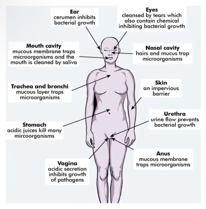
The body's external defence mechanisms against pathogens include features of the:
skin
digestive tract
urogenital tract
respiratory system
the ear
the eye
Pathogens that enter the body are targeted by non-specific immune responses of inflammation & fever.
Non-specific defences
Body's first line of defence against pathogens.
Not directed at a particular/specific pathogen.
Work against all pathogens in the same way.
Can be classified into external and internal.
External defences: skin
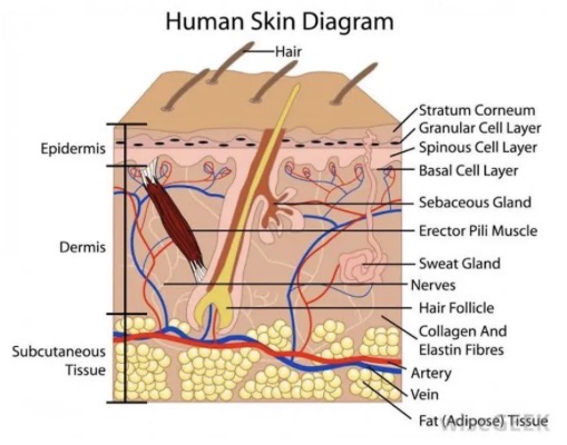
Effective barrier covering all outside of the body.
Prevents entry of pathogens into the body.
Sebum produced by the sebaceous glands in the skin contains substances that inhibit the growth of bacteria. Also helps to keep the skin from drying out and cracking.
Sweat secreted onto the skin contains lysozymes which is a substance that kills pathogens.
Contains harmless and useful bacteria that occupy the skin surface and prevent pathogens from gaining space on the skin.
External defences: mucous membranes
Body contains cavities that open to the outside such as mouth, nose and urethra.
These cavities are lined with mucus membranes.
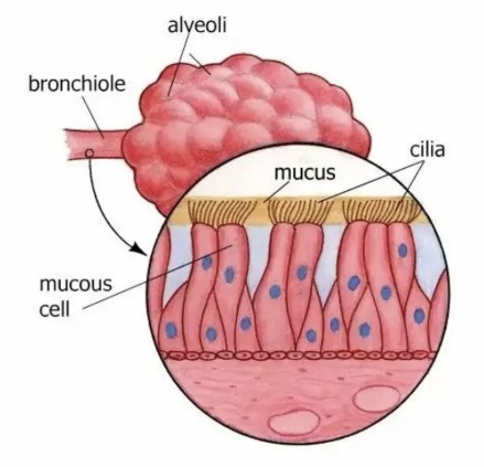
Mucus membranes secrete mucus which inhibits the entry of pathogens into the body by trapping them in their sticky fluid.
External defences: hair
Found in the nose and ears.
Help to trap larger pathogens and prevent them from gaining entry to the body.
Hair in the nasal cavity also helps to slow air movement to trap pathogens.
External defences: cilia
Tiny finger-like projections from cells.
Waft in the same direction to (with move mucus (with trapped pathogens) to the throat where it can be swallowed.
External defences: acids
Stomach juices contain hydrochloric acid that kills pathogens.
Vagina has acidic secretions that kill pathogens.
Sweat is slightly acidic to prevent the growth of pathogens.
External defences: lysozyme
Enzyme that kills bacteria.
Found in sweat, tears, saliva, nasal cavity and cerumen.
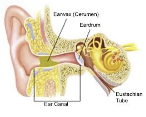
External defences: cerumen
Found in the ear (earwax).
Provides a physical barrier to pathogens entering the body.
External defences: flushing action of body fluids
Urine, sweat, saliva and tears help to flush and remove pathogens from the body.
Protective reflexes
Automatic, involuntary response to a stimulus.
Help to protect against pathogens.
reflexes help to protect against infection
4 reflexes against infection
Sneezing
Coughing
Vomiting
Diarrhoea
Reflexes: sneezing
irritation of the walls of the nasal cavity can stimulate a sneeze. This forceful expulsion of air from the lungs carries mucus out of body which may carry pathogens.
Reflexes: coughing
irritation of the bronchi or bronchioles can cause coughing. This will forcefully remove mucus from the lungs which may carry pathogens.
Reflexes: vomiting
contraction of the muscles of the abdomen and diaphragm expels the contents of the stomach. Stimulus can be excessive stretching of the stomach or bacterial toxins.
Reflexes: diarrhoea
caused by irritation of the small or large intestine by a pathogen. Irritation causes increased contractions of the muscles in the wall of the intestines. This will move the pathogen more quickly through the intestines.
Internal non-specific defences
If pathogens get past the external defences then the internal non-specifc responses is the next line of defence.
Cells included in internal non-specific defences
phagocytes
Leukocytes
Macrophages
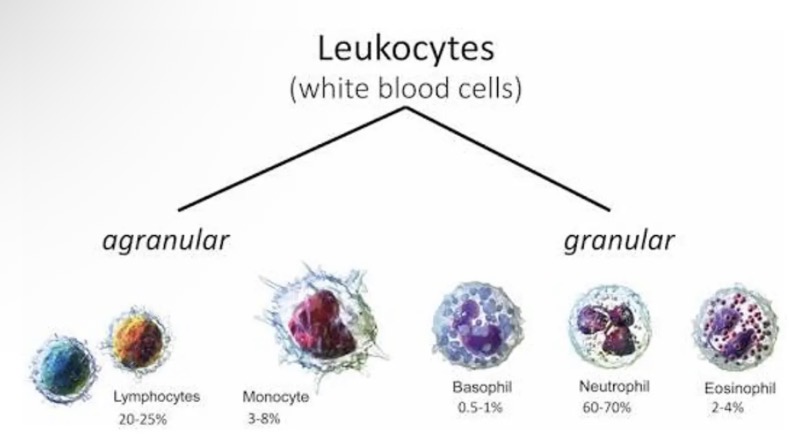
Internal non-specific defences: leukocytes
Several different types
Leave the blood capillaries and migrate to site of infection/injury.
Will either engulf and digest bacteria or secrete substances that destroy bacteria.
Internal non-specific defences: macrophages
Develop from leucocytes.
Some remain fixed in place and destroy pathogens that come past.
Other move around looking for pathogens to destroy.
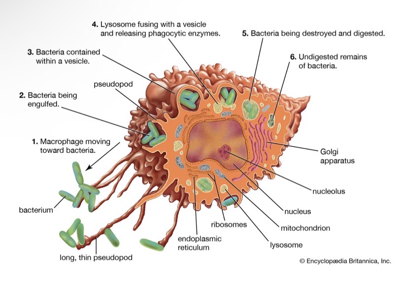
Internal non-specific defences: phagocytes
are a type of white blood cells that can engulf and digest micro organisms
Two types: leukocytes and macrophages
Function of the inflammatory response
reduce the spread of pathogens
Destroy and prevent entry of additional pathogens
Remove damaged tissue and cell debris
Begin repair of damaged tissue
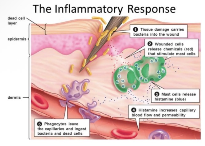
Signs of the inflammatory response
redness
Heat
Swelling
Pain
Main events in the inflammatory response PART 1
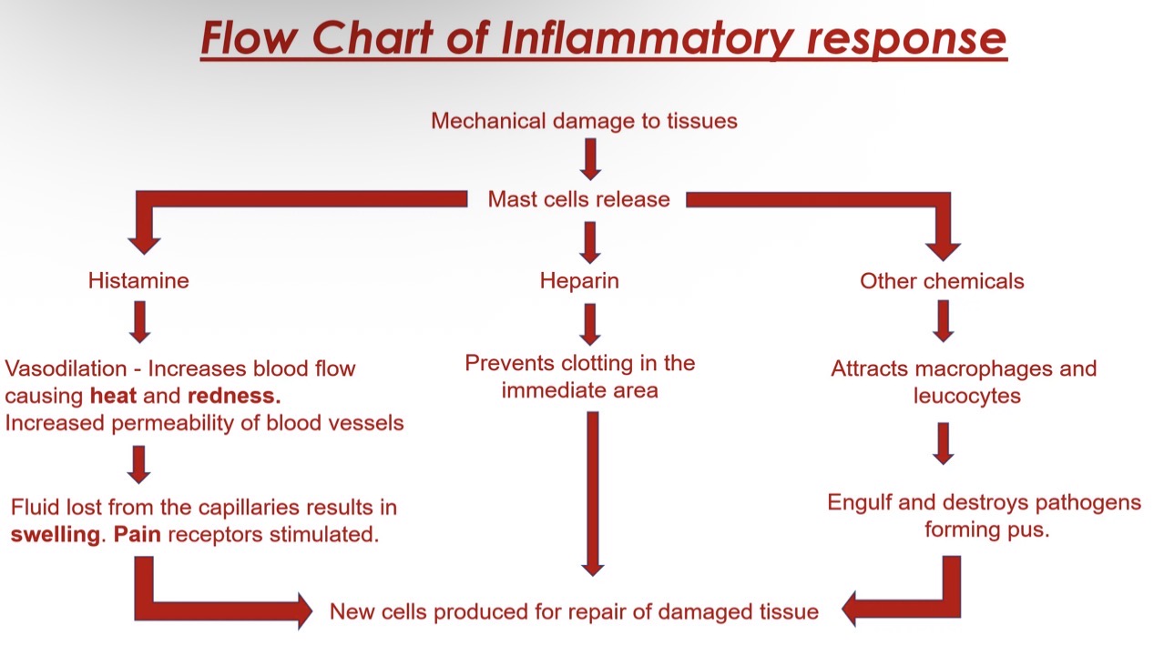
Damage to tissue stimulates mast cell (type of WBC) to release histamine, heparin and other chemicals into the area.
A clot of fluid forms around the damaged area to slow the spread of pathogens
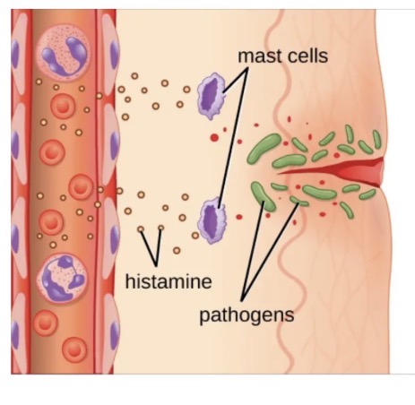
Inflammatory response: histamine
causes vasodilation of the blood vessels which leads to increased blood flow to the area = redness/heat.
It also causes increased permeability of the capillaries. Fluid is lost from the capillaries = swelling.
Inflammatory response: heparin
prevents blood from clotting in the immediate area. This ensures adequate amounts of blood carrying phagocytes will flow to area.
Main events in the inflammatory response PART 2
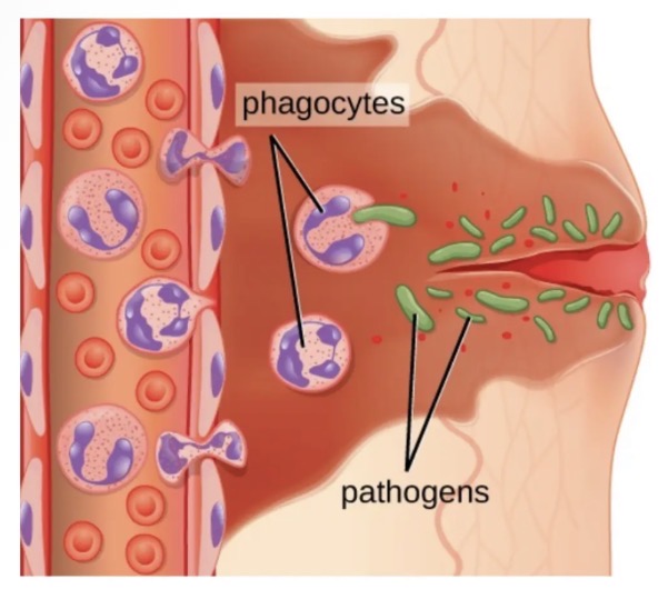
Complement system proteins and other chemicals released by the mast cells will attract macrophages and leucocytes(mainly neutrophils) to the area. They will engulf and destroy the pathogens by phagocytosis.
Changes in the tissues will stimulate the pain receptors causing the person to feel pain.
Leucocytes (mainly neutrophils) will die as they fill with bacteria / debris = yellow fluid called pus forms.
When the response is successful in removing the pathogen = tissue repair and healing will occur via mitosis.
Flow chart of inflammatory response
Fever
During the course of an infection a person may experience an increase in body temperature.
A body temperature above 37.2 degrees Celsius is considered having a
temperature (febrile).
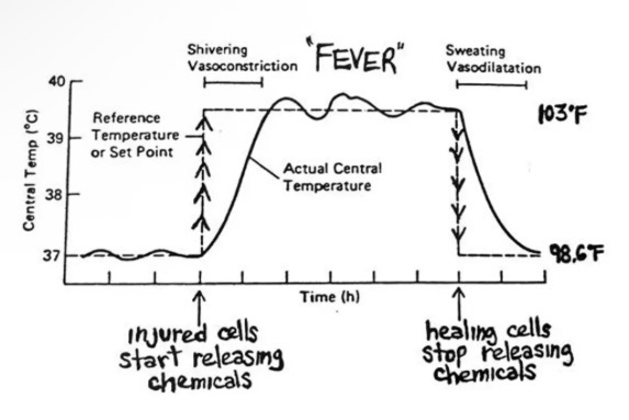
How a fever can assist the body in fighting pathogens
Interfering with viral replication
Inhibit growth and reproduction of some bacteria
Increasing production of killer T cells which speeds up the specific mediated immune response.
Increases heart rate and blood flow which enable lymphocytes to reach sites of infection quicker.
Increase metabolic activity which assists tissue repair.
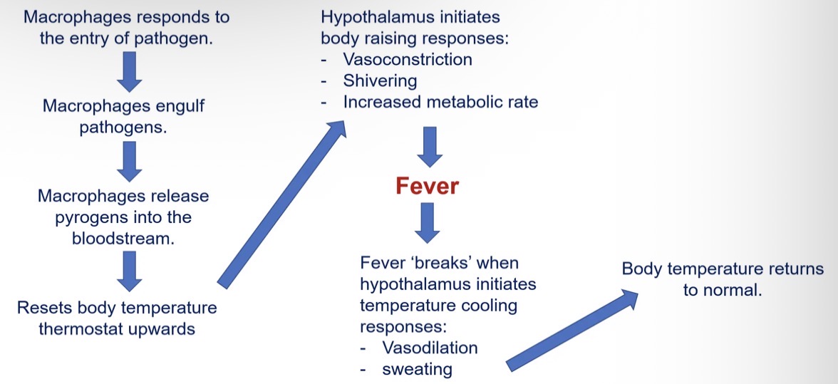
How a fever occurs
Macrophages are attracted to the infected area by release of chemicals from the mast cells.
Macrophages engulf and destroy pathogens. • Macrophages secretes cytokines/chemicals that produce chemical pyrogen(interleukin 1).
Pyrogen (interleukin 1) is released into the bloodstream. • Alters the body thermostat to make the body think it is cooler than it actually is.
Hypothalamus responds by initiating vasoconstriction and shivering.
Body temperature rises = fever.
Fever mechanism flow chart
Lymphatic system
Is an important part of the body's internal defence against pathogens.
Consists of lymph nodes.
All lymph will pass through a lymph nodes before entering back into the blood circulation.
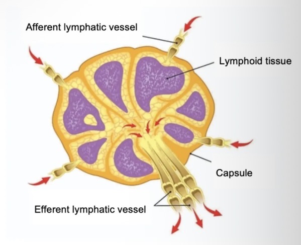
Lymph nodes function
remove debris and pathogens from the lymph and are thus sometimes referred to as the “filters of the lymph”
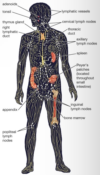
Bacteria in lymph
Any bacteria that infects the interstitial fluid are taken up by the lymphatic capillaries and transported to a lymph node.
Lymph nodes contain..
contains lymphoid tissue
Cells are arranged in a network of fibres - this helps to trap pathogens
Lymphatic system: macrophages
macrophages in the lymph node will destroy the foreign particles by phagocytosis
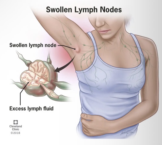
Lymphatic system: when an infection occurs
the formation of lymphocytes increases and more macrophages rush to the area to kill pathogens
This results in the lymph nodes becoming swollen and sore