11. Left vs Right Heart disease
 Call Kai
 Learn
Learn Practice Test
 Spaced Repetition
 Match
Match Flashcards
 Knowt Play
1/72

There's no tags or description
Looks like no tags are added yet.
Name | Mastery | Learn | Test | Matching | Spaced | Call with Kai | Chat |
|---|
No analytics yet
Send a link to your students to track their progress
73 Terms
left atrial enlargement, left ventricular enlargement, pulmonary edema, pulmonary venous enlargement, tracheal deviation, compression of mainstem bronchus
Which signs are consistent with left heart disease/failure?
15-20
normal tracheal position on lateral radiographs is at a _______ degree angle
right or left atria
Enlargement of which part(s) of the heart can deviate the trachea dorsally?
left ventricular border
In early feline heart disease the tracheal position is not as helpful, but the rounding of the ______________________ is a significant finding

normal indented cardiac waist
Is this heart normal or abnormal?

systemic hypertension, heart base mass
What are some differentials for a loss of cardiac waist that are NOT from an enlarged heart
enlarged right atrium, right auricular appendage, main pulmonary artery, ascending aorta, cranial vena cava
What structures can be enlarged that will lead to a loss of cardiac waist in canines?
aorta, ascending aorta, descending
In the dog, the size and shape of the ________ is a sentinel sign of cardiovascular disease. If the __________ is much bigger than the _________, we likely have disease.
12-1 oclock
Where on a VD would you see the aorta?
aortic bulge
The presence of an __________ on VD images can assist with the diagnosis of congenital heart disease like patent ductus arteriosus or with chronic hypertension
loss of caudal cardiac waist; L atrial enlargement
What is happening here? And what does this indicate?

dorsal deviation and compression of the left caudal mainstem bronchus
What sign of left atrial enlargement is seen here?

cool
Here is that dorsal deviation of the left caudal mainstem bronchus outlined if you struggled to see it before
cool?

backpack sign, severe left atrial enlargement
What sign do we see here and what does it mean?

between the mainstem bronchi, between 5-7 oclock
Where does the normal left atrium live on a VD view of the heart?

caudal border of the atrium has a double opacity sign
With mild left atrial enlargment in a the canine, you will be able to see what change on the VD/DV view?
double opacity; left atrial enlargement
So, what sign do we see here and what does it indicate?

normal
We can see the main stem bronchus at the bottom of the heart in this VD view... is this normal or abnormal placement?

bow-legged cowboy
As the left atrium enlarges, copression and deviation of the left caudal mainstem bronchus will cause what sign to be observed?

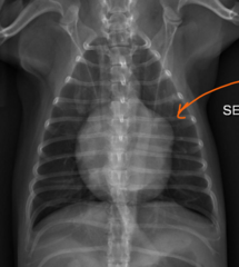
left auricle, atrium double opacity
As left heart disease in a dog advances, what else will enlarge? Seen here as the bulge at about 2-3 oclock. And what original sign becomes more clear?
false, the left atrium is the same on laterals, but is located at 1-2 oclock on the VD/DV images
true/false; cat radiographs have the left atrium located in the same place on both laterals and VD/DV images
heart base widens
Okay, so cats hearts are different on VD. How does the cat's VD image look when you start to get left atrial enlargement?
aorta
What is located at 12-1 oclock on the canine VD heart image?

main pulmonary artery
What is located at 1-2 oclock on the canine VD heart image?

left auricular appendage
What is located at 2-3 oclock on the canine VD heart image?

left ventricle
What is located at 3-6 oclock on the canine VD heart image?

right ventricle
What is located at 6-9 oclock on the canine VD heart image?

right atrium
What is located at 9-12 oclock on the canine VD heart image?

descending aorta
What is outlined here?
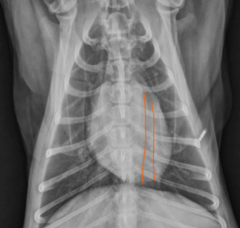
left ventricle slopes right, and takes over the right side
When there is mild enlargment of the left ventricle in dogs, what does the VD image look like?

ventral, central
Lobar VEINS are located _______ and _______ compared to the bronchus and arteries
left
There are two sets of artery/bronchus/veins, is the left or right set located more cranially on a lateral image?
4th rib
normal cranial pulmonary vessels should be less than 3/4 the width of the ________________ on a lateral view
caudal vena cava
The right caudal lobar vein is normally overlying the ____________ on the VD view, so its difficult to determine size
9th rib
caudal lobar arteries should be less than the width of the _______ on VD images
true
true/false: dogs with pulmonary hypertension can have very enlarged arteries that taper to normal size by the time they cross the 9th rib... so make sure to compare the whole visible vessel
left to right shunts (PDA, VSD, ASD), fluid retention from heart failure, fluid overload
What are differentials if both caudal lobar arteries AND veins are enlarged?
undercirculation, pulmonic stenosis, hypovolemia, shock, right to left shunts(tetrology of fallot, VSD, PS)
If both the caudal lobar artery and veins are SMALL, what are differentials?
mitral regurge, primary myocardial disease (cardiomyopathy), noncardiac left atrium obstruction, mass at heart base, thrombus in left atrium
if the caudal lobar vein is larger than the artery, what are differentials?
pulmonary hypertension, heartworms, COPD, pulmonary thormboembolism
If the caudal lobar artery is larger than the vein, what are differentials?
both enlarged
describe the lobar vessels

both small
describe the lobar vessels

vein larger than artery
describe the lobar vessels

compensated
____________________ heart disease is defined as the presence of left heart disease without evidence of pulmonary edema
pulmonary edema, failure
Left heart disease is DECOMPENSATED when __________________ is present and the heart is now considered to be in ___________
pulmonary venous congestion
________ will precede pulmonary edema if the heart is progressing into left failure
hilar regions,
usually, pulmonary edema in a dog starts where?
interstitial, alveolar
pulmonary edema is a continuum, begining as a ____________ lung pattern and progressing into a ________ pattern.
caudal lung lobes
In dogs, generally pulmonary edema is more visible where?
variable in location, patchy
Where is pulmonary edema in cats? and what does it look like?
dorsally deviated trachea, double opacity on VD, compensated disease, not failure
What heart signs does this dog have and if there are signs, is he in heart failure?

straightened cardiac waist, interstitial pattern, yes failure, because edema present
What heart signs does this dog have and is he in heart failure?
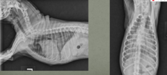
yes, variable edema
Is this CAT in heart failure?

right atrial enlargement, pulmonary arterial enlargement, ascites, hepatomegaly, CVC enlargement, tracheal deviation, (sometimes enlargement of main pulmonary artery)
What are the signs consistent with right heart disease/failure?
left (double opacity sign)
left or right heart disease?

right (reverse D sign)
left or right heart disease
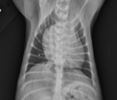
true
true/false: the reverse D sign does NOT taper, it stays about the same width for the full length of the heart

artery, vein
In some dogs with right heart disease, the [artery/vein] is larger than the [artery/vein]
they should be the same size
In a normal heart, which should be larger, the caudal vena cava or the descending aorta?
caudal vena cava
Okay, so if the caudal vena cava and the descending aorta are supposed to be the same size, when the ______________ starts to become larger, this marks the progression to right heart failure in a dog
liver, stomach axis
In the very beginning stages of right heart failure, which organ is mildly increased in size? This is due to a subtle shift in the _________
parallel to ribs
The normal stomach axis is where?
caudal vena cava, hepatomegaly, ascites
For right heart failure, an enlarged ______________ will precede ________ and ___________________
abdominal effusion
decompensated right heart failure is when there is any amount of ____________________ present.
right side
Which of these images has abdominal effusion
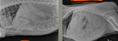
falciform fat pad
What is the structure outlined in this NORMAL radiograph?

cant see falciform fat, abdominal fluid, right heart failure
What is the abnormality here? Whats it caused by?
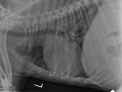
left atrial enlargement (if the L atrium is normal, you dont have L sided disease)
What is kind of the KEY thing Dr. Holland told us she uses to determine right vs left heart diease/failure?
cool
Here is that full slilde of differentiations, Cool

mitral regurgitation, dilated cardiomyopathy
Long standing left heart disease is commonly seen with _________ and rarely, can also have _________ documented with it, leading to biventricular failure
left, right
If there is BIVENTRICULAR heart disease, you WILL see [right/left] heart disease signs accompanied by MINIMAL [right/left] heart changes
serial radiography
_____________ is used to evaluate the course of heart disease and to monitor treatment
diuretics
Heart size and vessel size will be altered by ____[which drug class?]_____