Lab Exam 2
 Call Kai
 Learn
Learn Practice Test
 Spaced Repetition
 Match
Match Flashcards
 Knowt Play
1/36

There's no tags or description
Looks like no tags are added yet.
Name | Mastery | Learn | Test | Matching | Spaced | Call with Kai | Chat |
|---|
No analytics yet
Send a link to your students to track their progress
37 Terms
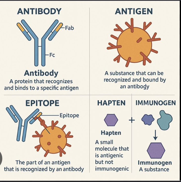
antigen (protein from a bacteria or virus)
has diff sites that will evoke an immune response
immune response results in the production of antibodies that bind to antigen specifically
sites on antigen is called an epitope
specific interaction btwn antibodies and antigens can be used in many ways in the design of diagnostic tests. major types of immune-based diagnostic tests that are used are agglutination tests and ELISA tests
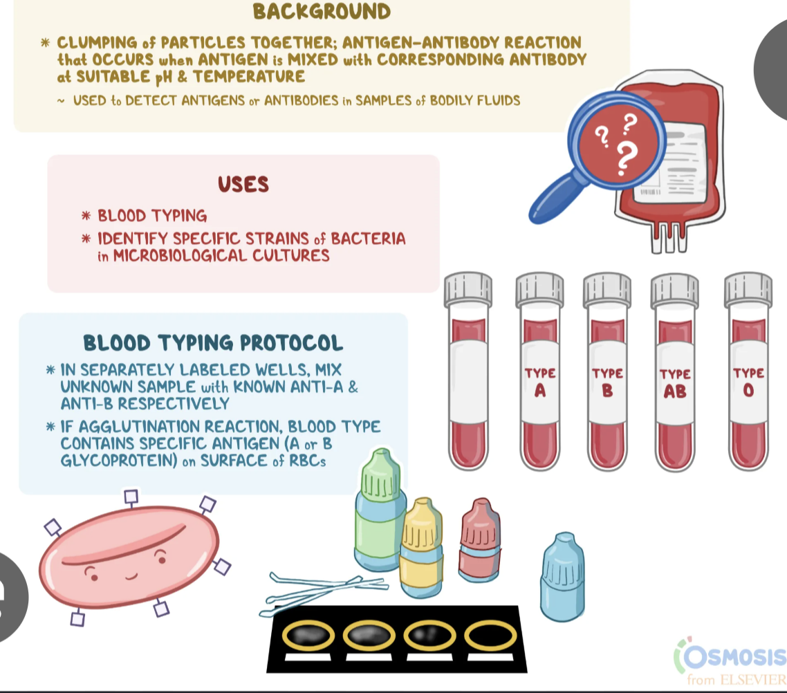
when antigen and antibodies are mixed, each antibody can bind two antigens = antibody-antigen complex, immune complex, or the clumps that can be seen = agglutination
serum
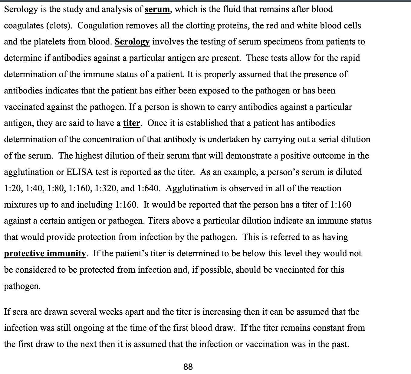
fluid that remains after blood coagulates, coagulation removes all the clotting proteins, RBC, platelets, and WBC
serology: testing of serum specimens from patients to determine if antibodies against a particular antigen are present
antibodies indicated the patient has been exposed to the pathogen or has been vaccinated against the pathogen
antibodies carried against a particular antigen means a person has a titer
titers above a particular dilution indicate an immune status that would provide protection from infection by the pathogen: protective immunity
if titer increases after several weeks, the infection was still ongoing at time of 1st blood draw
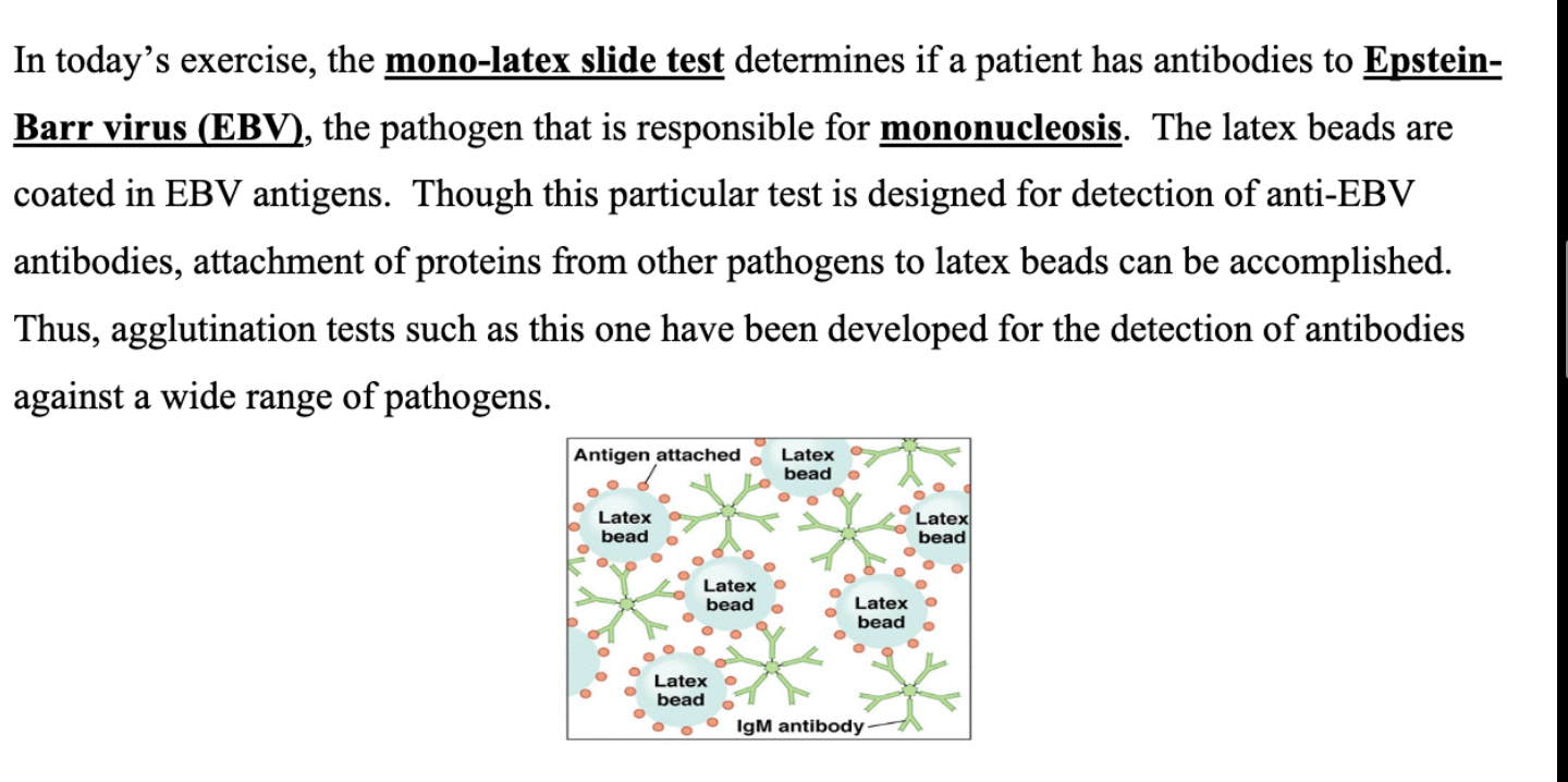
mono-latex slide test
determines if a patient has antibodies to Epstein-Barr virus (EBV), the pathogen that’s responsible for mononucleosis
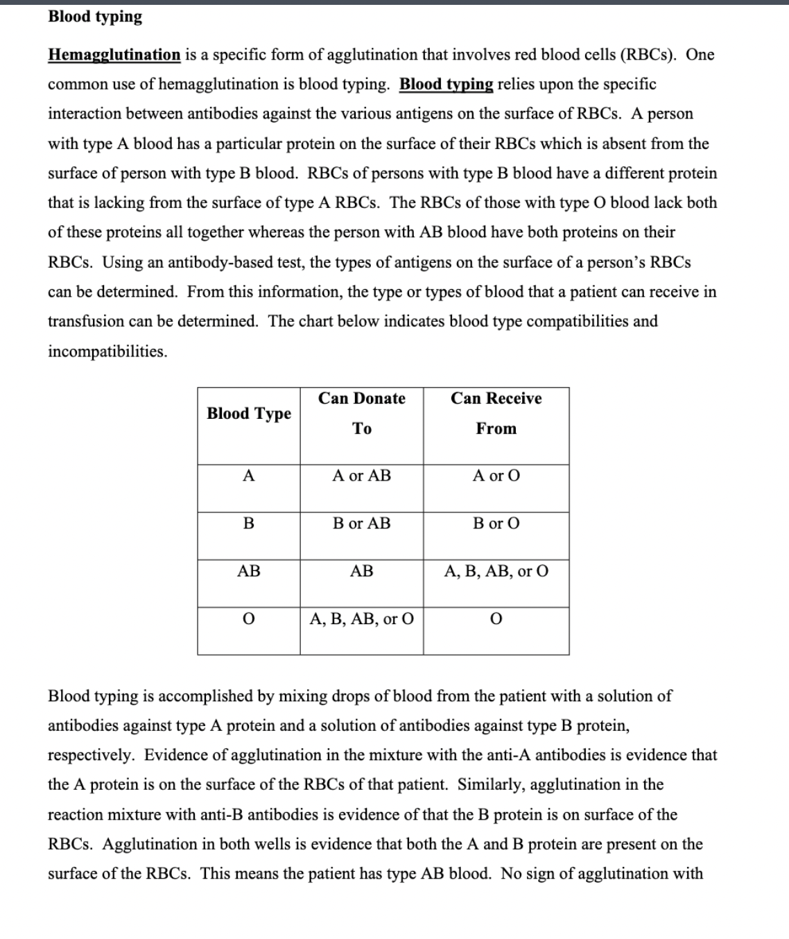
hemagglutination
involves RBCs and is used in blood typing:
specific interaction btwn antibodies against the various antigens on the surface of RBCs
blood typing is accomplished by mixing blood droplets with a solution of antibodies against type A protein and a solution of antibodies against B protein, respectively
Normally, persons will have a certain amount of antibody against the ABO antigens that they do not carry. The reasons for the presence of this antibody have never been discerned. If the wrong type of blood is given to a patient, the antibodies in their serum will bind to and cause the destruction of these newly introduced RBCs in the transfusion. When these RBCs are destroyed, chemicals are released that increase vasodilation and vascular permeability. This results in rapid loss of fluids into the tissues on a systemic basis and a systemic loss of vascular tone. Together, these two changes in the blood vessels will send the patient into shock. Use of the rapid blood typing reaction can eliminate blood type mismatches.
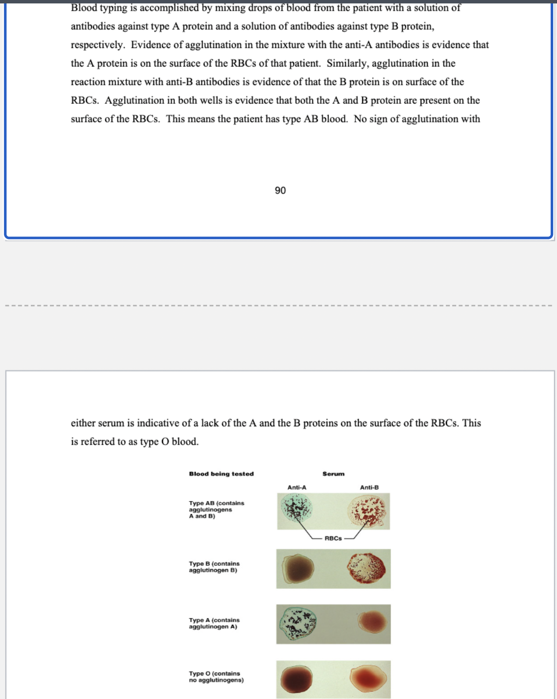
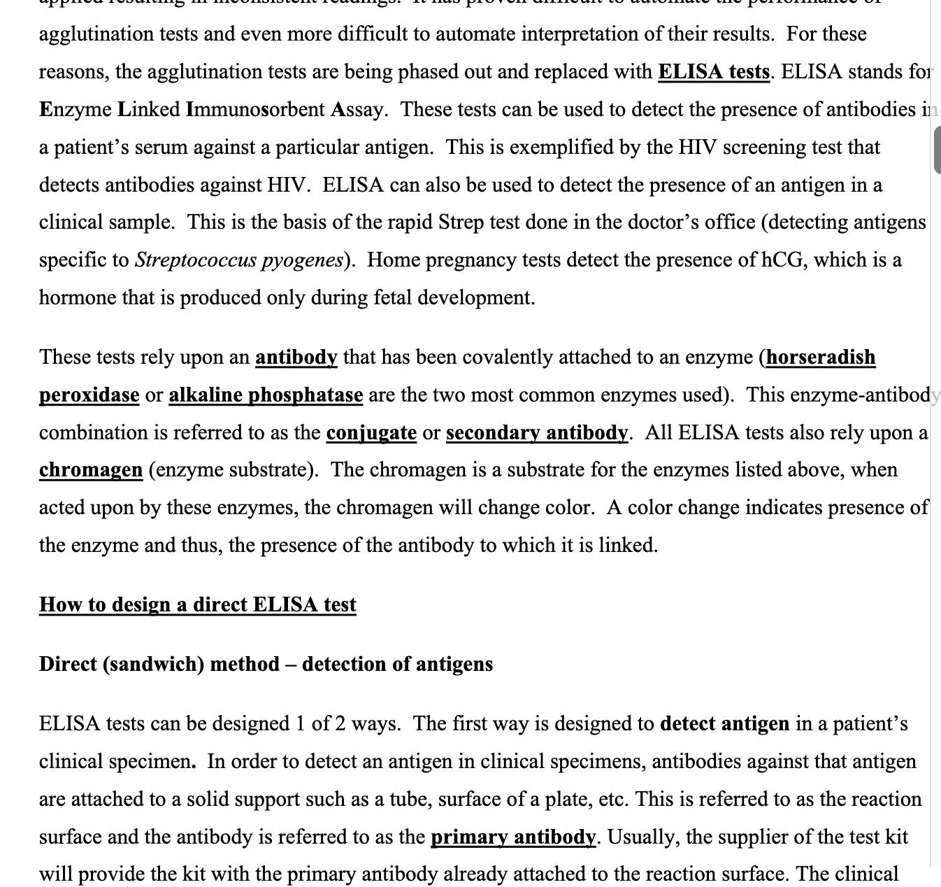
ELISA tests
ELISA= enzyme linked immunosorbent assay
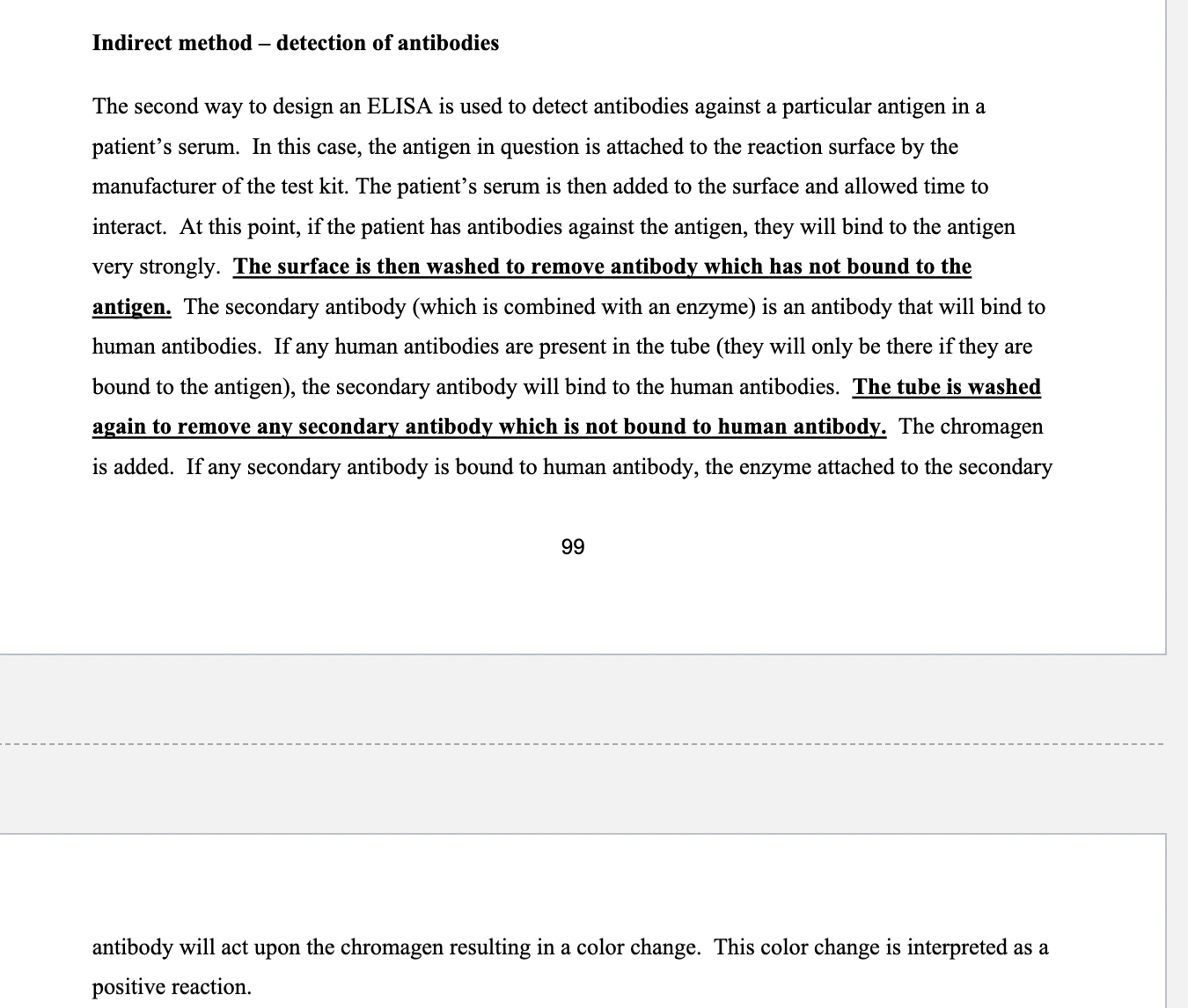
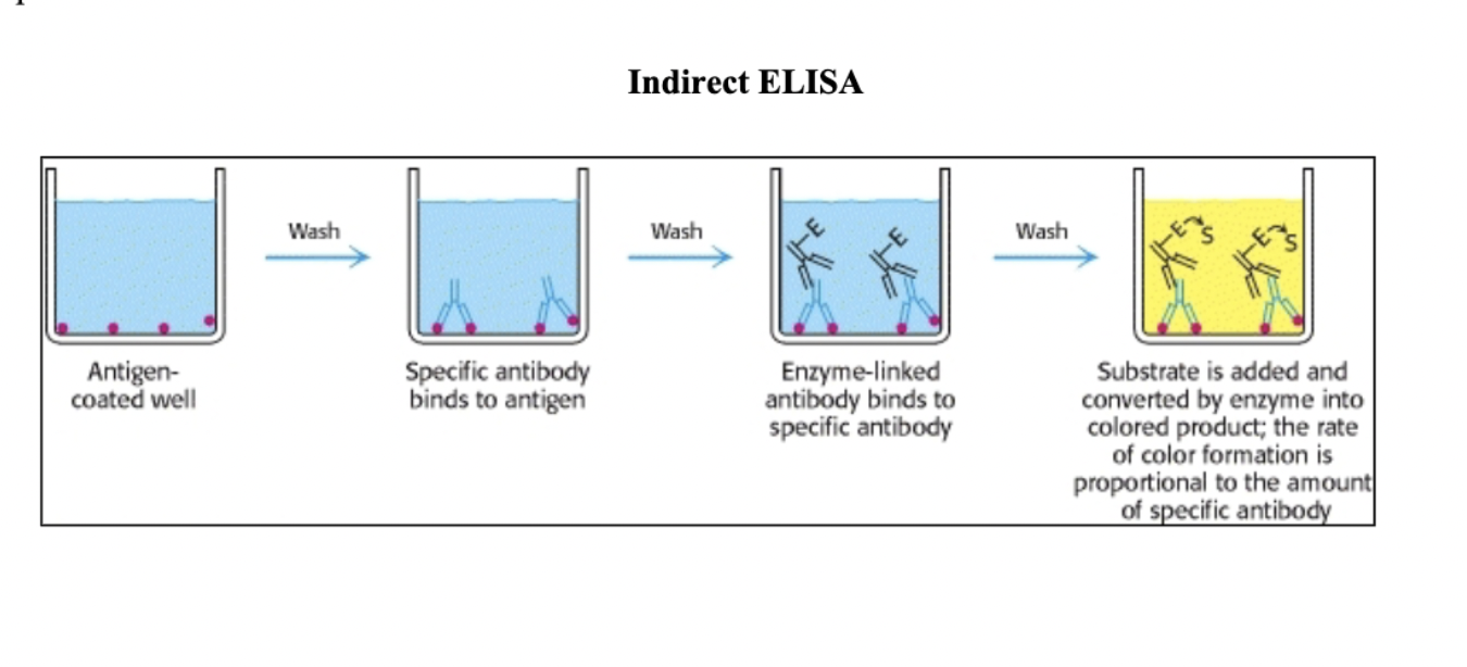
used to detect the presence of antibodies in a patient’s serum against a particular antigen and this is exemplified by HIV test.
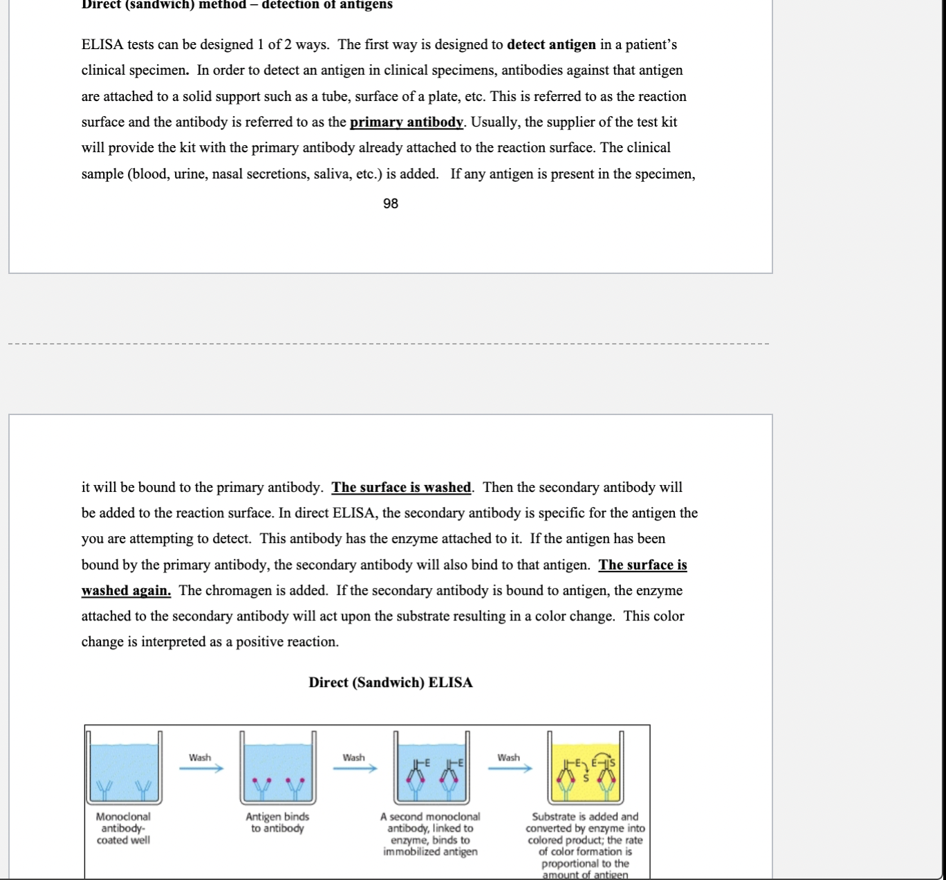
also used in clinical samples and is the basis of rapid Strep test
These tests rely upon an antibody that has been covalently attached to an enzyme (horseradish peroxidase or alkaline phosphatase are the two most common enzymes used). This enzyme-antibody combination is referred to as the conjugate or secondary antibody. All ELISA tests also rely upon a chromagen (enzyme substrate). The chromagen is a substrate for the enzymes listed above, when acted upon by these enzymes, the chromagen will change color. A color change indicates presence of the enzyme and thus, the presence of the antibody to which it is linked.
trophozoite stage
the active feeding stage for protozoan parasites
most of these organisms reproduce by simple cell fission while others are able to participate in a rudimentary form of sexual reproduction
Trophozoites lack the protective
covering of the cysts and are destroyed in the stomach after ingestion causing no illness. As the
disease resolves, normal rates of bowel movement return, and there is time for the encysted stage
of the organisms to develop.
Those protozoans that are acquired orally will pass through a stage known as the cyst; this stage of the organism’s life cycle is able to withstand severe environmental conditions, even stomach acid.
Entamoeba histolytica
This organism causes a condition known as amebiasis in humans. Ingestion of water or food contaminated with feces leads to the introduction of the cyst stage. Four free-swimming amoebas emerge from the cyst in the small intestine. They colonize the cecum (first part of the large intestine) and usually cause a mild condition typified by diarrhea and intestinal discomfort.
More serious cases involve bloody stools (dysentery), inflammation of the appendix and
perforation of the bowel. The organism can spread to other abdominal organs such as the spleen
or liver. It can penetrate the diaphragm leading to infection of the lungs.
Giardia intestinalis
causes a condition called giardiasis typified by diarrhea, cramps, flatulence and muscle weakness. Stools will often contain much unabsorbed fat. The protozoan is maintained in the environment because animals can also be infected.
Trichomonas vaginalis
trichomoniasis is a common sexually transmitted diseases. Trichomoniasis is caused by the organism Trichomonas vaginalis. Infection of the female usually involves the mucosa of the vagina, vulva and cervix. Symptoms of the infection in the female are vaginal itching, irritation, and a significant yellow-green, foul-smelling discharge 5 to 28 days after exposure. Lesions may be present on the vaginal mucosa. Infection of the male usually involves the urethra, prostate and seminal vesicles. Infection of the urethra can lead to pain when urinating (dysuria).
Diagnosis involves a vaginal or urethra swab and subsequent microscopic observation of the organism in a smear made from the swab. The trophozoite stage is pear-shaped with four flagella and an undulating membrane. This protozoan does not form cysts.
Plasmodium species
malaria
It is responsible for
millions of deaths annually. The etiologic agent of this disease is one of several species of the
genus Plasmodium. This agent is transmitted by the bite of certain types of mosquito.
The bite of an Anopheles mosquito, results in the injection of mosquito saliva containing
sporozoites into the bloodstream of a human. The sporozoite moves through the bloodstream
until they take up residence within cells of the liver. Within liver cells, the sporozoites undergo
division and form many cells referred to as merozoites. Rupture of the infected liver cells leads
to the release of thousands of merozoites into the bloodstream.
A portion of the merozoites in the red
blood cells will develop into male and female gametocytes. Red blood cells containing the
gametocytes are ingested when another mosquito bites an infected human for a blood meal. The
gametocytes are freed from the red blood cells in the intestine of the mosquito and bond together
to form a zygote. The zygote ultimately differentiates into a cyst. Sporozoites are released from
the cysts which then migrate to the salivary glands of the mosquito.
hosts
Organisms that host the development stages of the parasite are referred to as intermediate hosts. The organism that hosts the sexually mature stage of the parasite is referred to as the definitive host.
Flukes and tapeworms belong to the phyla Platyhelminthes and are commonly referred to as the
flatworms. These organisms have a complex life cycle, usually infecting different hosts at
different stages of their development.
Schistosoma mansoni
this organism is also referred to as a blood fluke. These organisms cause the disease
schistosomiasis. Initially, the entry of the schistosome through the skin causes dermatitis with a
pruritic papular (itchy and bumpy) rash. The acute phase of schistosomiasis, a condition referred
to as Katayama fever, may occur 2 to 4 weeks after heavy exposure.