Lecture 2b - Superficial Structures
 Call Kai
 Learn
Learn Practice Test
 Spaced Repetition
 Match
Match Flashcards
 Knowt Play
1/71

There's no tags or description
Looks like no tags are added yet.
Name | Mastery | Learn | Test | Matching | Spaced | Call with Kai | Chat |
|---|
No analytics yet
Send a link to your students to track their progress
72 Terms
Functions of the thyroid
Endocrine gland
Produces hormones (calcitonin - calcium homeostasis)
Controls metabolism
Controlled by the hypothalamus and pituitary
What connects the 2 lobes of the thyroid
Isthmus
Why is colour doppler preferred for thyroid scans
One of the most vascular organs of the body
Colour Doppler provides more diagnostic information in thyroid diseases
What transducer is used for thyroid scans
High resolution linear array transducer (7.5-17.0 MHz)
Thyroid characteristics on an US
Homogenous
Slightly hyperechoic
Butterfly shaped


Name this view
Transverse view of thyroid

Name this view
Longitudinal view of thyroid
How are thyroid volume measurements taken
Length of longitudinal view X width of lobe X depth of lobe

What’re the TI-RADS categories
Composition
Echogenicity
Shape
Margin
Echogenic foci
Where are the parathyroid glands
Posterior medial surface of thyroid gland

Parathyroids are only seen when…
They contain pathology
What is goitre
Swelling of the thyroid gland due to hyperplasia of normal tissue or thyroiditis
May be symmetric, asymmetric or multinodular
What is a thyroid nodule
Discrete lesion in the thyroid that is distinct from the surrounding thyroid parenchyma
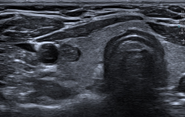
What is the pathology
Thyroid nodule


What is the pathology
Thyroid nodule

Name the pathology
Multinodular goitre
What is Hashimoto’s disease
Diffuse glandular enlargement
Autoimmune thyroiditis
How does Hasimoto’s diease appear on US
Giraffe pattern
More hypoechoic than normal thyroid tissue
May have any degree of vascular flow

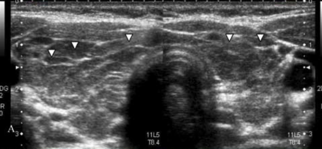
Name the pathology
Hashimoto’s thyroiditis

Name the pathology
Hashimoto’s thyroiditis
What is graves disease
Diffuse abnormality
Hyperfunctioning thyroid
Abnormal immune response that attacks the thyroid and causes it to overproduce hormone thyroxine
What are the characteristics of graves disease on US
Diffusely hypoechoic gland
Heterogeneous
Colour Doppler demonstrates hypervascularity (thyroid inferno)

What is this pathology
Graves disease
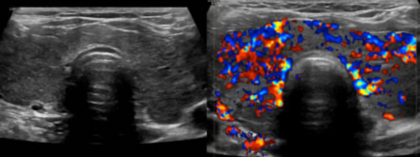
Name this pathology
Graves disease
Thyroid inferno
What does thyroid carcinoma look like on US
Solid hypoechoic mass
Irregular shaped
Heterogeneous
Microcalcifications with or without shadowing
Hypervascularity
Cervical lymph nose metastases
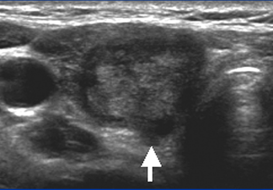
Name the pathology
Thyroid papillary carcinoma
How does a normal cervical lymph node appear on US
Cortex must be bigger than medulla

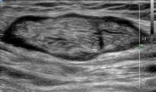
Name the pathology
Abnormal lymph node
What is a fine needle aspiration
Used for sus nodules or neck masses
US guided FNA allows for precise needle tip placement
What happens nodules that are >1cm
Biopsy
What happens to nodules that are <1cm
Fine needle aspiration
US to monitor progress
Layers of the breast
Skin
Subcutaneous layer
Mammary layer
Retromammary layer
Pectoralis

What does the subcutaneous layer of breast contain
Fatty tissue divided by thin connective tissue
What does the mammary layer of the breast contain
15-20 lobes
Divided by Cooper’s ligaments
Each lobe opens to the surface of the nipple through a lactiferous duct
Describe Cooper’s ligaments
Fibrous connective tissue in the mammary layer of the breast
Extends from the deep layer to the superficial layer
Echogenic (seen in fatty layer, hidden in glandular tissue)
What transducer is used for breast US exams
High resolution linear array transducer (7.5-17.0 MHz)
Patient positioning for breast examinations
Patient supine oblique
Shoulder and torso of the effected side elevated
Arm above head

Scanning technique for breast imaging
Transverse
Longitudinal
Radial
Antiradial
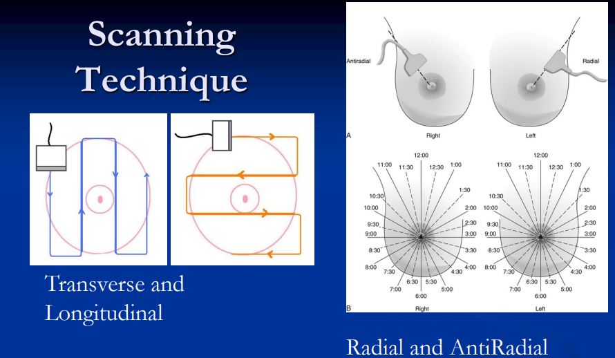
What’re the US characteristics of each layer of breast normally
Skin: 2 thin hyperechoic layers
Subcutaneous layer: hypoechoic separated by hyperechoic Cooper’s ligaments
Mammary layer: Hyperechoic compared to fat, homogenous
Retromammary: Hypoechoic pattern
Pec: Striated appearance

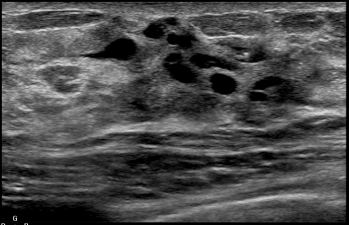
What is this
Milk ducts

Describe the pathology
Breast cyst
Posterior enhancement
Anechoic
Solid circumscribes boarders
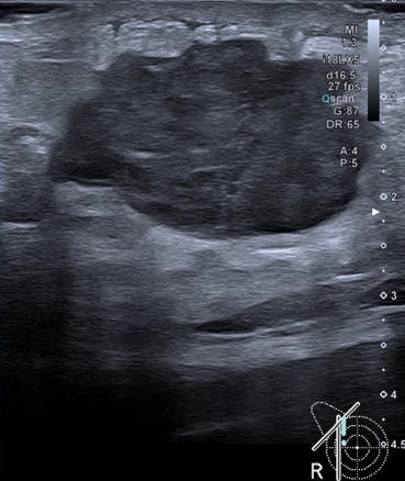
Describe the pathology
Breast abscess
Hypoechoic
Heterogeneous
Light posterior enhancement
What is fibroadenoma
Benign solid lesion of breast
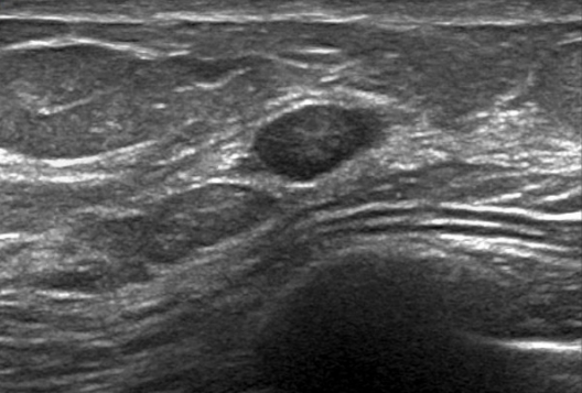
Describe this pathology
Fibroadenoma
Small firm nodule
Moves when palpated
Round with defined boarders
Hypoechoic with isoechoic fat lobules
What’re the characteristics of breast carcinoma
Irregular borders
Hypoechoic relative to fibroglandular/fatty tissue
Posterior acoustic shadow
E

Describe the pathology
Breast carcinoma in glandular mammary layer
Posterior enhancement
Heterogenous
Ill defined borders
Hypoechoic

What is the pathology
Breast carcinoma

What is the pathology
Breast carcinoma
Flood flow = cancer
Testicular anatomy
Scrotal wall
Tunica vaginalis
Tunica albuginea
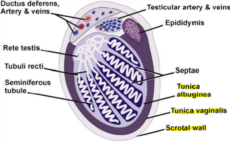
Describe the function of the tunica vaginalis
Creates space that contains a small amount of fluid hat prevents friction between the scrotal structures
Describe the function of the tunica albuginea
Composed of dense connective tissue
Encases each testis and covered by he tunica vaginalis
What is an intratesticular lesion
Malignant testicle lesion
What is an extratesticular lesion
Benign testicular lesion
What is cryptorchidism
Incomplete or partial failure of the intra-abdominal testes to decent into the scrotal sac


What is the pathology
Cryptorchidism in the inguinal canal
What transducer is used for scrotal exams
High resolution linear array transducer (7.5-17.0 MHz)
What shape is the testis in longitudinal view
Oval
What shape is the testis in a transverse view
Circle
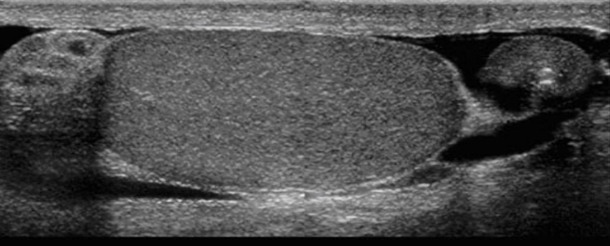
Label


What is the pathology
Cyst in epididymal head

Name the pathology
Testicular torsion
What is a hydrocele
Abnormal accumulation of serous fluid between the visceral and parietal layers of the tunica vaginalis

What is the pathology
Testicular hydrocele

What is the pathology
Testicular hydrocele
What is testicular varicocele
Collection of abnormal dilated. tortuous and elongated veins of the pampiniform plexus located posterior to the testis
Caused by incompetent valves in the testicular veins


What is the pathology
Epididymitis
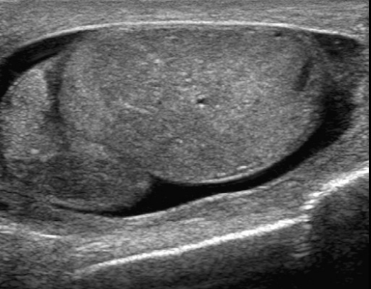
What is the pathology
Epididymitis
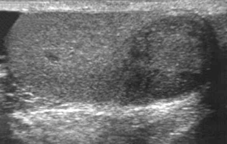
What is the pathology
Seminoma (testicular carcinoma)

What is the pathology
Testicular rupture
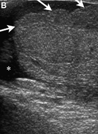
What is the pathology
Testicular rupture
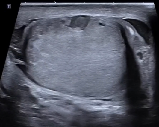
What is the pathology
Testicular fracture

