Seasons LG LO's Wk3 pt. 2
 Call Kai
 Learn
Learn Practice Test
 Spaced Repetition
 Match
Match Flashcards
 Knowt Play
1/39

Earn XP
Description and Tags
Introduction to Cardiac Pump Function
Name | Mastery | Learn | Test | Matching | Spaced | Call with Kai | Chat |
|---|
No analytics yet
Send a link to your students to track their progress
40 Terms
LO 1: how does blood move in the heart in relation to pressure gradients & valves
The cardiac cycle is a repeating loop of the heart filling with blood and then squeezing it out.
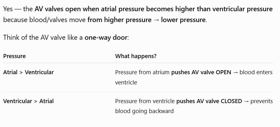
It relies on pressure gradients—blood moves from high pressure to low pressure—and valves act as one-way doors to ensure blood doesn’t go backward.
LO 1: what are the 4 stages in the cardiac cycle
ventricular filling (diastole)
isovolumetric contraction (early systole)
ventricular ejection (systole)
isovolumetric relaxation (early diastole)
LO 1: ventricular filling
what is the goal
mechanism: describe pressure differences & what valves open
volume/pressure: ventricular volume & pressure
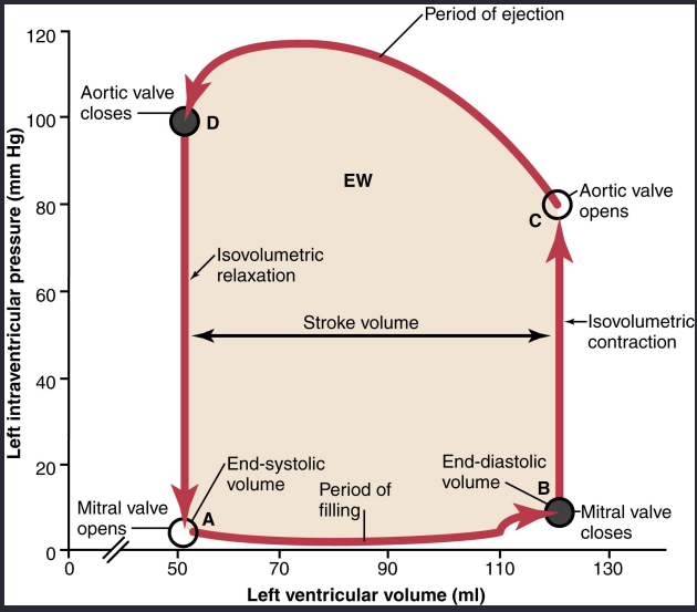
Ventricular Filling (Diastole):
The Goal: Get blood into the ventricle.
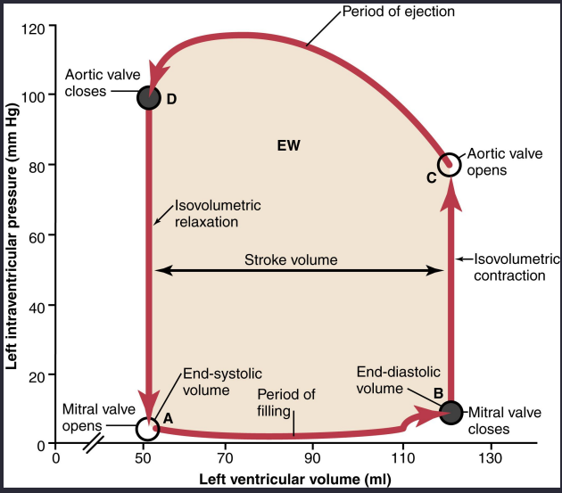
Mechanism: Atrial pressure becomes higher than ventricular pressure, forcing the Atrioventricular (AV) valves (Mitral/Tricuspid) to open.
Volume/Pressure: Blood flows in, increasing ventricular volume to the End-Diastolic Volume (EDV) (~120mL) while pressure stays relatively low.

LO 1: isovolumetric contraction: early systole
goal
mechanism: contraction, pressure differences, valves, heart sound
volume/pressure
Isovolumetric Contraction (Early Systole):
The Goal: Build up pressure to open the "exit door".
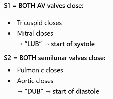
Mechanism: The ventricle begins to contract, and ventricular pressure immediately exceeds atrial pressure, causing the AV valves to snap shut (First Heart Sound, S1).
Volume/Pressure: Because both the AV and Semilunar valves are closed, the volume remains constant (isovolumetric) while the pressure "shoots up" rapidly → The right ventricle contracts while the tricuspid (AV) valve and pulmonic (semilunar) valve are both closed, so the blood has nowhere to go and ventricular pressure rises.

LO 1: ventricular ejection
goal
mechanism: pressure differences, valves
volume/pressure
Ventricular Ejection (Systole):
The Goal: Pump blood to the body/lungs.
Mechanism: Once ventricular pressure exceeds the pressure in the Aorta/Pulmonary artery, the Semilunar valves (Aortic/Pulmonic) pop open.
→ex.) blood (high pressure: R ventricle) to (low pressure: pulmonary artery)
Volume/Pressure: Blood is ejected, and ventricular volume drops to the End-Systolic Volume (ESV) (~50mL).
LO 1: isovolumetric relaxation (early diastole)
goal
mechanism: ventricle status, pressure differences, valves, heart sound
volume/pressure: valves status, volume & pressure
Isovolumetric Relaxation (Early Diastole):
The Goal: Drop pressure so the heart can fill again.
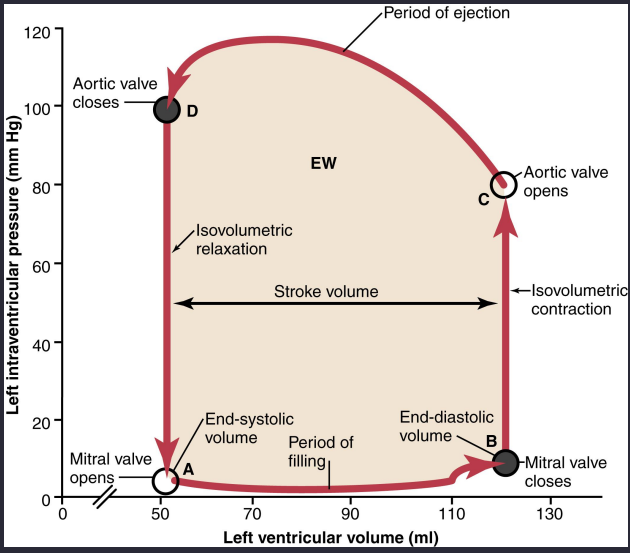
Mechanism: The ventricle relaxes. Pressure in the Aorta/Pulmonary artery becomes higher than in the ventricle, snapping the Semilunar valves shut (Second Heart Sound, S2).
→ During isovolumetric relaxation, the ventricles relax, so their pressure falls below the pressure in the pulmonary artery and aorta, causing the pulmonic and aortic valves to close; with all valves closed, the blood remaining in the ventricles cannot leave yet.
Volume/Pressure: All valves are closed again; the volume remains constant while the pressure falls back toward zero.
Ventricle was contracting hard → high pressure
Ventricle relaxes → pressure falls a lot
LO 1:
S1 marks the start of systole (______ closure)
S2 marks the start of diastole (_______ closure).
Isovolumetric phases occur when _______
S1 marks the start of systole (AV closure)
S2 marks the start of diastole (Semilunar closure).
Isovolumetric phases occur when all valves are closed.
LO 2: what is preload
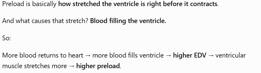
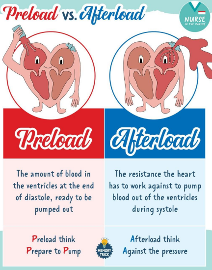
the amount of blood in the ventricles at the end of diastole ready to be pumped out
LO 2: what does the heart do when more blood is loaded into it
The heart has an intrinsic ability to handle whatever blood returns to it.
If you put more blood in, the heart stretches and then squeezes back harder to pump that extra blood out.
LO 2: what is the main idea of the Frank-Starling relationship
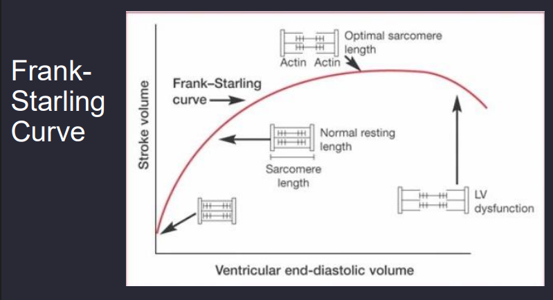
More blood fills the heart → cardiac muscle stretches more → stronger contraction → more blood pumped out.
LO 2: what is stroke volume
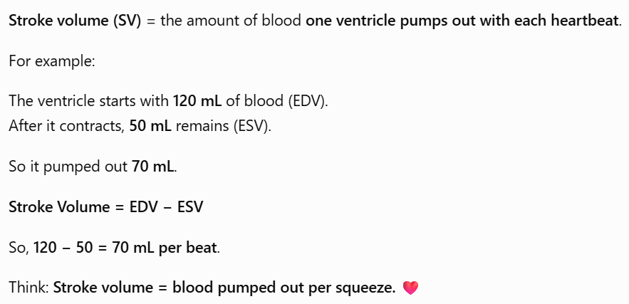
Stroke volume (SV) = the amount of blood one ventricle pumps out with each heartbeat.
LO 2: Franklin-Starling increased preload (EDV)
what happens when more blood returns to the ventricle
EDV: the amount of blood inside a ventricle at the END of diastole, after the ventricle has finished filling and right before it contracts.
Increased Preload (EDV): More blood returns to the ventricle, increasing the volume at the end of diastole
LO 2: Frank Starling sarcomere stretch
Sarcomere Stretch: This extra volume stretches the cardiac muscle fibers (sarcomeres).
sarcomere → basic muscle unit
⭐ Frank–Starling = “The more you fill it, the more you stretch it, the harder it squeezes.”
LO 2: Frank Starling optimal overlap
what happens to actin & myosin
Optimal Overlap: This stretch moves the actin and myosin filaments into a more optimal degree of overlap.

LO 2: Frank Starling stronger contraction
describe what happens with actin & myosin
Stronger contraction: Because more myosin heads can now bind to actin, the force of contraction increases.
LO 2: Frank Starling higher stroke volume
what occurs
what is SV
Higher Stroke Volume (SV): The resulting stronger contraction ejects a larger volume of blood.
stroke volume → the amount of blood a ventricle pushes out during one heart beat

LO 2: what is cardiac output & formula
Cardiac Output (CO): the total volume of blood the heart pumps per minute.
CO= HR x SV
CO=SV×HR
Example: 70mL/beat×70beats/min=4,900mL/min (~5 Liters).

LO 2: heart failure relation to Frank Starling relationship
In heart failure, the heart muscle is weakened, so increasing preload doesn’t increase contraction strength as effectively. If the ventricle becomes excessively dilated, the muscle fibers can also become overstretched, further reducing effective contraction.

LO 3: what are 3 variables that influence stroke volume independently
preload (how much you fill)
afterload (how much resistance you face)
contractility (how hard you can squeeze)
stroke volume → pumped out amount
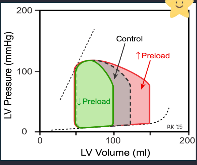
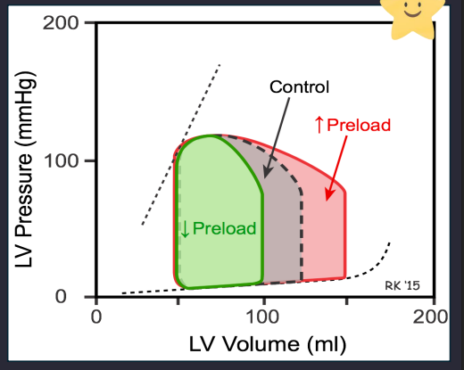
LO 3: stroke volume influence preload
what it is
effect on SV
pressure-volume loop
Preload (The "Fill"):
Definition: The degree of stretch on the heart at the end of diastole (often equated to EDV).
At the end of diastole, the ventricle has been relaxed and filling, and now it has reached its fullest point.
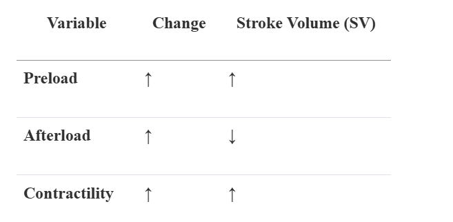
Effect: Increasing preload increases SV via the Frank-Starling mechanism.
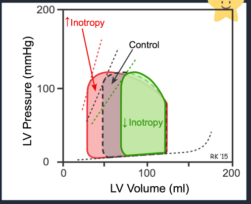
PV Loop: The loop widens to the right bc the x-axis of a pressure-volume (PV) loop is ventricular VOLUME.
So moving right = more blood volume in the ventricle.
LO 3: stroke volume influence afterload
definition
effect on SV
PV loop
Afterload (The "Resistance"):
Definition: The "load" the heart must pump against, primarily determined by aortic pressure aka how hard it is for the ventricle to push out blood
Effect: Increasing afterload decreases SV. Increasing afterload means there is more pressure resisting the ventricle from pumping blood out, so less blood is ejected and stroke volume decreases.
→ ex.) less blood comes out because the increased aortic pressure opposes the blood being pushed out of the left ventricle.
PV Loop: The loop becomes taller and narrower; the "exit" volume (ESV) increases.

LO 3: stroke volume influence contractility/inotropy
what is inotropy
effect on SV
PV loop
inotropy: Inotropy is essentially the exact same thing as myocardial contractility. It refers to the intrinsic, built-in ability of the heart muscle (the myocardium) to contract and generate force
3. Contractility/Inotropy (The "Squeeze"):
Definition: The intrinsic strength of the muscle, independent of stretch.
Effect: Increasing contractility increases SV. It allows the heart to squeeze down to a smaller volume left inside after (lower ESV)
PV Loop: The loop widens to the left (lower ESV).
LO 3: what happens to SV if preload, afterload, & contractility are increased
LO 4: what is coronary blood flow in simple terms
coronary blood flow: perfusion to the heart muscle itself
LO 4: left ventricle
systole: what occurs during contraction, pressure, coronary vessels status, & blood flow to the left ventricle
diastole: left ventricle muscle status, coronary arteries status
Left Ventricle (The High-Pressure Pump):
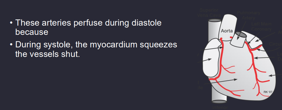
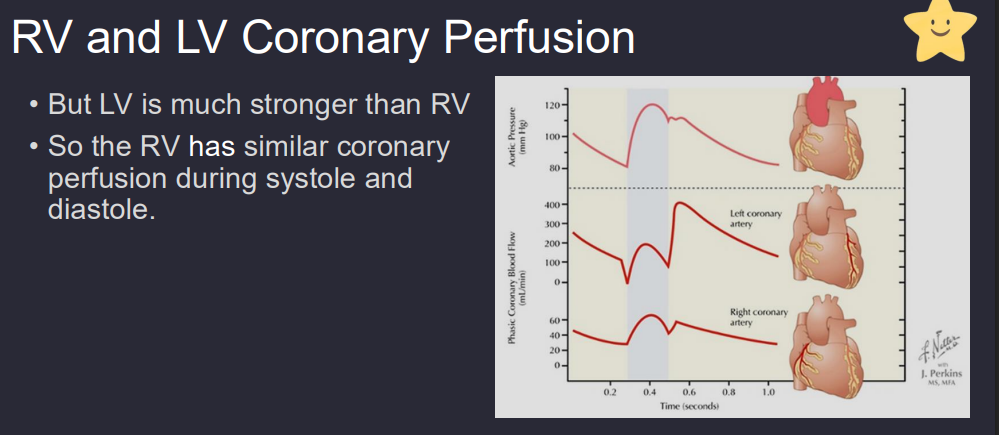
Systole: During contraction, the LV muscle is so strong and the pressure so high that it compresses its own coronary vessels shut. Blood flow to the LV muscle drops to nearly zero.
Diastole: This is the only time the LV muscle relaxes enough for blood to flow through the coronary arteries.
High-Yield: The LV is entirely diastole-dependent for its oxygen supply.
LO 4: right ventricle
systole & diastole: pressure, coronary vessel status during contraction
result: describe the blood flow during both phases
Right Ventricle (The Low-Pressure Pump):
Systole & Diastole: Because the RV is a much thinner, lower-pressure pump, it does not squeeze its vessels completely shut during contraction.
Result: The RV receives relatively continuous blood flow during both phases of the cardiac cycle.
LO 4: why is tachycardia dangerous to the left ventricle
Because the LV only receives blood during diastole, a very high heart rate (tachycardia) is dangerous.
Tachycardia shortens diastole disproportionately, meaning the hard-working LV has less time to get the oxygen it needs, potentially leading to ischemia (chest pain).
LO 4:
LV perfusion occurs primarily in ______.
RV perfusion is ______.
LV perfusion occurs primarily in diastole.
RV perfusion is continuous.
LO 5: what is the only way to give the heart more oxygen in simple terms
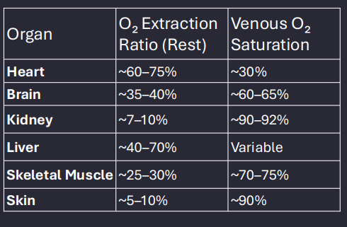
The heart is an "oxygen hog." Unlike skeletal muscle, which can "rest" and only takes some oxygen from blood, the heart always extracts almost all the oxygen available (~70-75%) even at rest.
Therefore, the only way to give the heart more oxygen is to increase blood flow.
LO 5: determinants of myocardial oxygen
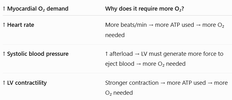
what are the demanders
increased HR
increased systolic blood pressure
increased LV contractility
LO 5: determinants of myocardial oxygen demanders
heart rate
Heart Rate (HR): More beats per minute = more energy used.

LO: determinants of myocardial oxygen demanders
increased systolic blood pressure
Wall Tension (Afterload/Pressure): Based on the Law of Laplace, higher pressure or a more dilated heart (larger radius) dramatically increases the tension/workload and oxygen need.
Higher pressure: If aortic pressure is high, the left ventricle has to squeeze harder to eject blood → ↑ wall tension → ↑ oxygen demand.
Larger ventricular radius: If the ventricle becomes dilated (stretched bigger), its walls have to generate more tension to squeeze → ↑ oxygen demand.

LO 5: determinants of myocardial oxygen demanders
increased LV contractility
Contractility: Squeezing harder requires more ATP.

LO 5: what does the heart do since there is a high oxygen need
Regulation of Supply (Matching Flow to Need):
Since oxygen extraction is already near maximum, the heart uses active hyperemia (increased flow due to metabolic activity).
LO 5: myogenic oxygen suppliers 2 main categories
autoregulation
oxygen carrying content
LO 5: myogenic oxygen suppliers flow
autoregulation: what are the sub-categories
oxygen carrying content: what are the sub-categories
autoregulation
myogenic control
metabolic control
endothelial control
neural control
oxygen carrying content
hemoglobin
oxygen saturation
LO 5: myocardial oxygen suppliers flow
autoregulation: myogenic control
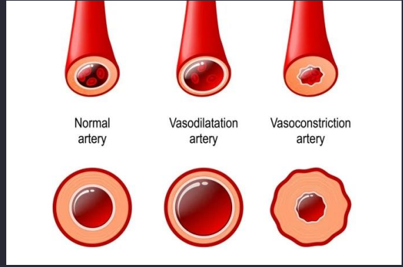
Coronary vessels automatically constrict when pressure rises and dilate when pressure falls to help maintain stable coronary blood flow.
↑ Blood pressure → vessel wall gets stretched → smooth muscle constricts → prevents too much blood flow.
↓ Blood pressure → less stretch → smooth muscle relaxes/dilates → helps maintain blood flow.
LO 5: myocardial oxygen suppliers flow
autoregulation: metabolic control
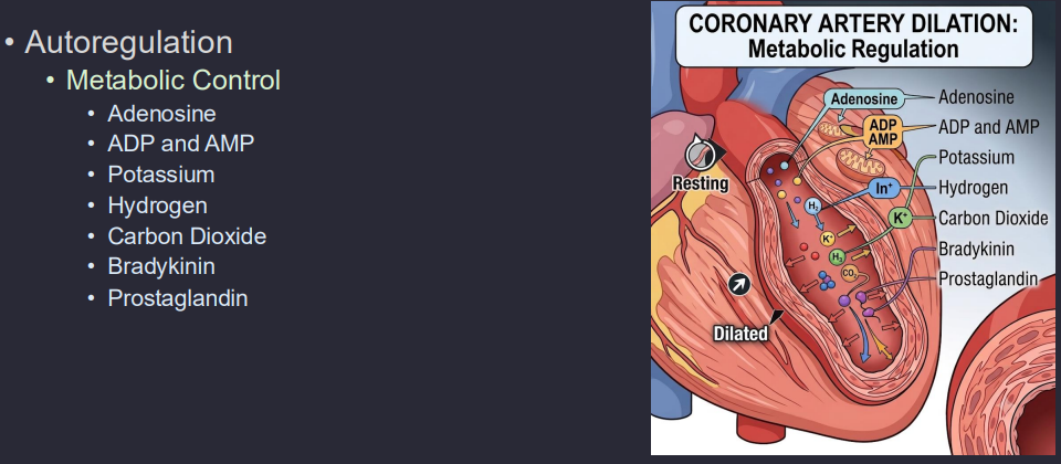
Metabolic Control (Primary): When the heart works harder, it uses up ATP, releasing Adenosine. Adenosine is a potent vasodilator that tells the coronary arteries to "open wide" to let more blood in. Other factors include CO2, H+, and K+.
When the heart works harder, metabolites build up → coronary vasodilation → ↑ blood flow
LO 5: myocardial oxygen suppliers flow
autoregulation: neural control
Neural Control: Sympathetic stimulation primarily increases flow indirectly by increasing HR and contractility, which then triggers the metabolic "demand" for more flow.
Sympathetic activity → ↑ HR + ↑ contractility → ↑ O₂ demand → indirectly ↑ coronary flow
Sympathetic → heart works harder → needs more O₂ → coronary dilation → more blood flow.

LO 5: myocardial oxygen suppliers flow
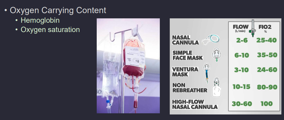
oxygen carrying content: hemoglobin & oxygen saturation
Depends mainly on hemoglobin + O₂ saturation, which determine how much oxygen the blood can deliver
More hemoglobin + high O₂ saturation → more O₂ carried in blood → more O₂ available to the heart.
Myocardial oxygen supply depends on coronary artery blood flow and the amount of oxygen carried in that blood (hemoglobin × O₂ saturation)