Inflammation and the Immune System
 Call Kai
 Learn
Learn Practice Test
 Spaced Repetition
 Match
Match Flashcards
 Knowt Play
1/103

Earn XP
Description and Tags
9 / 4 lecture
Name | Mastery | Learn | Test | Matching | Spaced | Call with Kai | Chat |
|---|
No analytics yet
Send a link to your students to track their progress
104 Terms
how does the body protect itself against disease
innate immune system
inflammation
adaptive immune system
chronic inflammation
innate immunity
first line of defense
the natural defenses in place to provide protection against pathogens and stressors from the environment
include physical, mechanical, and biochemical mechanisms designed to mitigate or prevent growth of pathogens and exposure to external stressors
in place at birth
fast acting
innate immunity: physical barriers
epithelial cells
tightly bound
line the surfaces of the skin and the GI tract
prevent deep penetration by pathogens
low temperature of the skin
inhibits pathogen growth
low pH of the skin and stomach
inhibits pathogen growth
innate immunity: mechanical barriers
mechanisms for cleaning pathogens from the surfaces of epithelial cells
vomiting → clears pathogens from the GI tract
urination → clears pathogens from the GU tract
goblet cells of upper respiratory tract → secrete mucous that traps pathogens
ciliated cells then brush out the mucous and pathogens
innate immunity: biochemical barriers
substances synthesized by and secreted by epithelial cells designed to trap or destroy pathogens
antimicrobial fatty acids and lactic acid → secreted by sebaceous glands of skin
antimicrobial peptides → secreted by epithelial cells
lysozyme → component of perspiration, tears, and saliva
innate immunity: normal microbiome
bacteria and fungi colonize the surfaces of the body
unique to a given area
non-pathogenic under normal circumstances
compete with other microorganisms for resources and space (block attachment to the epithelium)
secrete chemicals (ammonia, etc.) that inhibit growth of pathogens
immune system also suppresses growth of normal microbiome (aka normal flora)
innate immunity: failure due to medications
use of broad-spectrum antibiotics can kill the body’s normal microbiome
organisms that are resistant to the antibiotic have the opportunity to replicate and may lead to opportunistic infections
candida albicans = thrush
clostridium difficile = pseudomembranous colitis
immunosuppressant medications (meds that block the immune system) can also lead to opportunistic infections
innate immunity: the genitourinary tract
urinary tract is normally sterile
urination flushes bacteria from system and disrupts adhesion
increased risk of bacterial growth if there is:
short urethra
reflux/retrograde flow
obstruction
innate immunity: respiratory tract
mucociliary blanket lining nose and upper respiratory tract trap microbes (smoking damages this)
goblet cells secrete mucous and trap microbes
cilia in upper airway move encapsulated bacteria to back of throat where they are swallowed or expelled
alveolar macrophages destroy small organisms that travel down airway
innate immunity: gastrointestinal tract
gastric acid destroys pathogens in stomach (creates low pH)
viscous mucous layer coats gut and entraps microbes
pancreatic enzymes and bile detergents destroy organisms
IgA secreted by mucous membranes in gut
normal bacterial flora compete with pathogens for nutrients
inflammation
second line of defense
programmed response to tissue injury
integrated system of humoral (dissolved in blood) and cellular responses designed to:
limit tissue damage
destroy pathogens
initiate adaptive immune system
begin healing
rapid response in vascularized tissue (begins in seconds)
broken into vascular and cellular components
cells of inflammation: endothelial cells
layer of cells lining blood vessels
connected to underlying connective tissue
release nitric oxide (NO) to promote vasodilation
release inflammatory mediators (interleukins, prostaglandins, etc.) to regulate cellular changes during inflammation
regulate movement of cells through endothelial layer
release tissue factor in response to injury (activates extrinsic pathway of clotting cascade)
cells of inflammation: mast cells
cells that lie in connective tissue near blood vessels
when activated, mast cells:
degranulate (release inflammatory mediators stored in cellular granules)
immediate
primarily histamine
synthesize new inflammatory mediators
delayed
what is histamine responsible for
(released by mast cells)
vasodilation
increased capillary permeability
bronchoconstriction
cells of inflammation: neutrophils
first phagocytic cells to arrive in inflammation**
main function
phagocytosis of pathogens
these aren’t able to survive the acidic environment of inflammation for long
they become a component of purulent exudate (pus)
what is the first inflammatory mediator in a wound
histamine
what is the first cell in a wound
neutrophils
cells of inflammation: eosinophils
two main functions
defense against parasites
regulate changes associated with allergic reactions (mast cells, eosinophils, histamine, etc.)
mildly phagocytic
worms, wheezes, and weird diseases
cells of inflammation: dendritic cells
aka Langerhan cells
link between innate and adaptive immune system
two main functions
function as antigen-presenting cells (APCs) to initiate adaptive immunity
phagocytosis (mild)
cells of inflammation: monocytes/macrophages
monocytes are the precursor to macrophages
capable of surviving in acidic environments
last longer than neutrophils
if it’s in your blood, it is a monocyte
if it comes out into the tissues, it is a macrophage
main functions of monocytes / macrophages
clear pathogens and debris from injured tissue/wounds
function as antigen-presenting cells (APCs) to initiate adaptive immunity
chemical mediators of inflammation
responsible for coordination of vascular and cellular aspects of inflammation
tightly regulated
chemical mediators of inflammation: histamine
first inflammatory mediator released
produced by mast cells
responsible for
vasodilation
increased vascular permeability
bronchoconstriction (contraction of bronchial smooth muscle)
temporary
leukotrienes will take over once synthesized
chemical mediators of inflammation: bradykinin
initiated by activation of Hageman factor (factor XII)
responsible for
vasodilation
increased vascular permeability
bronchoconstriction
pain **
chemical mediators of inflammation: clotting factors
produced by liver
induces the clotting cascade
chain of events that leads to production of fibrin clot
chemical mediators of inflammation: complement proteins
plasma proteins
present in inactive form
complement proteins as part of inflammation
increase vascular permeability
promote chemotaxis (movement of cells following concentration gradient)
like when u float bc u smell a pie <3
complement proteins as part of immune system
act as opsonins and facilitate phagocytosis
create holes in cell membrane of pathogens (membrane attack complex → MAC)
basically sprinkles on a pathogen which attract the body’s response to eat the pathogen
chemical mediators of inflammation: arachidonic acid metabolites
fatty acids present in the cell membrane
metabolized by one of two pathways
cyclooxygenase (COX) pathway
produces prostaglandins and thromboxane
prostaglandins are responsible for pain
thromboxane = platelet aggregation
aspirin/NSAIDs block COX pathway
will stop you from feeling pain and blood coagulation
lipoxygenase pathway
produces leukotrienes
chemical mediators of inflammation: prostaglandins and leukotrienes
promote:
vasodilation
increased capillary permeability
bronchoconstriction
chemical mediators of inflammation: thromboxane
promotes:
platelet aggregation
chemical mediators of inflammation: IL and TNF
interleukins and tumor necrosis factor
many kinds
some are pro inflammatory (IL-1β, IL-6) **
some are anti inflammatory (IL-4, IL-10) **
many overlapping functions
recruitment and activation of leukocytes (WBCs)
induce acute-phase responses of systemic inflammation
fever, increased HR, anorexia, increased neutrophils, increased cortisol)
which IL and TNF are pro inflammatory
(IL-1β, IL-6)
which IL and TNF are anti inflammatory
(IL-4, IL-10)
chemical mediators of inflammation: interferons
produced by virus-infected cells
enhances defense against viruses
inhibits DNA/RNA synthesis
some active immune cells to destroy viruses
chemical mediators of inflammation: nitric oxide
produced by endothelial cells
promotes smooth muscle relaxation and vasodilation
what are the two types of inflammation
acute
chronic
acute inflammation
early, short term and self limiting
occurs before adaptive immunity can exert its effect
designed to remove injurious agent and limit extent of damage
neutrophils predominate in first 24 hours
chronic inflammation
late, long-term and self-perpetuating
usually the result of recurrent inflammation/irritation or slow processes that falls to induce an acute response
macrophages and lymphocytes are more common
acute inflammation: vascular response
following tissue injury
initial vasoconstriction (controls loss of blood)
vasodilation
increase flow of blood to the area
makes area warm and red **
increased vascular permability
endothelial cells contract
fluid leaks through gaps between cells → meant to dilute the pathogen
initially plasma with little protein (transudate)
quickly followed by movement of protein-rich fluid (exudate)
outflow of protein draws water from vessels to surrounding tissue (edema)
what are the stages of cellular response in acute inflammation
endothelial activation
margination
tethering
rolling
firm adhesion
diapedesis/transmigration
chemotaxis
leukocyte activation
phagocytosis
every month thunderstorms rolling from Dallas, Texas can leave puddles
endothelial activation
first stage of acute inflammation cellular response
inflammatory mediators (histamine, interleukins, etc.) promote expression of integrins on surface of leukocytes (mostly neutrophils → first responder)
also promotes expression of selectins and intracellular adhesion molecules (ICAM) on surface of endothelial cells
Endothelial cells have selectins and leukocytes have integrins
margination
second stage of acute inflammation cellular response
circulating leukocytes are swept against blood vessel wall
tethering
third stage of acute inflammation cellular response
integrins on leukocytes bind loosely to selectins on endothelium
rolling
fourth stage of acute inflammation cellular response
leukocytes move from selectin to selectin
just rolling across the endothelial cells
firm adhesion
fifth stage of acute inflammation cellular response
integrins on leukocyte attach firmly to ICAM
after rolling, the leukocyte will eventually come into contact with an intracellular adhesion molecule which traps the leukocyte and stop the rolling
diapedesis/transmigration
sixth stage of acute inflammation cellular response
leukocyte moves through gap between endothelial cells
chemotaxis
seventh stage of acute inflammation cellular response
leukocytes follow chemical gradient to the site of injury
chemicals are released by immune and non-immune cells
the smell of pie
leukocyte activation
eighth stage of acute inflammation cellular response
a chain of events that leads to attachment of a leukocyte to a pathogen and phagocytosis
through one of two ways
pattern recognition receptors (PRRs)
opsonization
leukocyte activation via PRRs and PAMPs
a way for phagocytic cells to recognize pathogens
pattern recognition receptors (PRRs) are toll-like receptors
molecules on the surface of phagocytic cells that recognize pathogen-associated molecular patterns (PAMPs) on surface of pathogens
PAMPs are not present on mammalian cells
PRR attachment to PAMP stimulates activation of the phagocytic cell and phagocytosis

leukocyte activation via opsonization
ex. complement proteins, cytokines, c-reactive protein
soluble molecules that bind to particles on surface of pathogens
phagocytic cells bind to opsonins on the surface of pathogen
opsonin attachment stimulates activation of the phagocytic cell and phagocytosis
phagocytosis
ninth stage of acute inflammation cellular response
engulfment of pathogens by leukocytes
engulfment: once activated, pseudopods extend around pathogen and enclose it in a phagocytic vacuole (phagosome)
fusion: phagosome merges with lysosome
killing of ingested pathogen: enzymes inside of the lysosome degrade the pathogen
local manifestations of inflammation
occur as the result of vasodilation and increased vascular permeability
heat → due to vasodilation
redness → due to vasodilation
swelling → due to increased capillary permeability
pain → due to increased capillary permeability
exudate compresses nerves in tissue
presence of prostaglandins and bradykinin
loss of function
what are the systemic manifestations of inflammation
fever → early response induced primarily by IL-1 acting on the hypothalamus
leukocytosis
increase in leukocytes to fight infection
may be accompanied by “left shift” in severe infections
most of your WBC get killed off and are replaced by immature cells that aren’t able to fight off pathogens as well
plasma protein synthesis
fibrinogen, prothrombin, clotting factors, plasminogen, complement proteins, etc. (produced by liver)
chronic inflammation
persistent infections (>2 weeks)
acute response may have been unable to control the infection or clear foreign objects (splinter, dirt, asbestos, etc.)
also may occur independently
some microorganisms can survive and replicate inside phagocytic cells
activates chronic inflammation
what organisms can survive and replicate inside phagocytic cells
mycobacterium tuberculosis, mycobacterium leprae, salmonella typhi
acute vs. chronic inflammation (cell types)
acute: characterized by high numbers of neutrophils (but they can’t survive very long)
chronic: neutrophils are replaced by macrophages and lymphocytes
take over for neutrophils (bc they are stronger)
what is a granuloma
happens during chronic inflammation
if macrophages cannot destroy the organism, the body attempts to contain the organism in a granuloma
core of macrophages surrounded by lymphocytes
may contain fibroblasts that produce collagen
may become calcified
center of granuloma can become necrotic
acute inflammation key points
fast, early
characterized by:
infiltration by neutrophils
protein rich exudates
chronic inflammation key points
slow, late
characterized by:
infiltration by macrophages and lymphocytes
presence of fibroblasts that secrete collagen
can become calcified
core can become necrotic
adaptive immunity
third line of defense
process designed to create a specialized immune response against a particular pathogen
augments protection already in place and prepares long term protection
major components
cells of adaptive immune system
antibodies
what are the two branches of adaptive immunity
cell mediated → mediated by T-lymphocytes
humoral → mediated immunoglobulins (antibodies) produced by plasma cells
antigen presenting cells (APC)
immune cells that initiate adaptive immunity
macrophages
dendritic cells
some B lymphocytes
bind to an antigen, engulf them, break them down into protein fragments
attach a protein fragment to a molecule of major histocompatibility complex (MHC)
presents the MHC/protein fragment to T lymphocytes for activation
major histocompatibility complex (MHC)
aka human leukocyte antigen (HLA)
glycoproteins on the surface of cells responsible for antigen presentation and helping the immune system differentiate native cells from foreign cells
has a groove that accommodates the protein fragment derived from an invading pathogen
when the MHC/protein fragment complex of an APC is presented to T-cells, the T-cells become activated
what are the two main MHC glycoproteins
MHC II: on APCs
activate CD4+ cells to become T-helper cells
MHC I: on all nucleated cells
activate CD8+ cells to become cytotoxic T cells
B lymphocytes
part of the adaptive immune system
when activated, differentiate into plasma cells and make antibodies
T lymphocytes
part of the adaptive immune system
several types differentiated by “cluster of differentiation” (CD) protein on the surface (CD4+ vs CD8+)
CD4+ T lymphocytes
when presented with antigen peptide/MHC II complex by APCs they differentiate into T-helper cells
function: regulate adaptive immune system and create memory
CD8+ T lymphocytes
when presented with antigen peptide/MHC I complex by virus infected cells or cancer cells, they differentiate into cytotoxic T cells
function: release reactive oxygen species and enzymes to destroy infected cells
pathogen recognition by T helper cells: cell-mediated immunity
APCs engulf pathogen, break it down, bind a protein fragment to MHC II
antigens can be microbial (virus, bacteria, fungi) or non-microbial (plant pollen, poison ivy resin)
APCs present MHC II/antigen complex to CD4+ T lymphocyte
CD4+ cell becomes an active “T-helper cell”
what does a T-helper cell do
cytokines are released that activate B cells
B cells become plasma cells which make antibodies (these specific antibodies target the antigen that was presented by the MHC II)
cytokines are released to make memory T cells
cytokines are released to recruit CD8+ T cells
cytotoxic T-cell response: cell mediated immunity
recruited CD8+ cells bind to MHC I / antigen complex of infected cell
CD8+ cell becomes an active “cytotoxic T cell”
cytotoxic T cells releases enzymes/cytokines that destroy the infected cell
humoral immunity
dissolve in your blood
the immune response is mediated by antibodies
antibodies form complexes with antigens
complexes may result in precipitation of antigen-antibody complexes, agglutination of pathogens, phagocytosis or lysis of infected cells
humoral primary immune response
first exposure to an antigen
slow to develop
results in memory B cells

humoral secondary immune response
subsequent exposure to the antigen
much quicker response

immunoglobulins
aka antibodies
proteins produced by plasma cells in response to an antigen
IgG
IgA
IgM
IgD
IgE
IgG
most abundant (75%)
arrives late
but stay around after the pathogen is already gone (G=gone) + stays with you the rest of your life
binds to infected cells and promotes lysis of infected cells
only one that crosses placenta
IgA
common in mucous membranes and secretions
provides local immunity
IgM
first to appear in response to antigen
for when you are the most miserable
fade away and get replaced by IgG
promote agglutination of organisms for lysis or phagocytosis
IgD
on B cells
required for B cell maturation
IgE
binds to Fc receptors on mast cells releasing histamine
inflammation, allergic responses, and parasites
eosinophils
worms, wheezes, and weird diseases
active immunity
immunity developed by vaccination or having the disease
body exposed to antigen and develops IT’S OWN immunity
pathogen may be weakened or killed
immune system of the host responds by creating antibodies to the antigen (may be a vaccine)
long term
passive immunity
immunity is from another
fetus is protected by IgG of mother
hyperimmune serum (IVIg)
short term
what are the disorders of immune response
hypersensitivity disorders
transplant rejection
autoimmune disease
HIV
hypersensitivity disorders
excessive inappropriate activation of immune system
4 types
Type I: anaphylactic/atopic/allergic (IgE mediated)
Type II: antibody mediated
Type III: immune complex mediated
Type IV: delayed, t cell-mediated
type I hypersensitivity: initial exposure
IgE mediated, immediate, atopic and anaphylactic
rapid allergic reactions (minutes)
localized (atopic) → localized edema and vasodilation (rhinitis, food allergies)
systemic (anaphylaxis) → widespread edema, vasodilation
initial exposure (sensitization)
IgE produced by plasma cells in response to antigen
IgE binds to mast cells
Type I Hypersensitivity: subsequent exposure
allergen binds to IgE bound to mast cells
stimulates release of histamine and other mediators (prostaglandins, leukotrienes, etc.) from mast cells
histamine → vasodilation, increased capillary permeability, bronchoconstriction
type I hypersensitivity: primary/initial phase
within 5-30 mins
vasodilation
increased vascular permeability (leakage)
smooth muscle contraction (bronchoconstriction may occur)
type I hypersensitivity: secondary/late phase
onset 2-8 hrs after resolution of initial phase
continued vasodilation
more intense infiltration with eosinophils and other immune cells
chemicals released by eosinophils promote tissue damage
severity of reaction depends on degree of sensitization
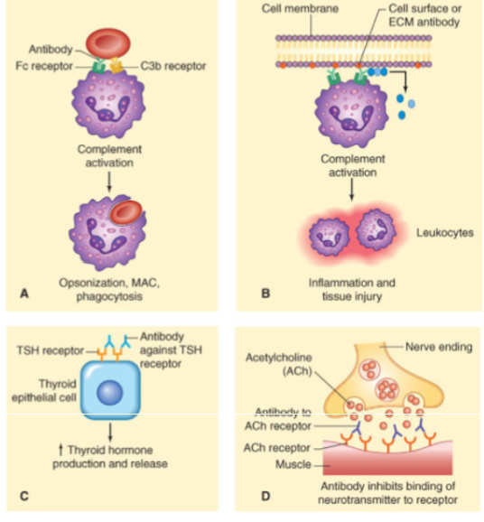
Type II hypersensitivity
antibody mediated
mediated by IgG or IgM (no IgE) directed against target antigens on cells
location of the target defines the response
examples
ABO/Rh incompatibility: antibody binds to cell-surface antigen and causes cell destruction
IgM/IgG binding activates complement cascade → lysis of RBC
graves disease: antibody binds to TSH receptor on thyroid follicle cells and activates it
myasthenia gravis: antibody binds to acetylcholine receptor on skeletal muscle cell and prevents activation
type III hypersensitivity
immune complex mediated
antigen/antibody complexes formed in bloodstream
complexes are eventually deposited in vascular endothelium or extravascular tissue
initiates inflammation
the location of the deposited antigen-antibody complex defines the response
EX
systemic lupus erythematosus (SLE)
glomerulonephritis
type IV hypersensitivity
delayed or t-cell mediated (no antibodies involved)
sensitized T lymphocytes
take time to develop, which is why it is “delayed’
e.g. contact dermatitis → poison ivy, nickel, etc
type IV hypersensitivity: initial exposure (sensitization)
APCs process antigen (e.g. poison ivy toxin, medication)
APCs present antigen to CD4 cells and activate them
CD4 cells become t-helper cells and produce memory t-cells
type IV hypersensitivity: subsequent exposure
rapid reactivation of memory t-cells
memory t-cells recruit phagocytic cells
phagocytic cells destroy tissue
memory t-cells activate cytotoxic t-cells
cytotoxic t-cells secrete enzymes that destroy tissue directly
stevens-johnson syndrome
rare cutaneous reaction to medications
type IV hypersensitivity
pathophysiology
APCs take a portion of medication, bind it to a molecule of MHC and express it on the surface
MHC I / medication complex activates cytotoxic T cells
cytotoxic T cells infiltrate tissue and release enzymes that promote cell destruction
MHC II / medication complex activates T helper cells
recruit macrophages/neutrophils that destroy cells
transplant rejection
process involving cell-mediated and humoral immunity
most common is t-cell mediated and is known as cellular rejection
donor antigens are presented to recipient T lymphocytes by APCs
antibodies will be created that target the donor tissue
may be expedited if previously sensitized (ABO mismatch or prior organ rejection)
APCs may come from recipient or from the donor
transplant rejection: direct pathway
antigen is pre-processed
APCs come from the DONOR
T cells (CD4 and CD8) of the RECIPIENT recognize foreign MHC molecules and antigens on the APCs of the DONOR TISSUE
T helper cells secrete cytokines that are responsible for 3 things
cytotoxic T cells release enzymes and kill foreign tissue
transplant rejection: indirect pathway
antigen needs to be processed first
APCs come from the RECIPIENT (host)
RECIPIENT APCs process the antigen from DONOR tissue
RECIPIENT APCs present antigen to T cells
T cells of the RECIPIENT recognize foreign antigen on the APCs and are activated
T helper cells secrete cytokines that are responsible for 3 things
cytotoxic T cells release enzymes and kill foreign tissue
this is a slower reaction
graft vs host disease
cells with functional immune capacity are transplanted into someone who is immunocompromised
the grafted tissue (the graft) rejects the recipient (the host)
3 requirements
tissue must have functional immune component
recipient must have antigens foreign to donated tissue
like if you were to get a transplant from someone related to you, it would be less likely for it to be foreign
recipient must be immunocompromised
t cell mediated
autoimmune disorders
tissue specific (graves disease) or may affect several systems
tolerance → the ability of the immune system to differentiate self from non-self
central tolerance: apoptosis of autoreactive cells prior to release into circulation
t cells removed in the thymus
b cells removed in the bone marrow
peripheral tolerance: mechanisms in place to eliminate autoreactive cells that escape the thymus or bone marrow