OMSI- FAAM V
 Call Kai
 Learn
Learn Practice Test
 Spaced Repetition
 Match
Match Flashcards
 Knowt Play
1/118

There's no tags or description
Looks like no tags are added yet.
Name | Mastery | Learn | Test | Matching | Spaced | Call with Kai | Chat |
|---|
No analytics yet
Send a link to your students to track their progress
119 Terms
muscle fibers
a muscle cell. Same as a myofiber
myoblast
immature muscle cell
myofibril
contractile unit of a muscle cell

myoepithelial cell
modified epithial cells found at the base of glandular elements to push product through duct
contracts to narrow the acinar and the early duct
flattened cells curving around acinar, may have elongated processes. Very eosinophilic staining, bright magenta in H and E

myofibroblast
contractile cells from a fibrocyte/fibroblast population in the area
used in wound healing, pulls it shut during healing
will de-differentiate or apoptose after wound is healed
smooth muscle cells
not very large, enlongated spindle shape
1 centrally located nucleous. will be elongated when relaxed, coiled when contracted
stains pale and lightly eosinophilic
surrounded by external lamina (similar to basal lamina)
external lamina
what smooth muscle cells attach to. composed of:
lamina rara- clear layer on electron micrograph
lamina densa- darker on electron micrograph
3 layers of rara-densa-rara
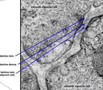
smooth muscle cells structures
communicate through gap junction because 1 nerve will control several muscle cells. shares contractile signals
sarcoplasmic riticulum is composed of finger-like tubules
myosin and actin contractile units. Ca concentration is regulated through calmodulin
dense bodies- membranous anchor points for contracile filliments. cell will contract from the plasma membrane
skeletal muscle cell
large, can be up to 4 cm in length
multiple nuclei, located on the periphery of the cell
will have fibroblasts and sateline precurser cells in the periphery
sarcomere parts
H band: only myosin, will be slightly darker on electron microscope
A band: myosin and actin, will be paler than H band and Z line
I band: only actin, includes Z line.
Z line: where actin attaches. will be very dark on electron microscope

cardiac muscle
smaller than skeletal muscle, similar to smooth muscle size
1, centrally located nucleus (may have 2)
striated.
dark bands are called interculated discs, connect cells together end to end with gap junctions. made of modified macula adherens
mesenchymal cells and muscle cells
in embryos, mesenchymal connetcive tissue will differentiate into myoblasts
will fuse to form a myotube, which will start synthesizing proteins for myofiliments
will start to form striations
some precursor cells will hang around in the periphery in case of damage. will look like a small, slightly rounder cell
sarcomere acessory structures
T-tubules, transduct signals from neuromuscular junction. WIll run over A-band I band junction
2 terminal cristernal surround T tubules, connected to sarcoplasmic reticulum surrounding sarcomere
transverse tubule system in cardiac muscle
similar to skeletal, but there’s only 1 T-tubule and 1 terminal cisternae.
Cross bridge cycle
Myosin is bound to an ADP
Calcium exposes actin binding sites, myosin associates
myosin dissasociate from ADP, causes a shape change where the myosin will pull actin
ATP will associate, myosin will release
ATP hydralized, now myosin can restart
how do smooth muscle contract
contracts around the cell instead of within, twisting it
calcium channels open and calcium binds to calmodulin
calmondulin activates myosin light chain kinase enzyme
MLC kinase will transfer phosphates to myosin, activating in
cross bridge initiates
sarcomere accessory proteins
maintain alignment and stabilizes sarcomere
alpha actin: attaches thin filiments and titin to Z line
titin: anchors thick filament to Z line. Can act as a tension gage
M line proteins and protein C: stabilize thick filaments and anchors titan
desmin: attaches Z lines to Z lines

costamere
striated muscle only. protein complex that connects cell cytoskeleton to sarcolemma and sarcolemma to extracellular matrix through dystophin.
how to scan for deep vein thrombosis
superior to inferior to prevent possibly dislodging the blood clot
common femoral artery and vein at inguinal ligament
at saphenous opening, where Great Saphenous drains into common femoral
where the lateral perforating vein drains into common femoral
where deep femoral vein drains into femoral vein
popliteal fossa
*and in between

deep vein thrombosis scan techniques
compression chain- compress and release at 1-2 cm intervals. just hold till the walls kiss
pt knee should be slightly flexed and externally rotated. probe marker is up
how to scan the patellar tendon
have pt flex knee about 20-30 degrees
transducer on interior aspect of the patella, marker towards pt head
can rotate probe marker to pt right for short axis
how to scan medial knee
flex about 20-30 degrees and externally rotate the leg
align probe with tibia on the medial aspect of knee joint
view with long axis, probe marker towards head
how to scan lateral knee
pt slightly flex and internally rotate knee
palpate head of fibula, put transducer on fibula head. probe marker superior and anterior
how to scan medial ankle
to view tarsal tunnel, place probe on medial malleolus. do not angle probe
can tilt probe to brighten image
What does the autonomic nervous system supply
both CNS and PNS elements. involuntary movement in:
smooth muscle
glands
cardiac muscle
*while not sensory, visceral sensory nerves will accompany ANS fibers
Autonomic pathway
2 neurons
preganglionic neuron (with cell body in spinal cord) will synapse postganglionic neuron (whose cell body is within a PNS ganglia)
postganglionic neurons will synapse with target organ
somatic motor pathway
1 neuron pathway
CNS to skeletal muscle
autonomic preganglionic cell bodies
found in intermediolateral section of the ventral horn gray matter. will exit ventrally

somatic motor cell bodies
found in the ventral horn of spinal cord
what structures are primarily sympathetic nervous
vessels
adrenal glands
skin
possibly adipose
where do parasympathetic nerves outflow
cranial
sacral
sympathetic nervous system anatomical characteristics
largest division of nervous system. Aka the thoracolumbar outflow (since that’s where the fibers exit spinal cord)
preganglionic cell bodies are in 12 throacic and 2 or 3 first lumbar
postganglionic cell bodies are close to the spinal cord. paravertebral ganglia or prevertebral ganglia
sympathetic chain: structure that runs from base of the skull to coccyx. One on right and left side. composed of connected paravertebral ganglia
sympathetic nervous system pregalglionic nerves
begin in gray matter of IML at T1-L2/3
preganglionic fibers exit through ventral root with the somatic motor
will go into ventral ramus to innervate viscera
preganglionic nerves will break off of ventral ramus as white ramus communicans. will run into sympathetic chain
they can:
Synapse at the paravertebral ganglion
fibers can ascend, snapse at with a prevertebral ganglion at another level
descend and synapse
can pass through chain and not synapse
ramus communicans
a nerve that connects to other nerves. Connects branch of spinal nerve and sympathetic trunk.
white- more myelin. only at T1-L2/3, made of preganglionic fibers
gray- less myelin. at all levels, postganglionic fibers
sympathetic nervous system postganglionic nerves
many possibilities
originate at paravertebral ganglia of the sympathetic chain. goes through gray ramus, joins with spinal nerve, and goes to target organ.
preganglionic will synapse at a higher paravertebral ganglion. postganglion will join with closest spinal nerve to target
preganglionic will decend to lower paravertebral. postganglion will join closest spinal nerve.
preganglionic cell will leave sympathetic chain and form splanchnic nerves. will synapse at prevertebral ganglion around major arteries. Supplies visceral pelvic.
cervical sympathetic chain
innervates thoracic viscera. 3 parts
superior: largest ganglia
middle
inferior
thoracic sympathetic chain
supplies thoracic viscera. receive white rami and give off gray rami. Can also give off thoracic splanchnic nerves that will synapse at prevertebral ganglion including:
greater splanchnic (T5-T9)
lesser splanchnic (T10-T11)
least splanchnic (T12)
lumbar sympathetic chain
includes 4 paravertebral ganglia
upper 2 or 3 receive white Rami
all give off gray rami
sacral sympathetic chain
no white Rami, only gray
ganglion impar
on the anterior aspect of coccyx. left and right sides of chain will fuse and terminate here.
parasympathetic anatomical characteristics
aka craniosacral outflow
preganglionic cell bodies are either in nuclei of 4 cranial nerves (II, VII, IX, X) or in ventral roots and rami of sacral spinal nerves (S2,3,4)
synapse at terminal ganglia close to target organ
postganglionic cell bodies at or near target organ. much shorter than preganglionic
types of visceral sensory fibers
sensation in the organs, accompaning ANS
pain afferent: sends pain inpulses to CNS. dull and difficult to localize. follows sympathetic nervous system.
monitoring afferent: a constant scanning pulse to organs, will accompany parasympathetic fibers. through vagus nerve. stretch, spasms, ischemia (not enough blood supply)
nissil substance
found in neuron cell bodies, the abundant rough ER and free ribosomes stains dark basophilic and splotchy

efferent neurons
motor. interpolar neurons (cell body at 1 end and synapse at other)
afferent neurons
sensory neurons. bipolar or unipolar
somatic: sensory info from skeletal muscle
visceral: sensory info from viscera, glands, and blood vessels
neuron cell bodies on light micrograph
large, euchromatic nucleus. large nucleolus
lots of nissil substance
Axon hillox (red arrow)- transition between axon and soma. where nissl substance stops.
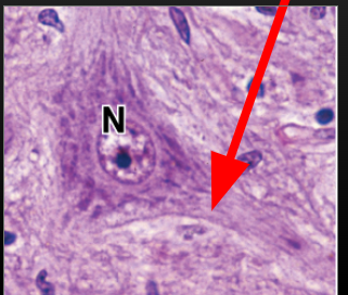
what triggers the release of neurotransmitters
voltage gated calcium channels. Calcium ions facilitate mediated exocytosis.

types of synapses
axosomatic: between cell axon and cell body
axodentritic: between axon and dendrites
axoaxonix: between two axons. usually inhibitory
cells in CNS gray matter
neurons
glial cells- smaller nuclei than neurons
neuropil- a network of neuronal and glial processes. the space between cell bodies
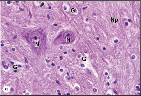
types of CNS glial cless
astrocyte- maintains envirnment. star shaped with protrusion wrapping around vessels and axons
oligodendrocyte- CNS mylination/protection. 1 cell can mylinate/encircle many. in light micrograms will have a halo due to myelin’s high fat content
microglia- a type of macrophage. small, flat appearance while inactivated (most of the time)
ependymal cells- epithelia that lines ventricles and central canal, produces CSF.
astrocyte
very abundant in CNS, maintains the envirnment, provides structure, blood-brain barrier, and will respond to CNS injury
protoplasmic astrocytes- gray matter
fibrous astrocytes- white matter
peripheral nerve linings
endoneurium- around individual neurons
perineurium- around fasciles. forms blood brain barrier, adhered by tight junctions. contains blood vesseles
epineurium- around nerve. dense irregular, contains larger vessels
ganglia vs gray matter
ganglia: grouped cells bodies, neuroglia is around somas orderly
gray matter: diapered cells bodies, ganglia is also more scattered
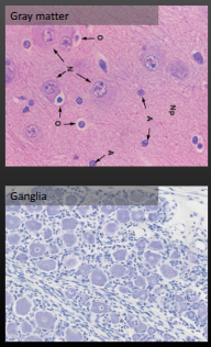
satelite cells
glia cells in the PNS, does the same thing as atrocytes in the CNS. surrounds neuronal cell bodies.
schwann cells
similar to oligocytes, surrounds the axon.
mylinating: produces myelin sheath. only 1 axon at a time
non-myelinating. simply encases the axon, can wrap around many axons this way
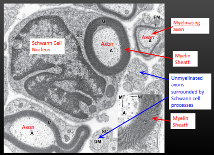
nerve conduction
aka action potential propogration. will be anterograde (towards terminal)
localized voltage gated channels will open
cell membrane potential will become more positive due to sodium entering cell
sodium ions will diffuse, depolarizing adjacent membrane
saltartory conduction (active)
in mylinated cells with nodes of ranvier. At each node there are a lot of voltage gated channels that will allow a large sodium ion influx. positive ion charge will have more force and spread down to the next node and and activate those channles
electronic conduction (passive)
describes how the sodium behaves once inside cell, not action potential itself. In unmyelinated cells. depolarization spread fueled by sodium ion passive diffusion though voltage gated sodium channel. creates “wave” of depolarization.
action potential steps
voltage gated sodium channels open.
ions rush in, membrane is depolarized through passive conduction. this opens adjacent channels
afterwards, Na channels inactivate (a state where they can’t be stimulated to reopen- ensures action potential only travels in one direction. creates the refractory period)
membrane is repolarized through potassium ion efflux. this will overshoot and hyperpolarize
resting membrane potential is restored, sodium channels will reactivate but remain closed
membrane resistance
prevents flow of ions across membrane
axonal resistance
makes flowing ions in axon more difficult. will conduct faster if axon has larger diameter.
how does myelin affect action potentials electronically
increases membrane resistance
decreases membrane permiability
decreases capacitance
* opposite properties to the nodes of ranvier
how will a nerve conduct an action potential
compound action potential of all the nerve fibers. Will create a bell curve of stimuli between the slowest, average, and fastest neurons.
electrical synapse gap junctions
neurons are joined by pairs of connexons (channels of 6 connexin proteins)
no delay in communication, cytoplasm is continuous. For cells that need to fire at the same time.
reciprocal synapse- flows in both directions
rectifying synapse- flows in one direction
chemical synapse
neurotransmitters are released by the presynaptic terminal. typically triggered by a voltage gated calcium channel at the terminal.
will diffuse across synaptic cleft
activate receptors on the receiving cell
ionotropic receptors- triggers an ion influx
metabotropic receptors- triggers cell signaling cascade
inhibitory vs exitatory neurtransmitters
inhibitory: induces an inhibitory post-synaptic potential. lowers membrane.
exitatory: induces a exitatory post-synaptin potential. raises membrane potential.
aceytlcholinesterase
clears acetylcholine from neuromuscular junction
how does stimulation of a muscle cells end
ACh is destroyed by enzyme
presynaptic re-uptake of acetylcholine
ACh simply diffuses away
what does Ach do at neuromuscular junction of MSK
will bind to nicotinic receptors on motor end plate
sodium influx into muscle cells, has it’s own action potential and threshold
action potential travels down membrane and enter through T-tubules. will trigger SR to release calcium through dihydropyradine receptor
why does cardiac action potential have a plateu
cell repolarization is delayed by an influx of calcium ion
types of reflexes
monosynaptic: sensory => motor
polysynaptic: sensory => interneuron => motor
both pass through spinal cord
where are proprioceptive receptors
in muscles, tendons, joint
proprioceptor types
Muscle spindles: embedded in skeletal muscle fiber at the belly
golgi tendon organs: interface of muscles and tendons
joint receptors: in joint capsules, low threshold
muscle spindle
reacts to lengthening by contracting, made of specialized intrafusal muscle fibers
spindle activation
1a sensory nerve fiber gives into to spinal cord
synapse directly with gamma motor neuron, causes contraction. innervate spindle, not extrafusal fibers.
*know structure
golgi tendon organ
reacts to force production by causing relaxation
1b sensory fiber carries info to spinal cord
synapse with interneuon- is inhibitory to alpha motor because it can only induce contactions
synapse with alpha motor neuron, neuron is deactivated to relax
types of intrafusal fibers
nuclear bag- the many nuclei of a muscle fiber is all clustered together
nuclear chain- the nuclei are in a row
1a sensory fiber wraps around nucleus area, called annulospial ending. Group II fibers will branch and form many synapses with the muscle cells but not wrap around, called flower spray ending
spindle stimulus response- 1a
when length increases, more action potentials become more clustered.
frequency will level out once length has plateu.
Will stop during shortening, and continue sporadically once held at shorter length
spindle stimulus response- II
always firing, tells only if the muscle is moving or not. 1a is more detailed
example- patellar tendon reflex
monosyaptic reflex, an example of a muscle stretch reflex
tendon is tapped, the deformation makes quad muscle quickly increase in length
sensed by primary spindle (1a)
synapse directly with alpha motor neuron
induces contraction
why does the muscle spindle work like it does?
regulates muscle stiffness and proprioception, muscle must stay taught for spindle to be effective
protects against over stretching and tearing
golgi tendon reflex
will sense force changes, too much force causes golgi tendon to start relaxation by inhibiting the alpha motor neuron. protects against tearing muscle
withdrawl (flexor) reflex
involves whole limb, needs many neruons to fire simultaneously
a sensory neuron synapses with interneuron in the spinal cord
interneuron activates several interneurons to activate motor neurons or inhibit antagonist muscles
triggers other arm/leg to do the opposite action to counterbalance harsh movements
Glutamate
excitatory neurotransmitter
GABA
inhibitory neurotransmitter, opens chloride channel
how does myelin decrease capacitance
capacitance is the interaction of two charges across a space. So, with the thick myelin, the membrane charge is not able to interact with the extracellular space, getting rid of the capacitance factor.
extracellular positive concentrations will not effect the sodium depolarizing the membrane. makes sodium diffuse faster
length constant
quantified the distance a signal will travel until 37% of the max value.
axon with more channels => smaller length constant
will be equal for retrograde and anterograde, but retrograde would not do anything since the sodium channels are inactivated
time constant
time it takes for the response to a stimuli to reach 63% of it’s maximum response
in a neuron, it’s the depolarization in membrane potential after a stimulus
larger tc => slower neuronal responses
how does summation work
considered by time and length constants. A change in voltage at the soma has to be strong enough to propogate towards the axon hillox. with multiple stimuli, they can compete or amplify the voltage.
longer time constant means stimuli will build on eachother. short means the first will dissipate before the next comes.
longer length constant also means they can build on eachother
what do joint receptors sense
angle
what can visualize unstained cells
bright field microscopy- hard to see detail
phase contrast microscopy- enhances contrast, dense regions are dark
differential interface contrast- enhances contrast somewhat, gives 3D appearance. best for imaging surface
acid/base stains
eosinophilic (basic structures), most cytoplasmic proteins and extracellular fibers
basophilic (acidic structures), neuclaic acids such as DNA, RNA, and ribosomes
trichrome stains
mix of 3 dyes in series, ment to increase contrast
helps distinguish cells, especially the nuclei, from ECM
types of trichrome stains
masson’s trichrome stain- nuclei are black, muscle is pink, collagen/cartilage is blue/greem
Mallory trichrome stain: red nuclei and muscle, RBC and keratin is orange. Collagen, cartilage, and bone are blue
periodic acid schiff
over an H and E stain, makes pollysaccharides bright pink. good for highliting GAGs in basement membrane
write (giemsa) stains
good for blood or bone marrow smears
stains leukocyte granules
nuclei are purple
erythrocytes are pink
silver/gold preparations
good for very thin fibers like neurofilaments, reticular fibers, basement membranes
elastinc stains
highlights elastic fibers brown or purple.
osmium tetroxide
stains lipids and myelin black, need to use a special type of prep that doesn’t remove lipids.
luxol fast blue
myelin stain, gray matter is purple while white matter is dark blue/violet
cresyl violet
nissil stain, basic stain for RER in neuron somas