Unit 4 - Skeletal System
1/17
There's no tags or description
Looks like no tags are added yet.
Name | Mastery | Learn | Test | Matching | Spaced |
|---|
No study sessions yet.
18 Terms
Functions of the skeletal system
💪🏻support (bear the weight of the body)
🛡️protection (encase essential organs)
🏃🏻♀️movement (from joints)
📦storage (store minerals to be released into the bloodstream)
🏭manufacturing (production of red and white blood cells from red bone marrow)
*storage and manufacturing done by bone marrow
!!! Bones = solid matrix of living cells and fibers surrounded by calcium deposits, classified by shape
Bone is created/dissolved to maintain a certain level of calcium in the blood at all times
Bones/teeth are like storage tanks for calcium for later use
calcium used in movement of muscles + transportation of electrical impulses thru the nerves + more
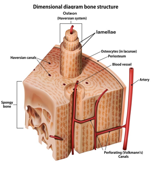
(flip to see compact bone structure)
Broken bone repair process:
Hematoma forms (blood enters wound, cells begin to die, phagocytes ingest dead bone cells/debris)
Callus forms (blood vessels grow, cartilage forms to hold bone tgt)
Callus ossifies (spongy bone forms to replace the cartilage)
Compact bone forms (osteoclasts form a larger medullary cavity, spongy bone is converted to compact bone)
^(1blood enters, 2cartilage, 3spongy bone, 4osteoclasts form larger medullary cavity, 4compact bone)
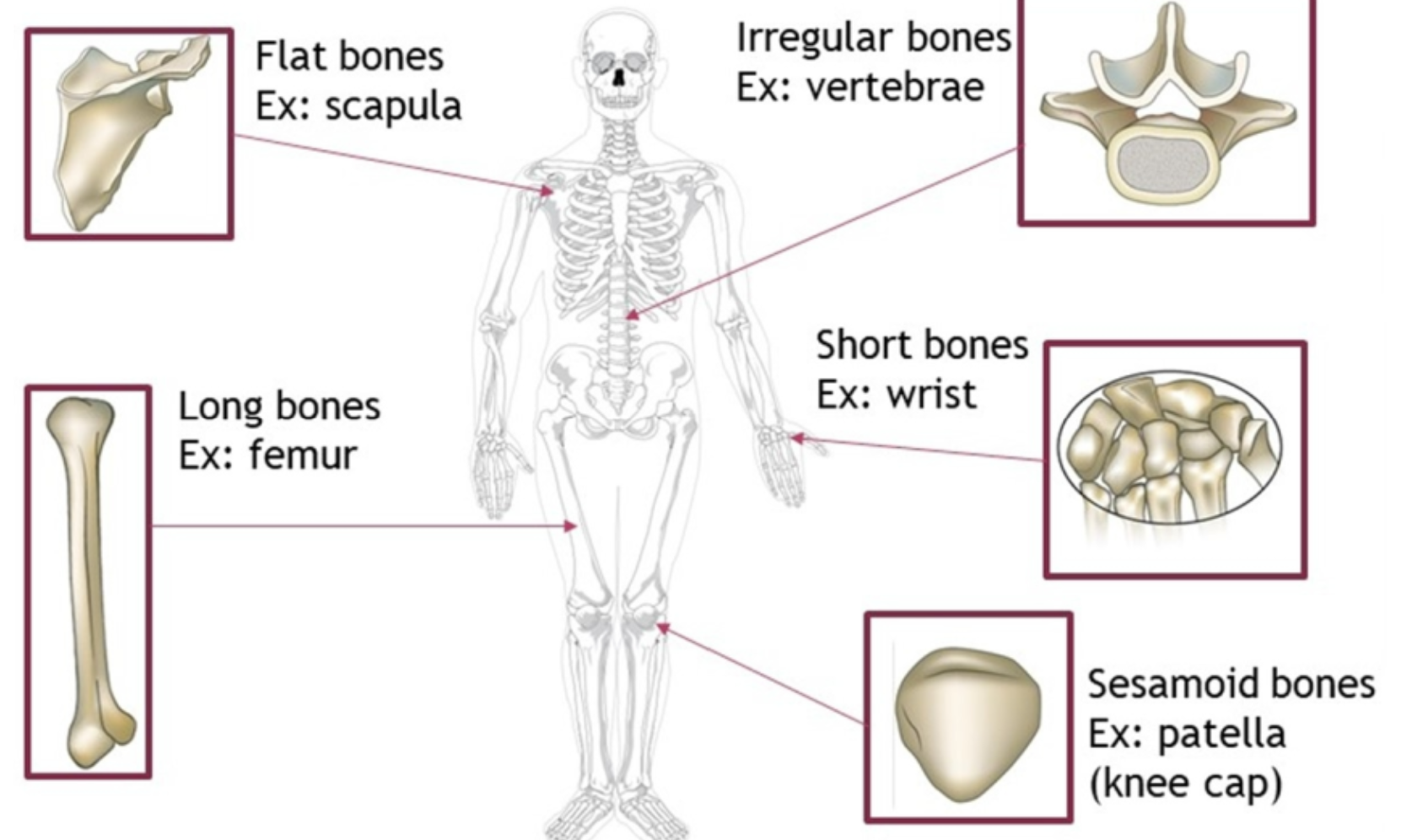
Bone shapes
flat (ex: scapula)
irregular (ex: vertebrae)
short
long
sesamoid
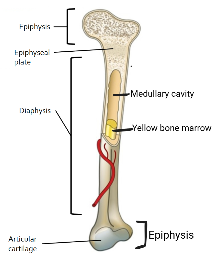
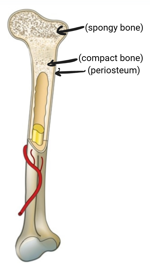
Features of a long bone
Epiphyses: ends of bone
Articular cartilage: external layer of cartilage on the ends of epiphyses, provides smooth movement of joints and cushioning from shock
Diaphysis: shaft, long part of bone
Medullary cavity: hollow, filled w/ yellow bone marrow (when young starts out red) where new blood cells are produced (hematopoiesis)
Epiphyseal/growth plate: thin layer of internal cartilage
Layers of bone
Periosteum
outer layer made of tough connective tissue
location of muscle attachment and bone repair
Compact bone
thick layer
arranged in cylinders called osteons, which are arranged in concentric circles called lamellae, which surround a central (Haversian) canal that has blood vessels and nerves
central canals are connected by perforating (Volkmann’s) canals running perpendicularly (connect to arteries)
Spongy bone
at the ends of long bones
lattice of trabeculae that are found along lines of stress (perfect to resist compression) - btwn trabeculae are spaces filled w/ marrow or blood vessels
Specialized bone cells and their functions
Osteocytes
mature bone cells that are a majority of bone structure
connected w/ tentacle-like canaliculi
Osteoclasts
break down bone (think crack)
secrete acid to enlarge medullary cavity as bone grows so that marrow is available for all cells
Osteoblasts
build/produce bone
^Osteoclasts/osteoblasts line Haversian canals and the surfaces of the compact and spongy bone
Canaliculi
connect all bone cells, allowing them to receive nutrients and remove wastes
How bone is formed
Ossification - process of incorporating calcium and minerals into cartilage to calcify (harden) and become bone
embryo’s skeleton made of cartilage
osteoblasts secrete mineral deposits to replace the cartilage
osteoblasts mature into osteocytes
How bones grow as a child ages
Tall columns of chondrocytes (cartilage cells) at the epiphyseal plate divide then deteriorate as the matrix around them calcifies (hardens) → osteoblasts, forming spongy bone
Bone composition
Osteoid (organic)
35% (made of ground substance & collagen)
provides flexibility/tensile strength so bones aren’t constantly breaking (lack of collagen causes ‘Brittle Bone Disease’)
Mineral salts (inorganic)
65%
crystalline salts made of hydroxyapatites (Ca10(PO4)(OH)2) (lack of hydroxyapatites causes ‘Rickets’ - soft/weak bones)
provides strength/hardness
2 hormones that regulate blood calcium level/trigger bone remodeling
Calcitonin
from thyroid gland
stimulates osteoblasts to deposit extra calcium from blood into bones
to lower blood calcium levels when Ca in blood is too high
Parathyroid hormone (PTH)
from parathyroid gland
stimulates osteoclasts to break down bone so Ca goes to blood
to raise blood calcium levels when Ca in blood is too low
*Blood calcium levels too low → PTH released → Ca from bones is absorbed into blood → blood calcium levels increase
*Blood calcium levels too high → Calcitonin released → Ca from blood is absorbed into bones → blood calcium levels decrease
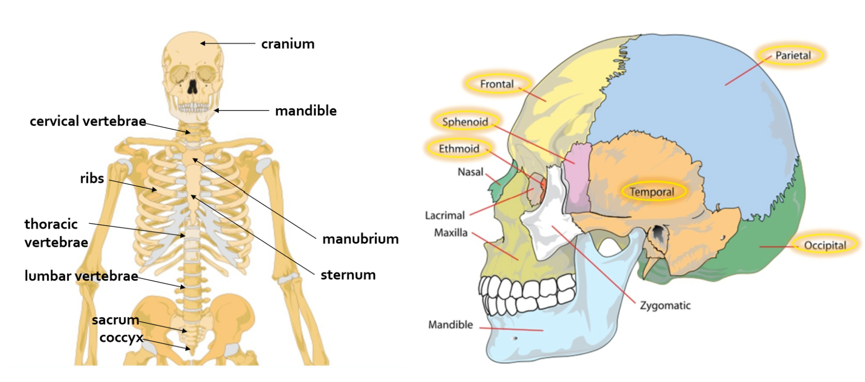
Major sections of the skeleton
Axial
central axis of the body
skull, ribs, sternum, vertebrae, manubrium, mandible, sacrum, coccyx; 80 bones
Appendicular
pectoral and pelvic girdles
bones of the arms, legs, pelvis, shoulders; 126 bones
Axial skeleton bones
Cranium
frontal
parietal
temporal
occipital
sphenoid
ethmoid
+nasal
+lacrimal
+zygomatic
+maxilla
Mandible
manubrium
sternum
ribs
Cervical vertebrae
thoracic vertebrae
lumbar vertebrae
sacrum
coccyx
Vertebral column structure
from skull to pelvis
provides support & protects the spinal cord
vertebrae (that aren’t fused) separated by intervertebral discs that provide cushioning and absorb shock
primary (convex) and secondary (concave) curvatures of the spine allow for better balance and distribution of weight
*convexly curved at birth, later 2 portions in the cervical and lumbar vertebrae develop concave curves; cervical 7, thoracic 12, lumbar 5, sacrum 5 fused, coccyx 4 fused
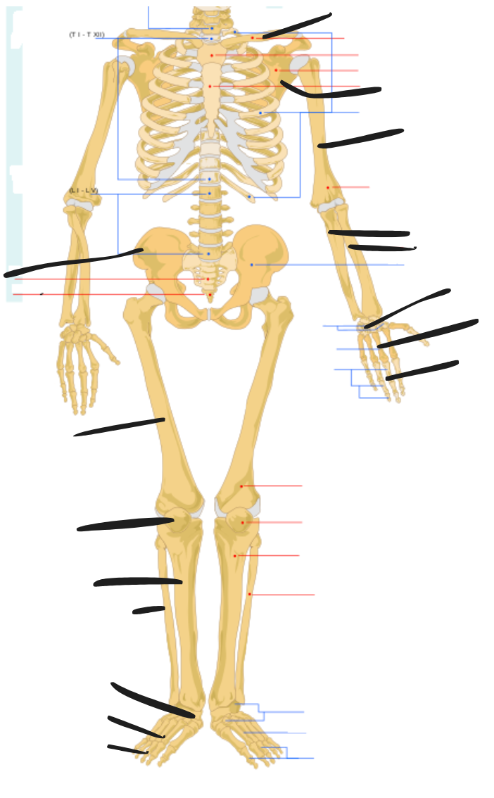
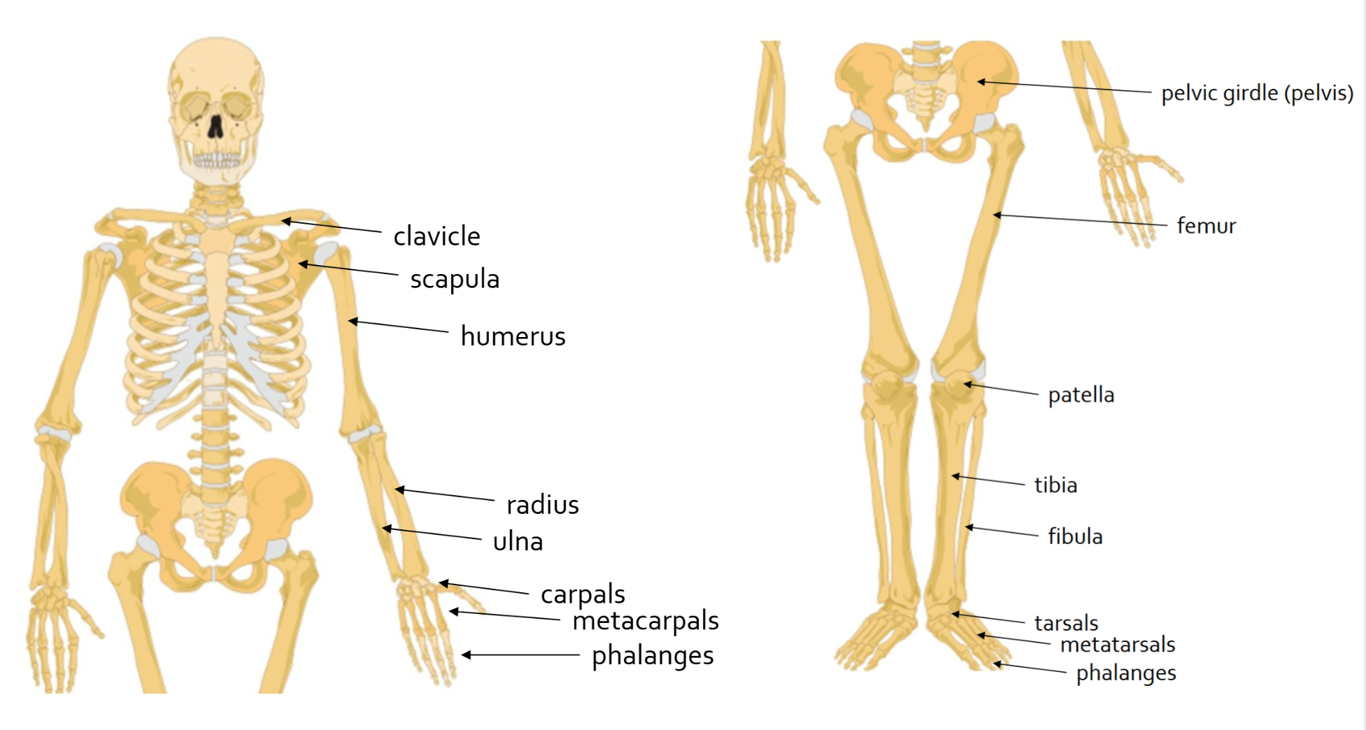
Appendicular skeleton bones
Clavicle
Scapula
humerus
ulna
radius
carpals
metacarpals
phalanges (hands)
pelvis
femur
patella
tibia
fibula
tarsals
metatarsals
phalanges (feet)
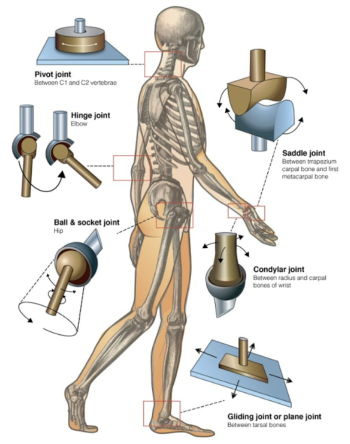
Types of joints
Fibrous
immovable or slightly movable
held tgt by fibrous connective tissue (ex: skull)
Cartilaginous
immovable or slightly movable
held tgt by cartilage (ex: ribs)
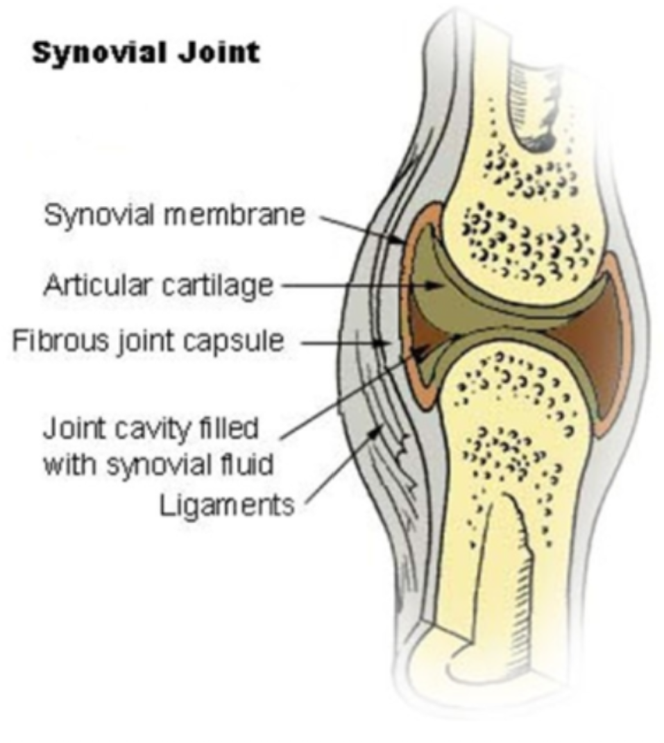
Synovial (see pic)
highly movable
joint capsule at ends of bones contains synovial fluid for frictionless movement (ex: knee)
synovial membrane and articular cartilage line the joint cavity
Ligaments vs. tendons
connect bones to bones
vs.
connect bones to muscle
Types of synovial joints
Pivot
saddle
hinge
condylar
ball & socket
gliding/plane