Micro 150 Ch 3.2 HW + Quiz WIP
1/39
There's no tags or description
Looks like no tags are added yet.
Name | Mastery | Learn | Test | Matching | Spaced | Call with Kai |
|---|
No analytics yet
Send a link to your students to track their progress
40 Terms
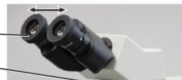
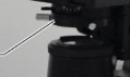
What lens is this?
Ocular

What connects the ocular lens to the microscope?
Body
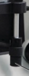
Component that holds the objective lens?
Nosepiece
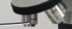
Type set of 3 lens used when the microscope is on viewed through the Ocular Lens
Objective lens
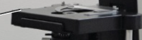
“Platform” used to hold glass slide for viewing
Mechanical Stage
Component that focuses light into Mechanical stage
Aperture diaphragm control
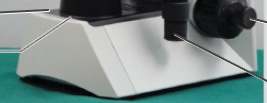
Component that is keeps the microscope level and flat on counter surface?
Base

Component that emits the light of the microscope
Field diaphragm lever
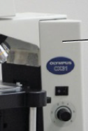
Component that holds the Ocular, Objective, Nosepiece, and or majority of the microscope’s staging components
Arm

Greater knob paired with a finer knob. Used in x4 an or x10
Coarse focus

A “Fine” knob used to focus and or adjust slide image at a higher magnification
Fine focus
Component used to adjust stage
Stage adjustment
Good magnified image consists of what concepts?
All of these options
Resolution is influenced by?
Wavelength & Numerical Aperture
Contrast is increased by
Staining
Type of stain that Stains the specimen
Positive Stain
Type of Stain that stains the Background
Negative Stain
Stain method of Negative Stain
Capsule Stain
Stain method(s) of Positive Stain
All of the three options
Differential Stain associates with what other Stain method
Gram Stain - Determines presence of LPS Layer
Acid Fast Stain - ID mycobacterium tuberculosis
Stain method Associated with Special Stain
Flagellar Stain - Defines means of motility
Differential Stains
Distinguishes organisms
The Gram Stain - permits the viewer to distinguish between gram-positive and gram-negative bacteria
Simple Stains
Easy ID of Shape and Arrangement
The methylene blue stain - Uses one dye
Special Stains
Illustrate certain structures
The flagellar stain
Magnification
The apparent increase in size of an image
Contrast
The ability to be distinguished from the surroundings
Resolution
The ability to distinguish between two separate structures that are very close to one another
Bacteria are larger than human cells. T/F
False
Bacteria are only visible with an electron microscope. T/F
False
The term used to describe a cluster of spherical bacteria is _______.
staphylococci
What general type of stain is used to separate types of bacteria based on their cellular structures?
Differential
Why must fresh cells be used when performing a Gram stain?
Old cells may not Gram stain properly Correct
After Gram's iodine is added, what color do the cells appear under a light microscope?
All cells appear purple
When ethanol is applied correctly, gram-positive cells appear ______ and gram-negative cells appear ______.
purple; colorless
The thinner peptidoglycan layer of gram-positive bacteria allows the crystal-violet-iodine complex to leave the cell. T/F
False
The differential stage of the Gram stain is the application of _______.
Ethanol
If the Gram's iodine step was skipped in Gram stain procedure, what color would the cells likely be when viewed under the microscope?
Most cells would appear red/pink
All bacteria can be classified as either gram-positive or gram-negative. T/F
False
The presence of flagella can be determined by a Gram stain. T/F
False
Rod-shaped bacteria _______.
can be either gram-positive or gram-negative