Anatomy Quiz #3
1/45
There's no tags or description
Looks like no tags are added yet.
Name | Mastery | Learn | Test | Matching | Spaced |
|---|
No study sessions yet.
46 Terms
3 types of functional classification of joints (based on allowed movement)
Synarthrosis → immovable
Amphiarthrosis → slightly movable
Diarthrosis → freely movable (all synovial joints)
3 types of structural classification (by the material that binds bones together & presence or absence of a joint cavity)
Fibrous → bones connected by dense connective tissue (fibers); tend to be immovable or only slightly moveable
Cartilaginous → bones connected by cartilage; highly moveable
Synovial → fluid-filled joint cavity between bones; highly moveable
Which joints have high mobility?
Diarthroses
Cartilaginous
Synovial
3 movements allowed by Synovial Joints
Gliding → one bone slides across the surface of another (e.g., at the wrists, ribs 2-7 & sternum)
Angular → movements change the angle between bones (e.g., flexion at the knee)
Rotation → movement around a bone’s long axis
3 types of Fibrous Joints
Suture → very short fibers (e.g., only in the skull)
Syndesmosis → ligaments that hold bones together
Gomphosis → peg-in-socket joint (e.g., teeth)
What kind of joints are cartilaginous joints?
Synarthroses (immovable)
Where are cartilaginous joints located?
Between epiphyseal plates & diaphysis
Joint between first rib & sternum
What is fibrocartilage?
Cartilage that unites bones; resists tension & compression
General Structure of Synovial Joints
All synovial joints are diarthrosis (most mobility)
Ligament → can also have extracapsular or intracapsular reinforcing ligaments
Joint cavity → space filled by synovial fluid
Synovial fluid → viscous-like egg white; acts as a slippery lubricant
Articular cartilage → hyaline cartilage containing synovial fluid; weeping lubricant
Fibrous layer → dense, irregular connective tissue
Synovial membrane → loose connective tissue; makes synovial fluid
Periosteum → thin sheet of connective tissue; delivers blood & nutrients to bones
What does the articular capsule contain?
Fibrous layer
Synovial membrane
Periosteum
Characteristics of Synovial Joints
Rich supply of sensory nerves (detects pain & monitors how much the articular capsule is being stretched)
Rich supply of blood vessels running to synovial membrane; is redundant, if some blood vessels are obstructed by certain joint positions, the joint keeps working
Filtration from the extensive capillary beds of the blood vessels gives rise to the base fluid for synovial fluid
Synovial fluid contains glycoproteins that are secreted by fibroblasts
What are the 4 functional types of Synovial Joints?
Nonaxial → movement does not occur around a particular axis
Uniaxial → movement occurs around a single axis (e.g., finger & elbow)
Biaxial → movement occurs around two axes (e.g., movement in both frontal & sagittal planes)
Multiaxial → movement occurs around all 3 axes or along all 3 planes (e.g., shoulder)
What are the 2 basic forms of Synovial Joints?
Bursa → a flattened fibrous sac lined by a synovial membrane
Tendon Sheath → an elongated bursa that wraps around a tendon
What is an example of a Synovial Joint?
Temporomandibular (jaw) joint
3 factors that contribute to stability in Synovial Joints
Articular surfaces - seldom deep enough to play a major role (exceptions: hip, elbow, & ankle)
Ligaments - more ligaments generally means a stronger, more stable joint, ligaments only stretch 6% in length & then snap; which side of the joint the ligaments are on determines what particular movements are resisted (and stable)
Muscle tone - constant, low levels of contractile force to keep tension on the tendons; especially important in the shoulders, knee, & arches of the foot
What terms accurately describe the elbow joints?
Diarthrotic & synovial
What terms accurately describe the joints between the bones of your skull?
Synarthrotic & fibrous
What are muscles?
a complex tissue containing both muscle cells & surrounding connective tissue, it makes up nearly half of the mass of the body
Function of Muscle Tissue
Produces movement: either body parts moving bones or squeezing substance through organs (ex. the heart)
Opens & closes body passageways: muscle sphincters function as valves
Maintains posture 7 stabilize joints: basic muscle tone stabilized many synovial joints, enabling sitting & standing
Heat generation: contraction of muscles produces heat that help maintain normal body temperature
Properties of Muscle Tissue
Contractibility: muscle tissue contracts due to the presence of myofilaments containing either actin or myosin (e.g., biceps brachii movement)
Excitability = irritable: nerve signals cause electrical impulses that initiate contraction
Extensibility: muscle tissue can be stretched — typically skeletal muscle by an opposing muscle & smooth muscle by substances in the organ containing the smooth muscle (doesn’t break) (e.g., elbow flexing motion)
Elasticity: after being stretched, muscle tissue passively recoils to resting length
What is sarcolemma?
Plasma membrane of muscle cells
What is sarcoplasm?
Cytoplasm of muscle cells
What are myofibrils?
Strings of actin & myosin filaments that allow your muscles to contract (fiber = cell)
What are skeletal muscles (muscle organs) composed of?
Connective tissue
Blood vessels
Highly innervated
Contraction
What types of tissue are contained in skeletal muscle (organ)?
Mostly muscle tissue
Connective tissue
Epithelial tissue lining blood vessels
Nerves
Role of connective tissue sheaths in Skeletal Muscle
Binds a skeletal muscle & its fibers together & extend from it to form a tendon
3 types of sheaths of connective tissue in Skeletal Muscle
Epimysium - dense regular connective tissue surrounding the entire muscle (G. epi = on)
Perimysium - surrounds each fascicle (group of muscle fibers) (G. peri = around)
Endomysium - a fine sheath of connective tissue wrapping each muscle cell (G. endo = inside) (G. mys = muscle mouse, mouse)
What is aponerosis?
Wide, flat tendon
Naming Skeletal Muscles
Shape (ex. the deltoid is triangular (like Greek delta symbol))
Relative size (maximus, minimus, & longus indicate size) (e.g., gluteus maximus & gluteus minimus))
Structure = number of origins (e.g., words “biceps”, “triceps”, & “quadriceps”)
Location (e.g., brachialis is located on the arm (brachium))
Attachments (name can reveal points of origin & insertion (e.g., brachiordialis))
Action (often the action is part of the muscle’s name (e.g., flexor, extensor, adductor. & abductor))
Direction of fascicles & muscle fibers (name tells direction in which fibers run (e.g., rectus abdominis & transverseus abdominis) (L. rect = straight, L. transvers = transverse))
What are synergists?
Helps the prime mover by adding extra force or by reducing undesirable movements such as controlling the position of intermediate joints (ex. extensors of wrist are synergists for making a fist; for adduction at the shoulder, latissimus dorsi is agonist & pectoralis synertist)
Example(s) of synergists
Extensor carpi ulnaris & flexor carpi ulnaris; they act together to stabilize the wrist
Example(s) of agonists/prime mover
Flexors of pollex (thumb) & digits when you clench your fist or pectoralis major when flexing arm at the shoulder
What are prime movers/agonists?
Muscles that produce the primary action
What are antagonists?
Opposes or reverses a movement
Example(s) of antagonists
Latissimus dorsi in flexing arm at the shoulder, extensors of the pollex (thumb) & digits are antagonists to flexors of the pollex & digits making a fist
What are fixators?
Holds one body part or bone in place while another part is moved
Example(s) of fixators
Rectus abdominal fixes the pelvis when you raise your legs either on the floor or when seated (rectus not involved in primary movement — it runs from the xiphoid & costal cartilages to pubic crests; never makes it to the legs)
What are the 2 types of muscle attachments?
Origin - less movable attachment (generally more proximal or nearer the axis of the body)
Insertion - more moveable attachment (generally more distal or farther from axis of the body)
Origin & insertion of brachialis
Origin: humerus (when flexing), radius (when doing chin-ups)
Insertion: radius (when flexing), humerus (when doing chin-ups)
Origin & insertion of sartorius
Origin: ilium
Insertion: proximal end of tibia
Origin & insertion of vastus lateralis
Origin: on the greater trochanter & intertrochanter line of the femur
Insertion: ligaments that are around the knee
Origin & insertion of vastus intermedius
Origin: on greater trochanter & intertrochanter line of the femur
Insertion: ligaments that are around the knee
Origin & insertion of vastus medialis
Origin: greater trochanter & intertrochanter line of the femur
Insertion: ligaments that are around the knee
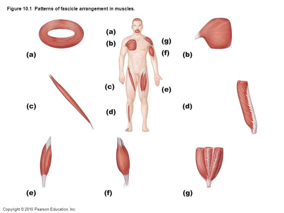
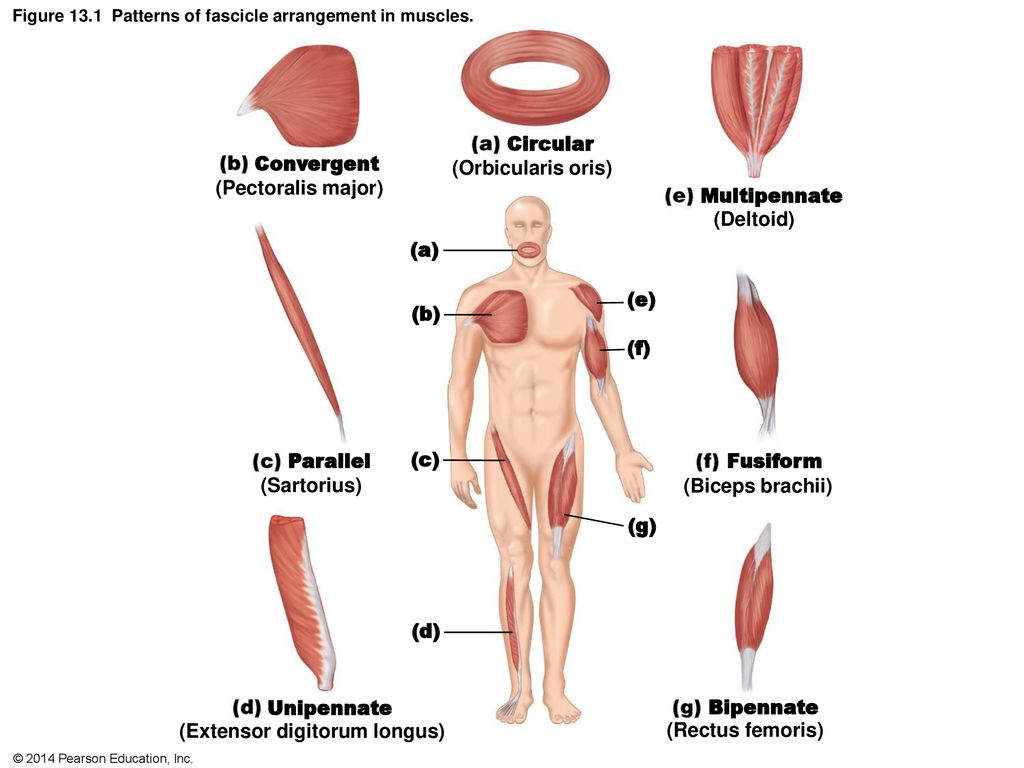
What are the different ways fascicles can be arranged?
Circular: fascicles in concentric rings, work as sphincters to close openings (e.g. muscles of the mouth)
Convergent: origin of muscle is broad & then fascicles converge toward tendon where muscle inserts to give an overall triangular or fan-shaped, fibers run full length of muscle (e.g. pectoralis major)
Parallel: fascicles run parallel to the long axis of the muscle & fibers run full length of muscle, muscles can be either fusiform with expanded belly (e.g. biceps brachii) or strap-like (e.g. sartoris)
Pennate: fascicles are short & attach obliquely to a tendon that runs the whole length of the whole muscle; making muscle look like a feather (L. penna = feather) (e.g. extensor digitorum longus)
Unipennate: if fascicles insert only on ONE SIDE of tendon (L. uni = one) (e.g. rectus femoris)
Bipennate: fascicles insert on BOTH sides of tendon (L. bi = two, double) (e.g. rectus femoris)
Multipennate: many feather-like arrangements all inserting on a single tendon (L. multi = many) (e.g. deltoid, origin: lateral third of clavicle, acromion & spine of scapula)
Muscle attachments to origins & insertions by connective tissue
Direct or fleshy attachments: connective tissue fibers are short so fascicles appear to attach to bone
Indirect attachments: tendon: form long connective fibers form cord-like structure or aponeurosis if they form flat
Example(s) where tendons meet bones (forms bone markings)
Tubercles, trochanters, crests, tuberosities