chapter 2 summary
1/102
There's no tags or description
Looks like no tags are added yet.
Name | Mastery | Learn | Test | Matching | Spaced | Call with Kai |
|---|
No analytics yet
Send a link to your students to track their progress
103 Terms
structure of the nucleus
1. surrounded by nuclear envelope (double membrane)
- the nuclear envelope contains many pores
2. granular, jelly-like substance inside the nucleus called nucleoplasm
3. contains chromosomes which are made from protein-bound, linear DNA
4. contains nucleolus which is a small sphere inside the nucleus:
- is the site of rRNA production
- makes ribosomes
function of nucleus
1. DNA replication and transcription site (mRNA)
2. contains genetic info
3. controls cell's activity
structure of smooth endoplasmic reticulum
1. has a folded membrane called cisternae
2. does NOT have ribosomes
function of smooth endoplasmic reticulum
Synthesises, processes, stores and transports lipids and carbohydrates
Structure of rough endoplasmic reticulum
1. has a folded membrane called cisternae
2. has ribosomes on the cisternae
function of rough endoplasmic reticulum
1. provides large SA for protein and glycoprotein synthesis
2. folds and processes proteins that have been made at the ribosomes (protein synthesis)
Structure of Golgi Apparatus
fluid-filled, membrane-bound flattened sacs (cisternae)
small, rounded, hollow structures called vesicles
Function of Golgi Apparatus
1. processes and packages lipids and proteins
2. makes lysosomes
3. produces secretory enzymes
structure of golgi vesicles
1. fluid-filled sac in the cytoplasm
2. produced by golgi apparatus
3. surrounded by a membrane
function of golgi vesicles
Stores lipids and proteins made by the Golgi apparatus and transports them out of the cell.
structure of lyozomes
1. a round organelles surrounded by a membrane
- no clear internal structure
2. a type of golgi vesicle
3. contains digestive enzymes called LYZOSYMES
function of lyosome
1. hydrolyses phagocytosis
2. autolysis (breaks down dead cells)
3. exocytosis (releases enzymes to outside cells to destroy materials)
4. digests/breaks down worn-out components of the cell for the re-use of the material
structure of mitochondria
1. oval shaped
2. double membrane
- inner membrane folded to form cristae
3. has a fluid centre called mitochondrial matrix
- contains enzymes involved in respiration
4. loop of mitochondrial DNA
function of mitochondria
1. site of aerobic respiration
2. site of ATP production
3. contains DNA to code for the enzymes needed in respiration
structure of ribosomes
1. very small organelle
either:
- floats freely in the cytoplasm
- is attached to RER
2. made up of 2 sub-units:
1. rRNA
2. Protein
3. NOT surrounded by a membrane
function of ribosomes
site of protein synthesis via translation:
- large sub-unit joins amino acids
- small subunit contains mRNA binding site
structure of vacuole
1. membrane-bound organelle found in the cytoplasm of plant cells
2. contains cell sap (weak solution of sugars and salts)
3. surrounded by membrane called tonoplast
function of vacuole
1. helps maintain pressure inside of cell and keep cell turgid/rigid
2. provides support and prevents plant from wilting
3. temporary store of amino acids and sugars
4. involved in the isolation of unwanted chemicals in the cells
5. the pigment could colour petals, which would attract pollinators
structure of chloroplast
1. small flattened structure
- found in plants and algal cells
2. surrounded by a double membrane
3. contains thykaloids (folded membranes embedded with pigment)
- thykaloids stack up to from grana
- grana are linked together by lamellae (thin flat peices of thykaloids)
4. fluid-filled stroma that contains enzymes for photosynthesis
function of chloroplast
Site of photosynthesis
structure of cell wall in plants and fungi
in plants:
1. made of microfibrils of cellulose polymer
in fungi:
1. made of chitin
function of cell wall
1. provides structural strength to the plant
2. prevents cell from changing shape
explain the role of cholesterol, glycoproteins and glycolipids in the cell-surface membrane
1. cholesterol: steroid molecule that connects phospholipids and reduces fluidity
2. glycoproteins: cell signalling, cell recognition and binding cells together
3. glycolipids: cell signalling and cell recognition
cell wall in prokaryotic cells
1. made of murein
- a glycoprotein
supports the cell
capsule in prokaryotic cells
only in some prokaryotes (e.g. bacteria)
made of secreted slime and helps prevent bacteria from attack by immune system cells
circular DNA in prokaryotic cells
1. floats freely in the cytoplasm
2. one long coiled-up strand
3. not attached to any histone proteins
plasmids in prokaryotic cells
1. small loops of DNA
- not part of the main circular DNA
2. contains genes for things like anti-biotic resistance
viral replication
attachment proteins attach to receptors
viral nucleic acid enters cell
reverse transcriptase makes DNA from RNA
virus assembled and released from cell
describe how prokaryotic cells replicate by binary fission
replication of circular DNA and plasmids
division of cytoplasm to produce 2 daughter cells, each with a single copy of circular DNA and a variable number of plasmids
Outline the role of organelles in the production, transport and release of proteins from eukaryotic cells
1. DNA in nucleus is the code for protein
2. Ribosomes produce protein
3. Golgi apparatus modifies proteins
4. Vesicles transprts protein
Name an organelle found in both a chloroplast and a prokaryotic cell.
ribosome
The detail shown in the diagram above would not be seenu using an optical microscope.
Explain why.
B/c optical microscopes have low resolution
- light has a longer wavelength
The cell surface membrane can be seen with a transmission electron microscope but not with an optical microscope.
Explain why.
Electron microscope has higher resolution (than optical microscope);
Before the cell was examined using the electron microscope, it was stained. This stain caused parts of the structure of the cell-surface membrane to appear as two dark lines.
Suggest an explanation for the appearance of the cell-surface membrane as two dark lines.
1. Membrane has phospholipid bilayer
2. Stain binds to phosphate / glycerol
3. On inside and outside of membrane.
Contrast how an optical microscope and a transmission electron microscope work and contrast the limitations of their use when studying cells.
TEM use electrons and optical use light
TEM allows a greater resolution
So with TEM smaller organelles can be seen and in greater detail
TEM view only dead specimens and optical can view live specimens
TEM does not show colour but optical can
TEM requires thinner specimens
TEM requires a more complex and time consuming preparation
TEM focuses using magnets and optical uses lenses
Describe how you could make a temporary mount of a piece of plant tissue to observe the position of starch grains in the cells when using an optical microscope.
Add a drop of water to a glass slide;
Obtain thin section of plant tissue and place on slide;
Stain with potassium iodide;
Lower cover slip using mounted needle
The scientists used an optical microscope to measure the number of capillaries in thin sections cut from samples of heart muscle.
Describe the method they would have used to find the mean number of capillaries per mm2.
measure diameter of the field of view and calculate the area
Use the micrometer slide and eyepiece graticule
Select fields of view randomly
Count the number of capillaries in a large number of fields of view and calculate the mean
mitotic index
number of cells undergoing mitosis ÷ total number of cells
how to calculate the numbers of minutes a cell spends in a particular phase of the cell cycle
(number of cells in that cell cycle ÷ total number of cells) × number of minutes spent in that particular phase of the cell cycle
explain why the tip of the root is used
because the cells in the tip of the root are actively dividing (by mitosis)
explain the purpose of incubating the root tip with hydrochloric acid
stops cell division from occurring
hydrolyses the middle of the lamella so that the cells can be seperated easily
describe why a stain is used
the stain binds to the chromosomes so that they become visible
describe how you would count the cells to ensure mitotic index is accurate
examine a large field of view to ensure a representative sample size
count only whole cells, to standardise counting
optical microscopes
uses light
lower resolution
can view live specimens
can show colour
smaller structures not visible
focused using glass lenses
electron microscopes
uses beam of electrons
higher resolution
can only view dead specimens
cant show colour
smaller structures visible
focused using magnets
describe TEMs (transmission electron microscopes)
higher resolution
produces image of internal structure (e.g. organelle structure)
produces 2D images
requires thinner specimen
describe SEMs (scanning electron microscope)
lower resolution
produces images of external structures only
produces 3D images
can use thicker specimens
How do you calibrate an eyepiece graticule?
place stage micrometer on the stage of the microscope
align the scales of the eye piece graticule and stage micrometer whilst looking through the eyepiece
count the number of eyepiece graticule divisions that fit into one division on the stage micrometer
divide the length of one micrometer by this answer
outline how a student could prepare a temporary mount of tissue for an optical microscope
add drop of water to glass slide
obtain thin section of plant tissue and place on the slide
stain with potassium iodide
lower cover slip using mounted needle
explain why fractionated cells are kept in a cold, buffered, isotonic solution
cold: to reduce the activity of enzymes that break down organelles
buffered: maintains pH to prevent damage to organelles and to prevent enzymes denaturing
isotonic: organelles must be the same water potential as the solution to prevent osmosis
osmosis could cause the organelles to shrivel or burst
describe what happens during cell fractionation
homogenisation
cells broken open in cold, buffered, isotonic solution by using a blender or by crushing them
filtration
solution is filtered through a gauze to remove large cells/ tissue debris
ultracentrifugation
filtered solution is spun at high speed in centrifuge
this separates organelles according to their density
this process is repeated several times at increasing speeds
remove supernatant and leave behind pellet
State the order of sedimentation of organelles during differential centrifugation.
most dense to least dense:
1. nucleus
2. mitochondria
3. lysosomes
4. RER
5. plasma membrane
6. SER
7. ribosomes
Define simple diffusion
movement of small, lipid-soluble, non-polar molecules, through a partially-permeable membrane, down a concentration gradient
passive process
Define facilitated diffusion
movement of molecules down a concentration gradient via channel (or carrier) proteins
passive process
Active transport
movement of molecules against a concentration gradient via a carrier protein
using ATP
active process
co-transport
movement of 2 different substances using a carrier protein
contrast active transport and facilitated diffusion
facilitated diffusion is passive but active transport is active (requires ATP)
facilitated diffusion involves either channel or carrier proteins, whereas active transport only involves carrier proteins
facilitated diffusion takes place down a concentration gradient but active transport can occur against a concentration gradient
compare and contrast how water and inorganic ions enter cells
both move down concentration gradient
both move through protein channel in membrane
ions can move against a concentration gradient by active transport
Features of a cell specialised for absorption
Large number of mitochondria
to release energy for AT in the form of ATP
Large number of channel and carrier proteins
for facilitated diffusion
folded membrane
so large SA
Describe how a carrier protein could facilitate the diffusion of a glucose molecule across a cell membrane
the glucose molecule attaches to the carrier protein
The carrier protein then changes shape and releases the glucose molecule on the opposite side of the membrane
How the movement of Na+ molecules out of the cell allows for the absorption of glucose into the cell
1. The movement of Na+ out of the cell maintains a concentration gradient
2. Na+ moving in by co-transport
- bringing glucose with it
How the movement of substances is affected by membrane structure
1. Phospholipid bilayer allows for the movement of non-polar substances
2. Phospholipid bilayer prevents movement of polar substances
3. Carrier proteins allow active transport and co-transport
4. Channel proteins allow facilitated diffusion
5. Shape of proteins channel and carriers determines how much movement
6. No. of proteins channels and carriers determines how much movement
7. Membrane SA determines how much movement
8. Cholesterol affects fluidity/permeability
Osmosis definition
Movement of water molecules down a water potential gradient
explain why the cell cycle does not occur in some cells
after differentiation some types of cells no longer have the ability to divide
describe what happens in interphase
G1:
- cell grows and doubles in size
- organelles double and proteins are made
S:
- DNA is replicated
G2:
- cells keep growing and proteins are made for cell division
- DNA is checked for errors e.g mutation
stages of mitosis
prophase
chromosomes condense and become visible
centrioles seperate
nuclear envelope breaks down
metaphase
chromosomes align along equator of cell
spindle fibres released and attached to chromatid by centriole
anaphase
spindle fibres retract and pull centromere and chromatid towards opposite poles
centromere divides into 2
individual chromatids are pulled to each opposite pole
telophase
chromosomes are now at each pole in the cell
chromosomes become longer and thinner again
spindle fibres disintegrate
nucleus starts to reform
cytoplasm divides into 2 (cytokinesis)
antigen
foreign protein/molecule that stimulates an immune response
Give 2 ways in which pathogens can cause disease
1.release toxins
2.kill cells
4 things antigens are used to identify
1. pathogens
2. toxins
3. abnormal body cells
4. cells from organisms of the same species
if physical barriers fail, what is the next line of defence in an organism?
phagocytosis
antibodies
a protein specific to an antigen produced by B cells
explain the structure of an antibody
4 polypeptide chains
2 heavy chains and 2 light chains
variable region
contains specific binding site
generic constant region
allows attachment to phagocytic cells
hinge
allows for flexibility
hence can bind to multiple antigens
antibody diagram

antigen-antibody complex
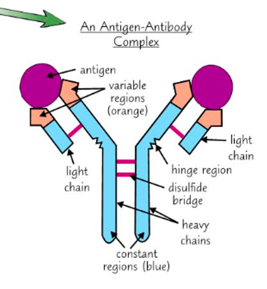
what is the role of the disulfide bridge in forming the quaternary structure of an antibody
joins 2 polypeptides
role of antibodies in stimulating phagocytosis
antibodies bind to antigens and cause agglutination
cells clump together and makes it easier for phagocytes to locate them
serve as markers
stimulates phagocytes to engulf cells
explain why antibodies are only effective against a specific pathogen
antigens have a specific tertiary 3D structure
shape of the antibody is complementary to the antigen
antibody binds to the antigen, forming an antibody-antigen complex
state how antibodies deal with infections via neutralisation
antibodies bind to toxins and prevents the binding of these toxins to the host cells
monoclonal antibody
an antibody produced from identical B cells
give one example of using monoclonal antibodies in medical treatment
carries medicine to specific cells
describe 3 ethical considerations in the use of monoclonal antibodies
1. animal testing
- involves use of mice in antibody production
2. informed consent
- patients must know all the benefits + risks of the drugs
3. drug trials
- testing on volunteers can be dangerous
- there can be issues over trial conduct
positive result in ELISA test
The first antibody binds to complementary antigen
A second antibody with enzyme attached is added
The second antibody attaches to the first antibody
Solution containing substrate is added and colour changes
phagocytosis
A phagocyte recognises a foreign antigen on a pathogen and moves towards pathogen via chemotaxis
pathogen is engulfed (via endocytosis) and enclosed in vacuole/phagosome
vacuole/phagosome fuses with lysosome, forming phagolysosome
lysosome contains enzyme called lysozyme
pathogen destroyed by lysozymes
phagocyte absorbs the products from pathogen hydrolysis
phagocyte presents the pathogens antigens
antigens from pathogens are displayed on cell surface membrane
in what case is an immune response disadvantageous
in organ transplants:
- immune system recognises the organ as 'non-self' and attempts to destroy it
how do doctors minimise the risk of organ rejection?
1. tissue type is matched
2. immunosuppressants are used
outline the process of the cell-mediated/cellular immune response?
1. complementary helper T cells bind to foreign antigen on antigen-presenting cell
2. releases cytokines
3. Helper T cells divide by mitosis (clonal expansion of complementary T helper cells)
- become memory cells or trigger humoral response
4. clonal expansion of cytotoxic T cells (TC)
How do Cytoxic T cells kill infected cells?
by producing an enzyme called perforin that makes holes in the cell surface membrane
describe the steps in the humoral response
1. B cells bind to complementary antigen
2. B cells divide via mitosis to produce plasma cells
3. plasma cells produce and secrete specific antibodies that are complementary to the antigen
4. some B cells develop into memory cells
- circulate the blood
- secondary response
in what cases may reinfection of the same pathogen occur?
1. different strains of the same pathogen
2. antigenic variability (different antigens)
3. when memory cells arent useful and produce an incorrectly shaped antibody
what is antigenic variation and what are its consequences?
When pathogens change their surface antigens
The immune system cannot recognise this new antigen, so the memory cells don’t recognise it
So a primary response must occur, which takes time and gives the individual symptoms
How do countries combat antigen variation
New vaccines are developed and chosen each year
Governments and health authorities have vaccinator programmes for the most effective vaccine that year
what are 2 issues with taking a vaccine orally?
there are enzymes in the gut that may break down the oral tablet
the molecules of the vaccine could be too large to be absorbed into the gut
Explain why antibiotics are ineffective against viruses
antibiotics work by preventing bacteria from making normal cell walls
but viruses rely on host cells for metabolic activities
viruses have a protein coat
so they dont have sites where the antibiotics can work
Describe how vaccines can lead to the production of antibodies against a disease-causing organism.
1. Vaccine contains antigen from pathogen
2. Macrophage presents antigen on its surface
3. T cell with complementary receptor protein binds to antigen
4. T cell stimulates B cell
5. B cell secrets large amounts of antibody
Why may vaccination not eliminate a disease?
1. fails to induce immunity in some people
2. vaccinated people may harbour the pathogen and infect others
3. antigenic variability (where antigens change frequently)
4. varieties of pathogen/strains
5. some pathogens 'hide' from the bodys immune system
6. medical/religious/ethical objections to taking the vaccine
what is herd immunity?
When a sufficiently large proportion of the population has been vaccinated to make it difficult for a pathogen to spread within that population
Why is herd immunity important?
its impossible to vaccinate everyone
- due to religious/medical/ethical objections
state some ethical issues with vaccinations
1. animal testing
2. side effects
3. vaccination testing
4. trialling new vaccines with unknown health risks
5. herd immunity
- should we make vaccines compulsory
6. balancing individual risk and community risk
Describe the difference between active and passive immunity.
1. Active involves memory cells, passive does not
2. Active involves production of antibody by plasma cells / memory cells
3. Passive involves antibody introduced into body from outside
4. Active long term, because antibody produced in response to antigen
5. Passive short term, because antibody is broken down
6. Active takes time to work, passive fast acting
Describe the structure of HIV
1. lipid envelope + attachment proteins embedded
2. Capsid encloses 2 single RNA strands + enzymes including reverse transcriptase
3. Its a retrovirus bc it contains RNA