A & P II LAB: Heart + Arteries
1/46
There's no tags or description
Looks like no tags are added yet.
Name | Mastery | Learn | Test | Matching | Spaced | Call with Kai |
|---|
No study sessions yet.
47 Terms
Right Atrium
receives deoxygenated blood from 3 veins
superior vena cava
inferior vena cava
coronary sinus
pushes blood down into the right ventricle

Left Atrium
receives freshly oxygenated blood from the pulmonary veins
pushes blood down into the left ventricle

Atria
receiving chambers
Ventricles
discharging chambers
Right Ventricle
discharges blood out the pulmonary trunk to get oxygen in the lungs

Left Ventricle
discharges blood out the aorta to the rest of the body

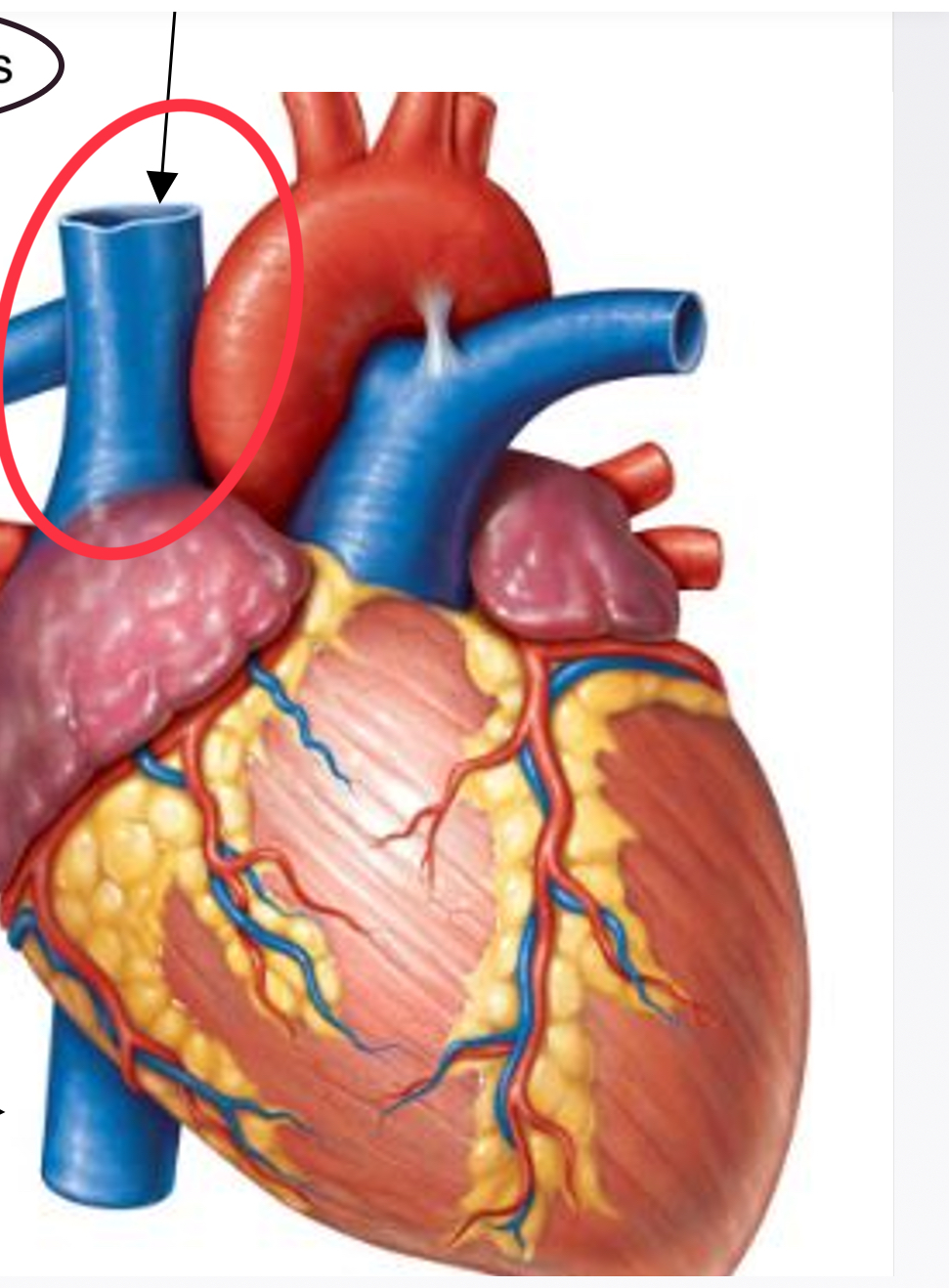
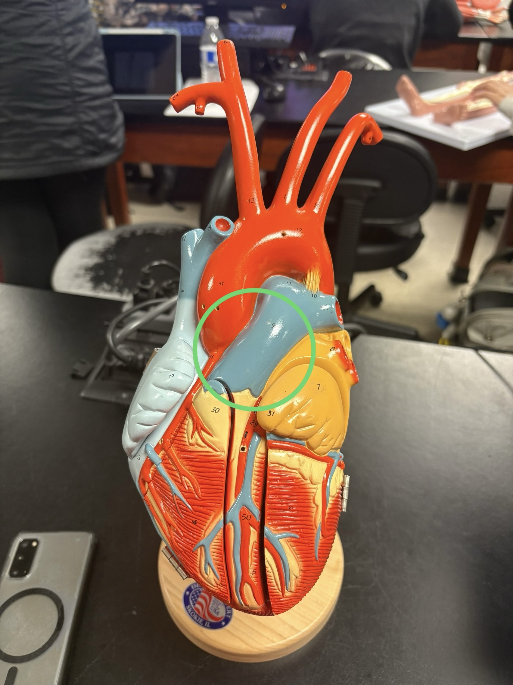
Superior Vena Cava
deoxygenated blood from tissues above the diaphragm
Inferior Vena Cava
deoxygenated blood from tissues below the diaphragm
Coronary Sinus
deoxygenated blood from the heart
returns deoxygenated blood to RA
Pulmonary Trunk
discharges deoxygenated blood out
Right & Left Pulmonary Arteries
where pulmonary trunk splits
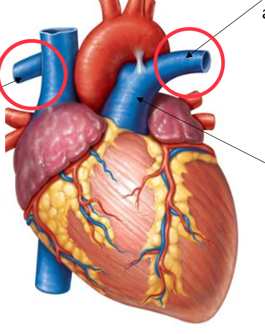
Right & Left Pulmonary Veins
oxygenated blood from lungs
Aorta
3 distinct regions around heart
ascending aorta
aortic arch
descending aorta
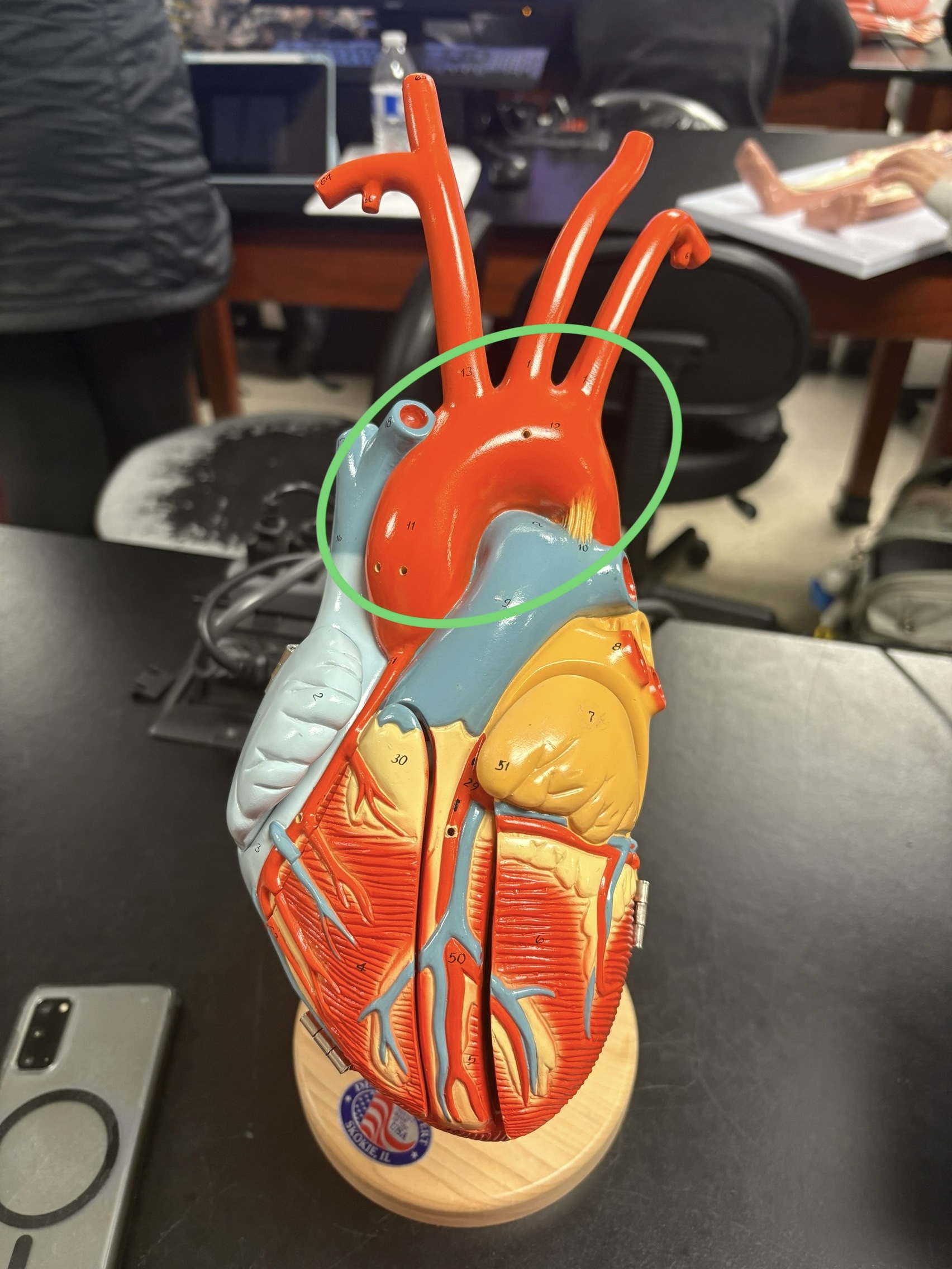
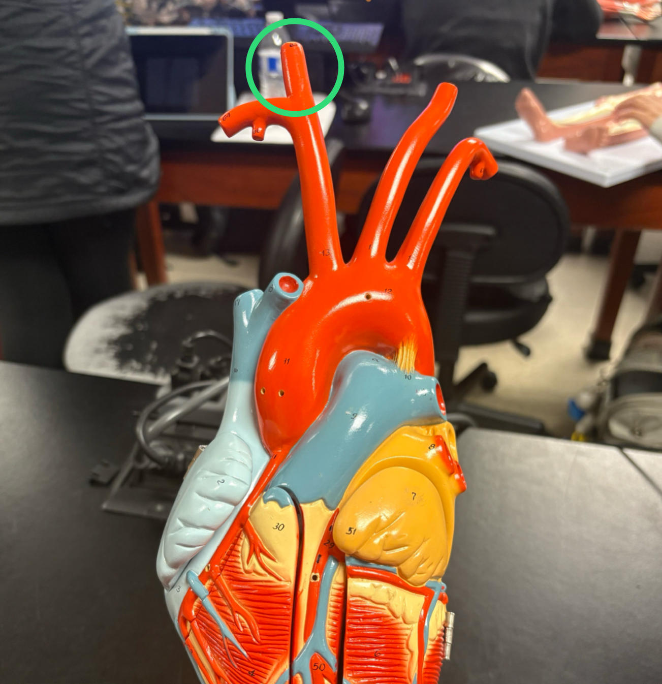
Brachiocephalic Trunk/Artery
splits into right common carotid artery & right subclavian artery
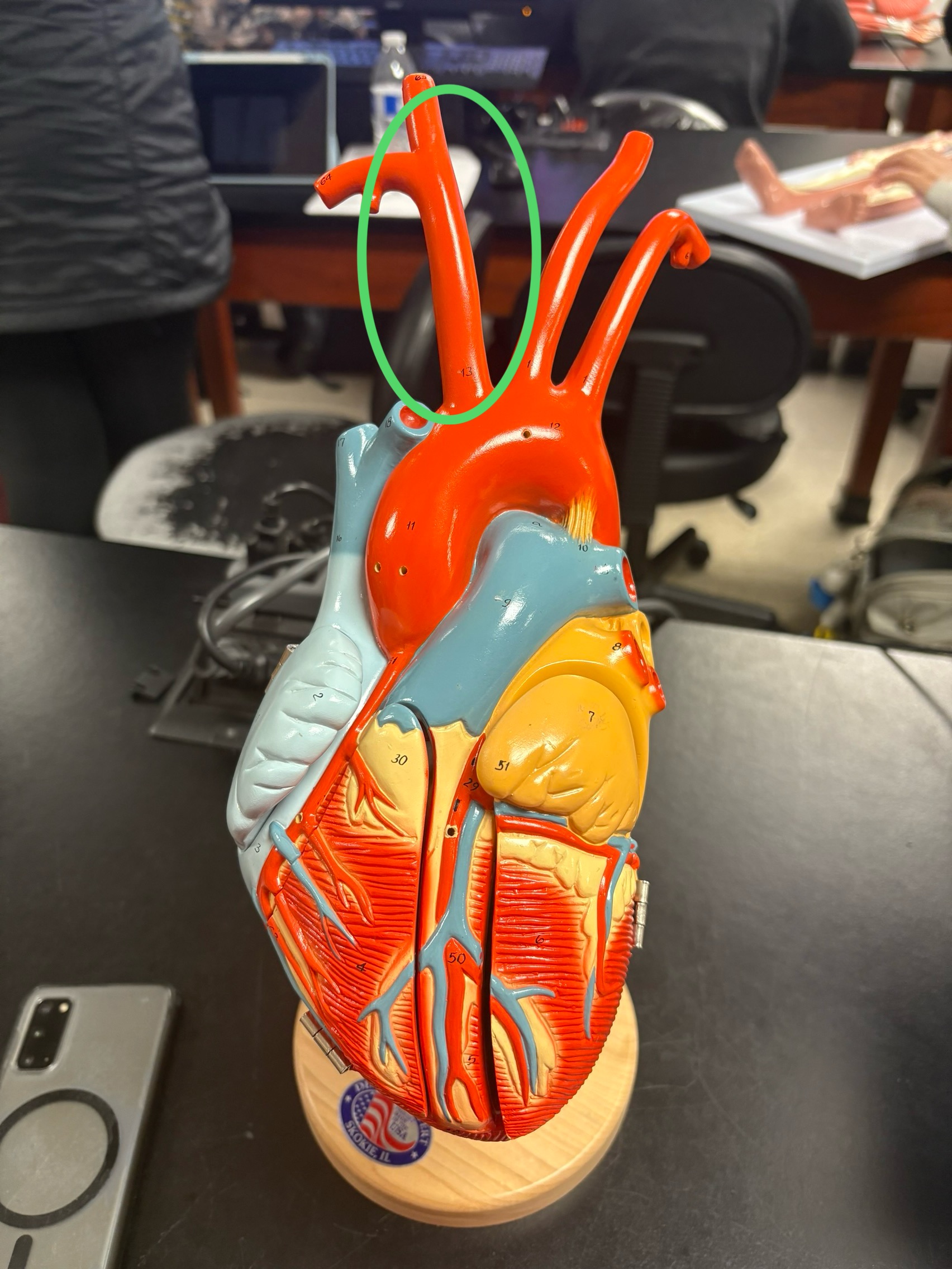
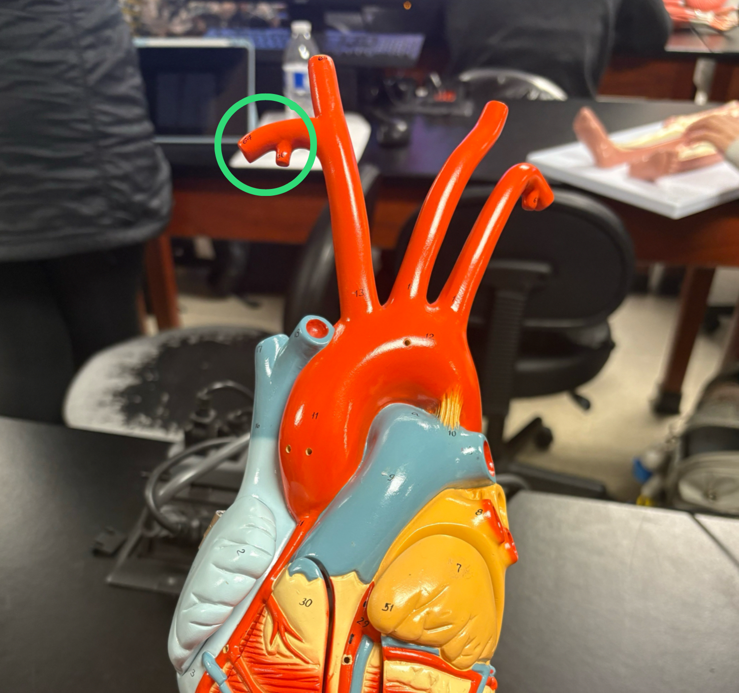
Left Common Carotid Artery
in the middle between brachiocephalic trunk & left subclavian artery
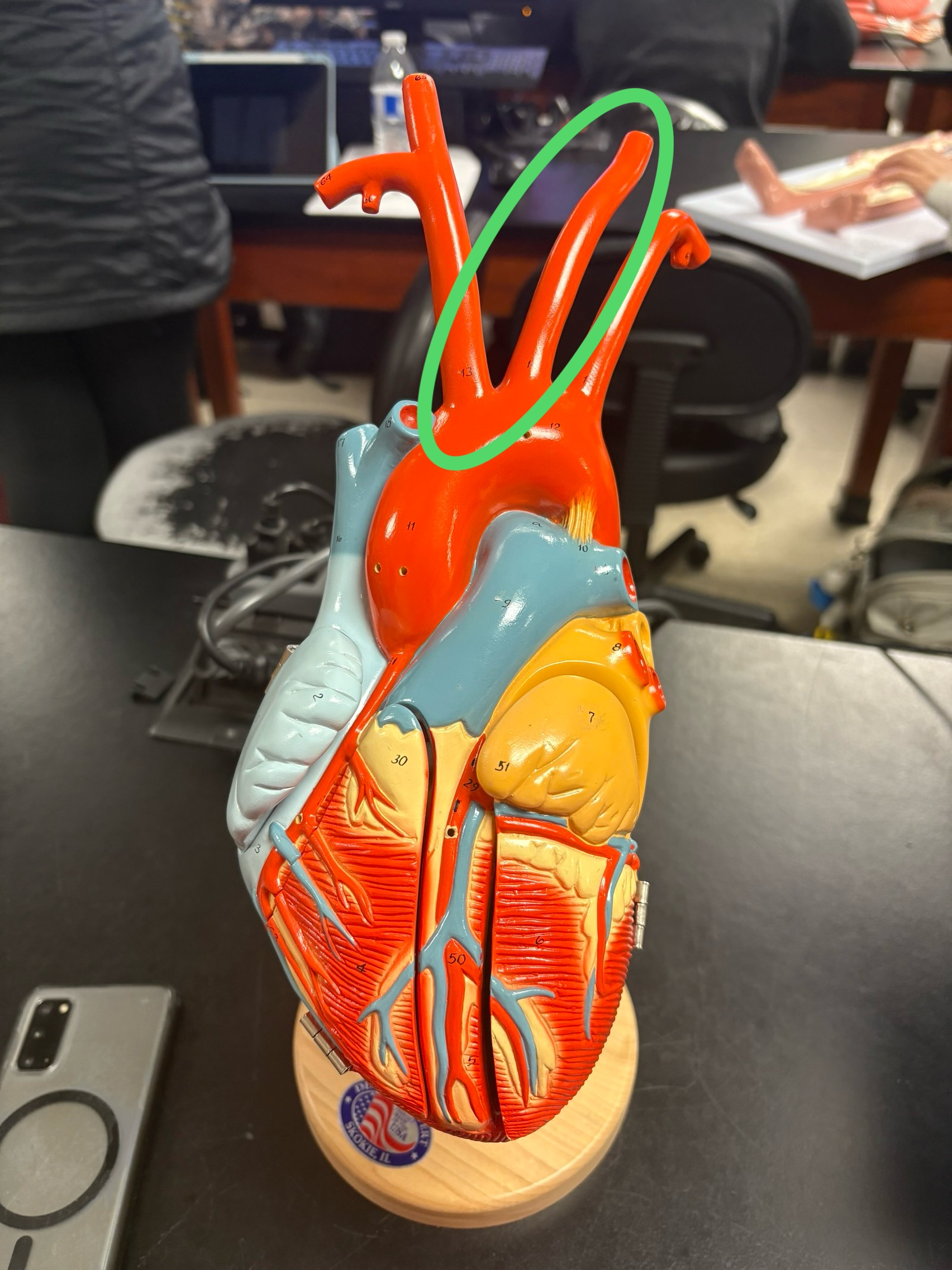
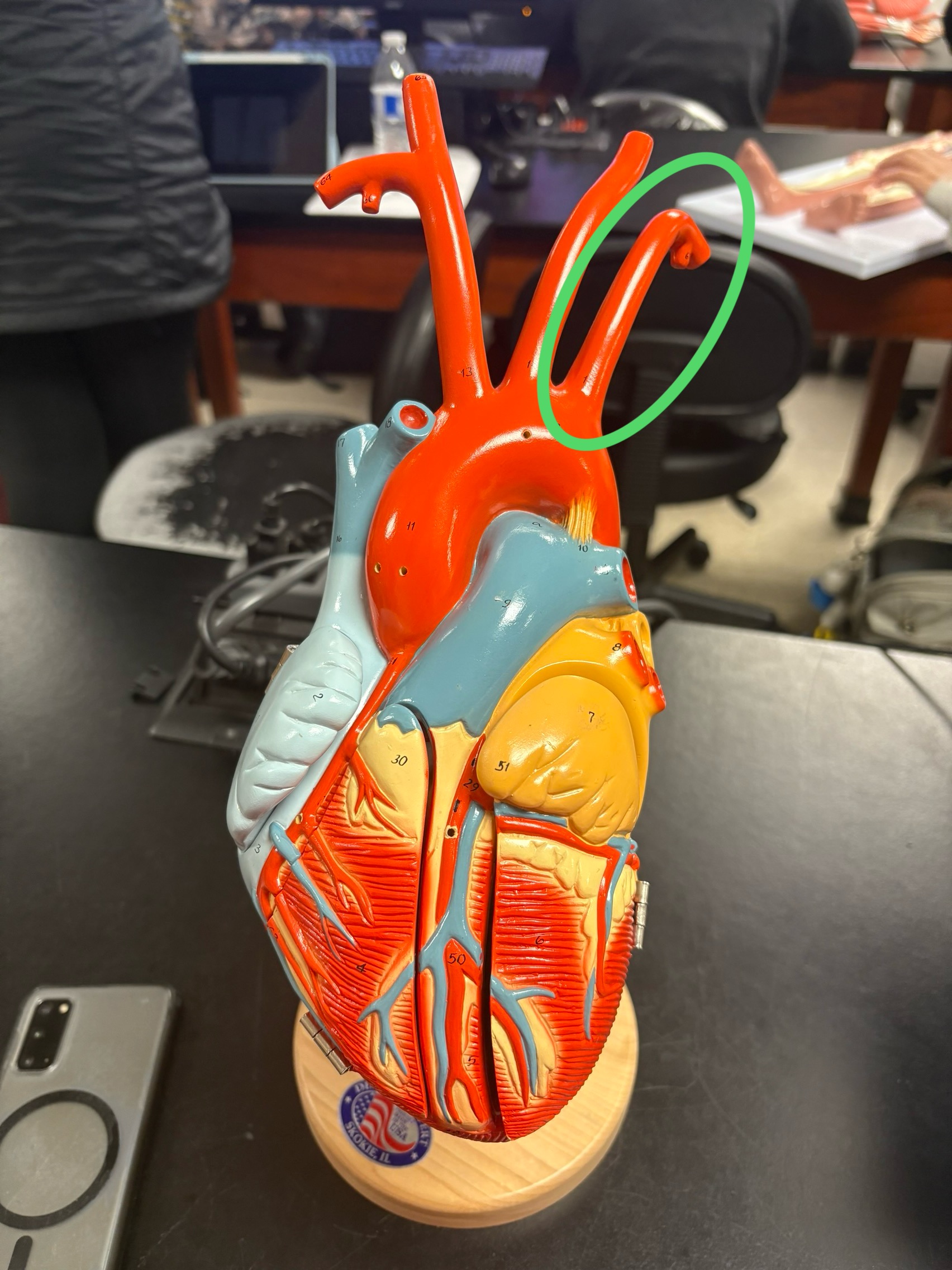
Left Subclavian Artery
next to left common carotid artery
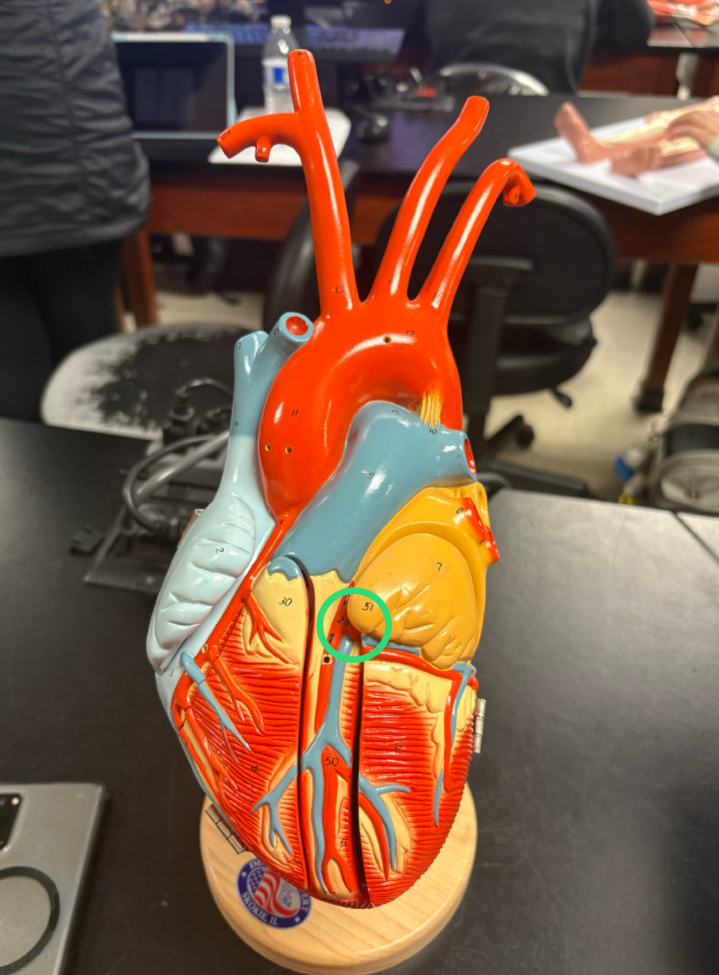
Fossa Ovalis
round depression in the wall of R. Atrium
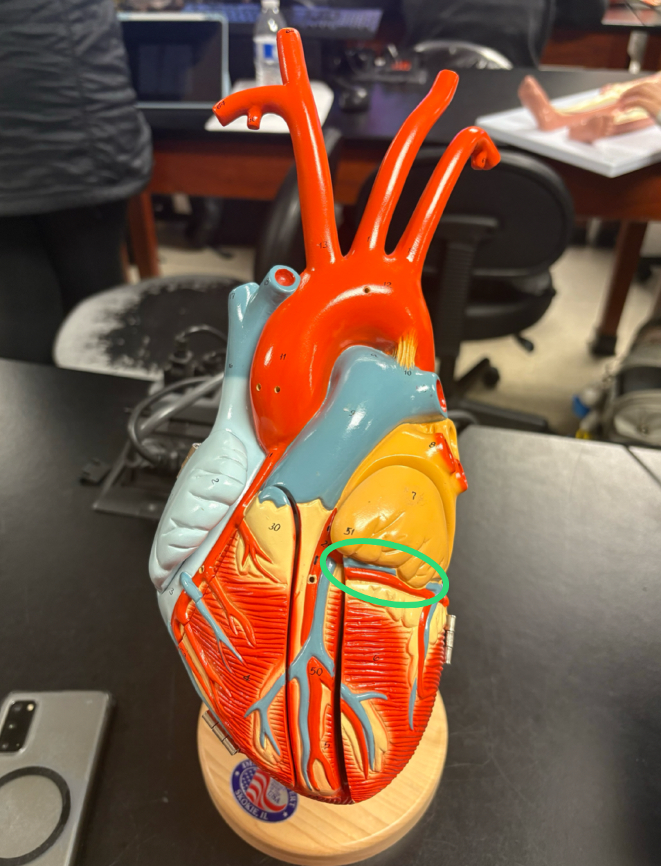
Tricuspid Valve (AV valve)
3 cusps, where deoxygenated blood enters the RV
Pulmonary Semilunar Valve
where deoxygenated blood exits the RV to go into the pulmonary trunk

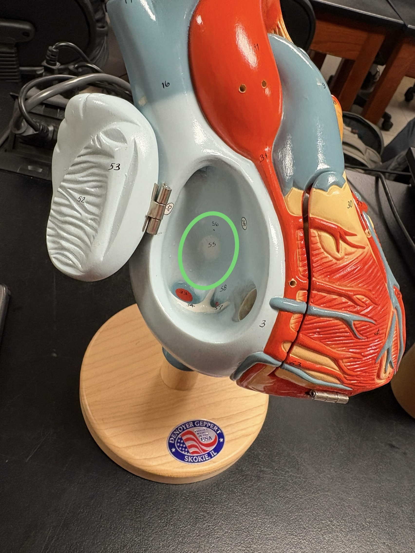
Bicuspid/Mitral Valve (AV valve)
2 cusps, where oxygenated blood enters the LV
Aortic Semilunar Valve
where oxygenated blood exits the LV to go into the aorta
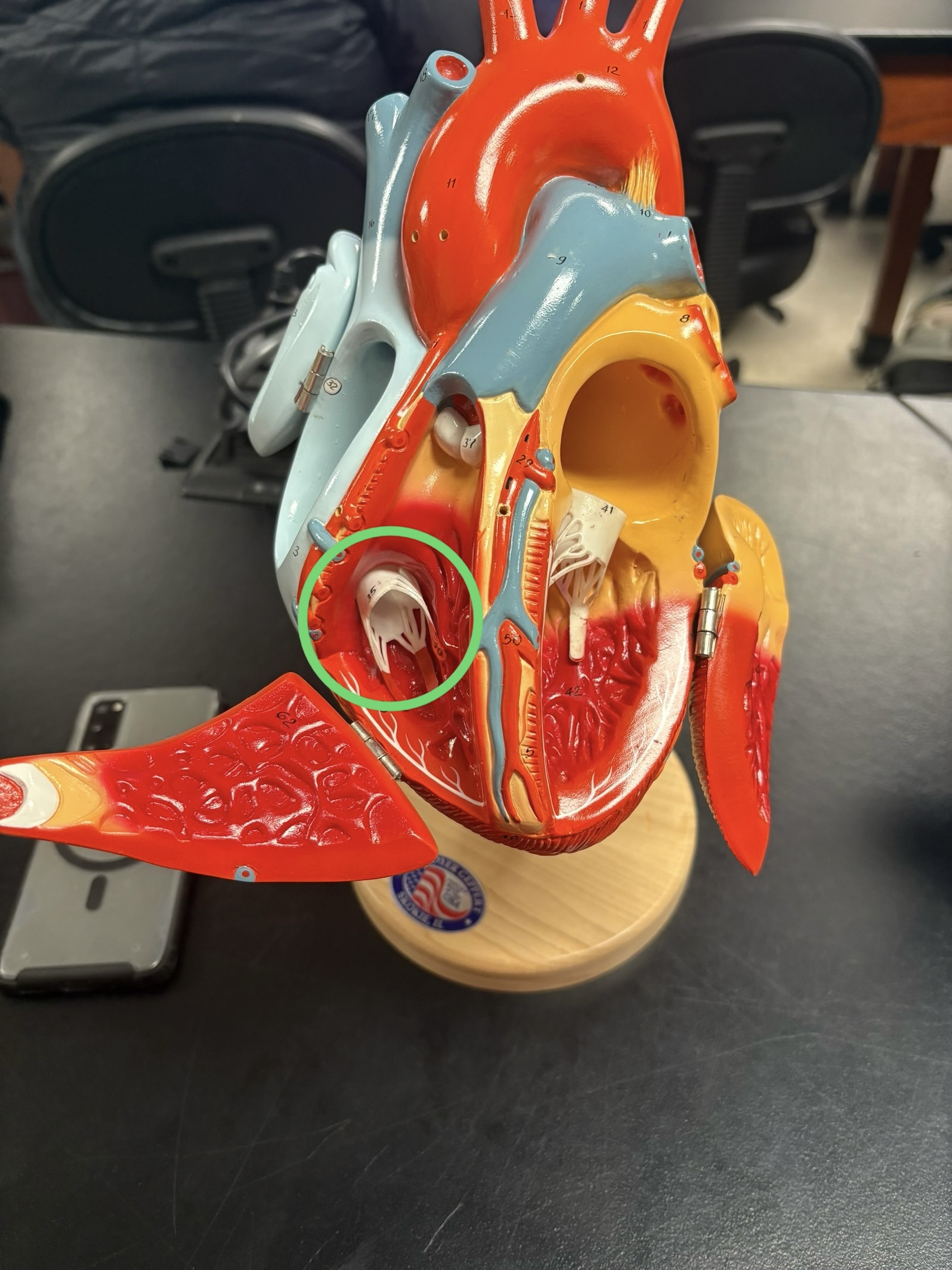
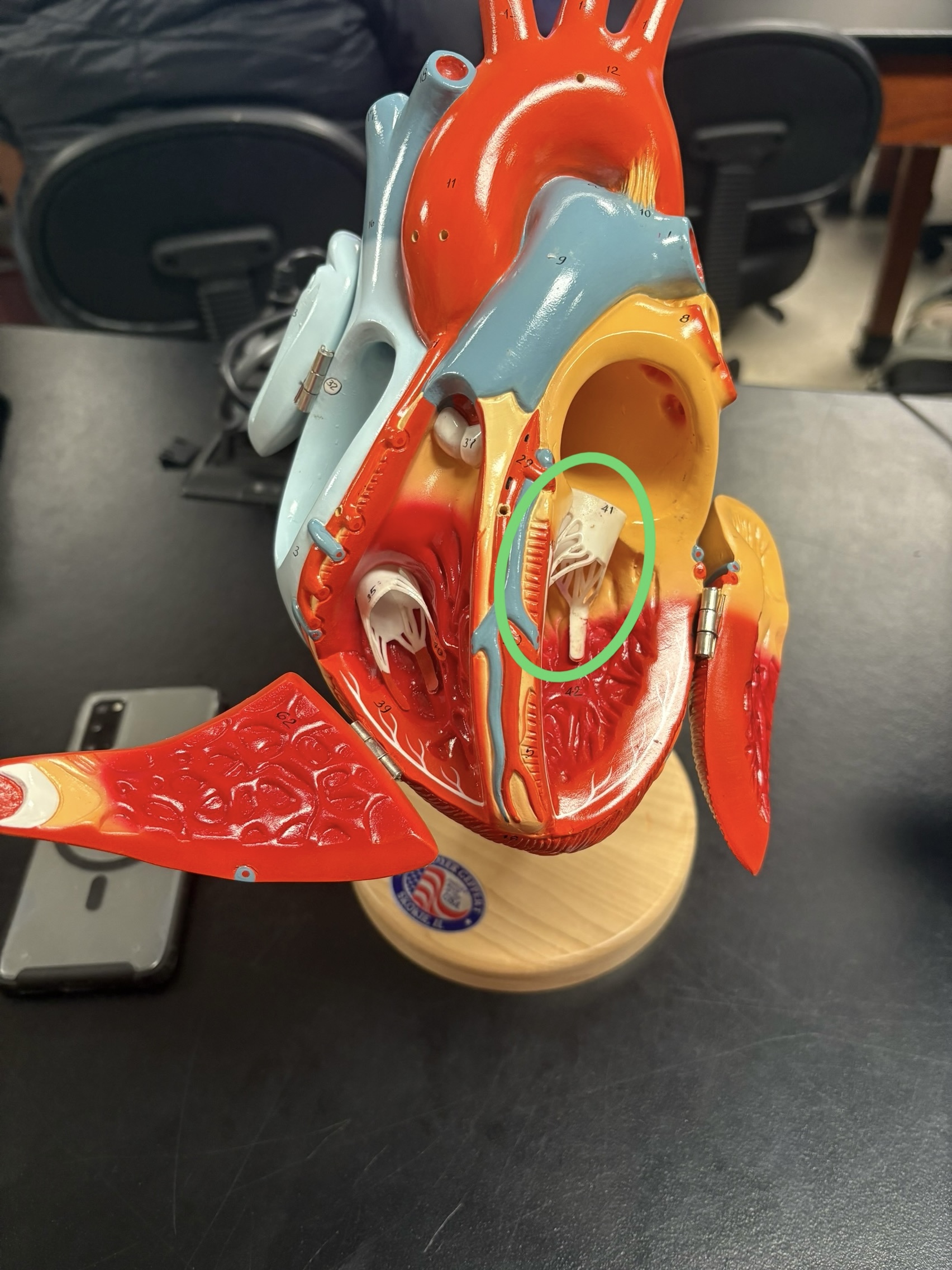
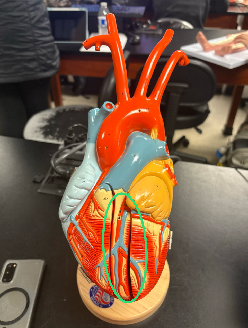
Choardae Tendineae
“heart strings” attached to the AV valves ]anchor the cusps of the valves to the papillary muscles
![<p>“heart strings” attached to the AV valves ]anchor the cusps of the valves to the papillary muscles</p>](https://knowt-user-attachments.s3.amazonaws.com/fc8b00d2-5fe1-4b28-884f-58ac0e9344f3.jpg)
Papillary Muscles
ridges of muscle attached to the choardae tendineae that help keep the valves closed when ventricles contract

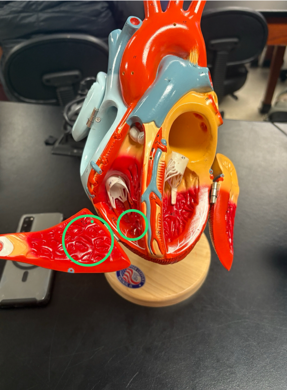
Trabeculae Carneae
irregular ridges of muscle inside the ventricles
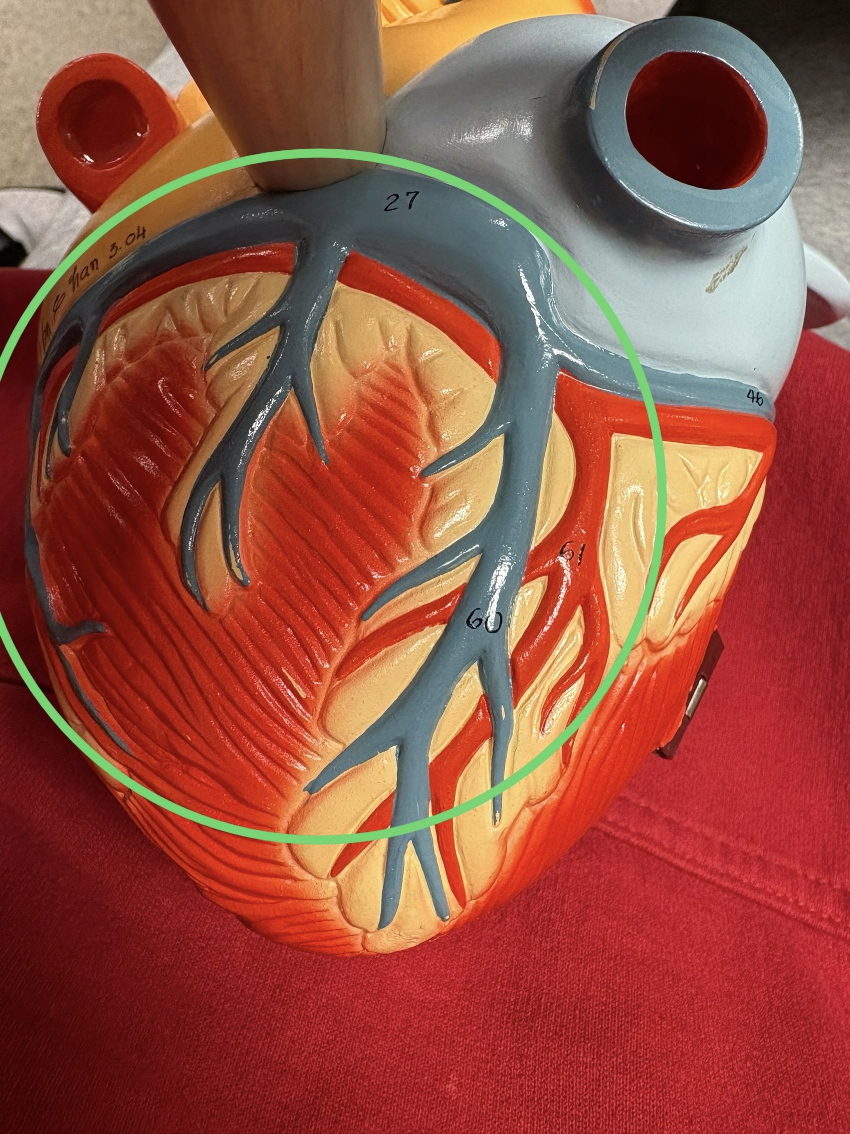
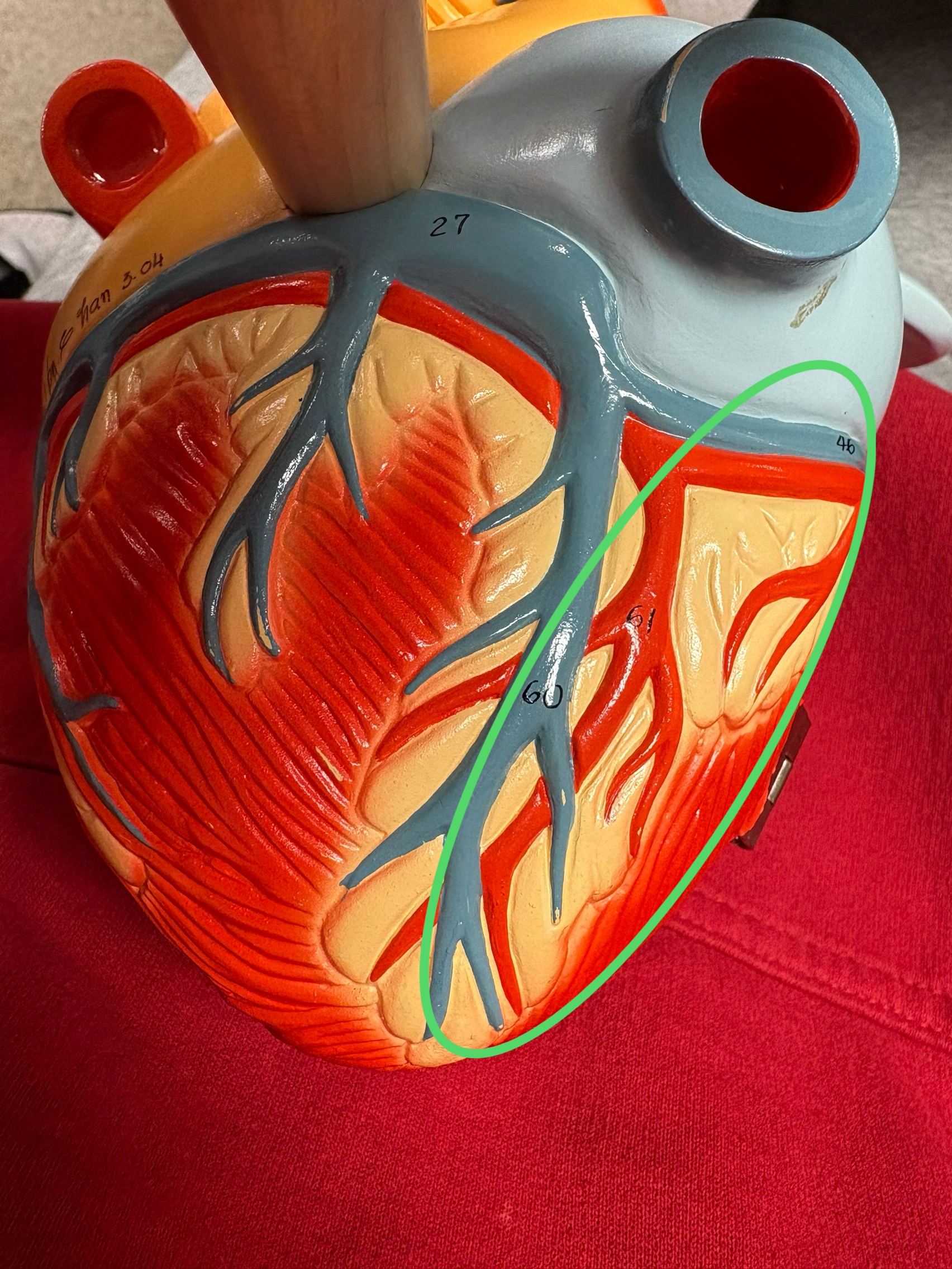
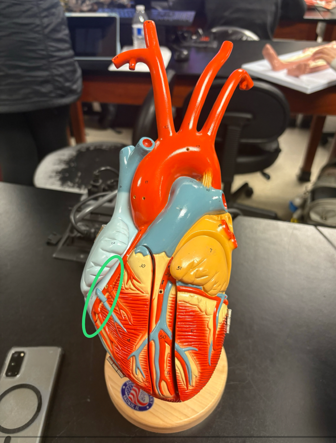
Left Coronary Artery
branch off ascending aorta
circumflex artery
anterior & posterior interventricular artery
Circumflex Artery
splits left atrium/ventricle
Anterior Interventricular Artery
runs between the ventricles anteriorly
Posterior Interventricular Artery
runs between the ventricles posteriorly
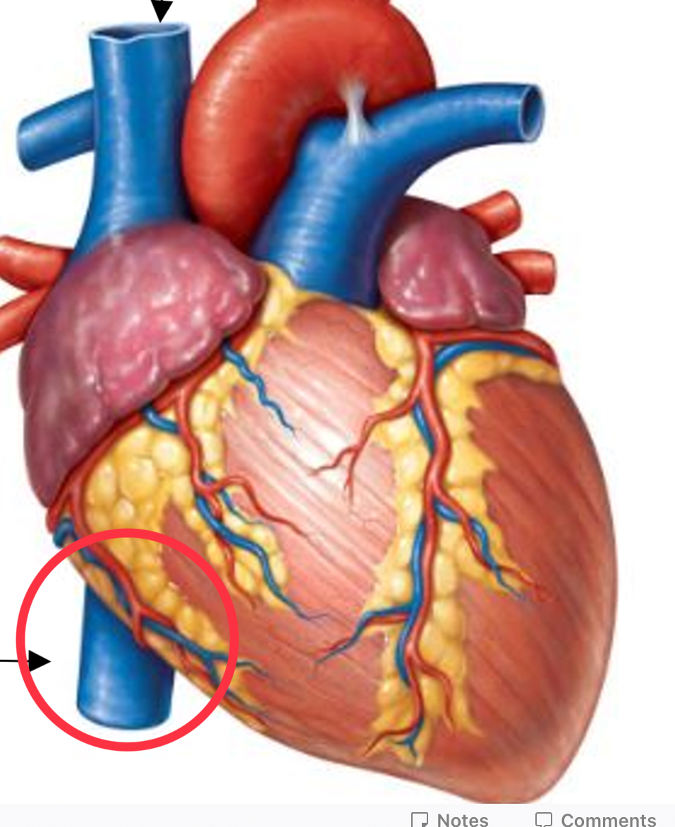
Right Coronary Artery
branch off ascending aorta
right marginal artery
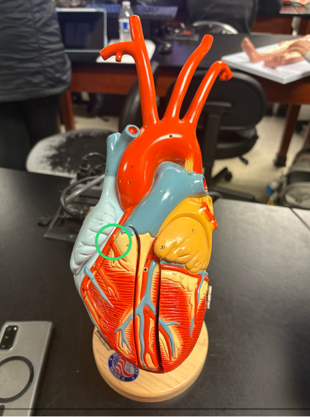
Right Marginal Artery
runs down lateral RV
Right Common Carotid Artery
artery that splits straight off of brachiocephalic artery
Right Subclavian Artery
artery that splits off the side of brachiocephalic artery
Abdominal Aorta
aorta cross diaphragm into abdomen
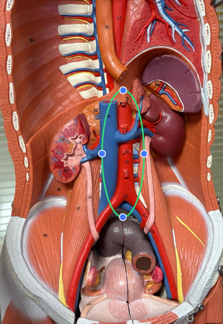
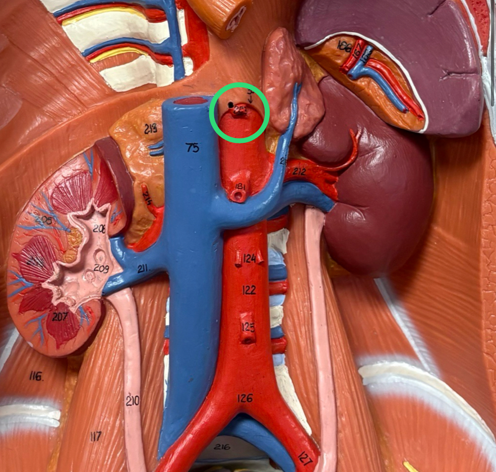
Celiac Trunk
large trunk off abdominal aorta that immediately splits into vessels
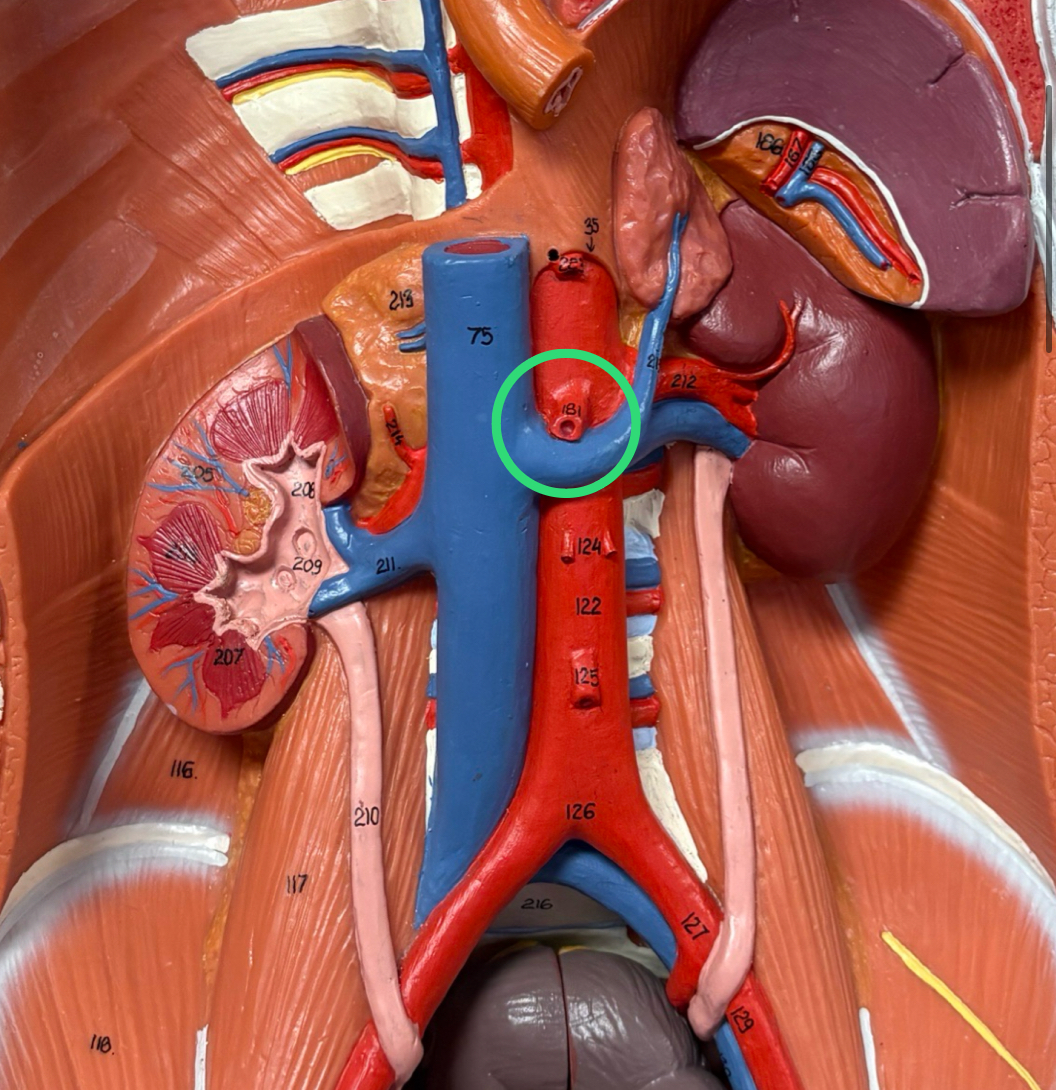
Superior Mesenteric Artery
large artery off abdominal aorta inferior to celiac trunk
Inferior Mesenteric Artery
large artery off abdominal aorta inferior to SMA
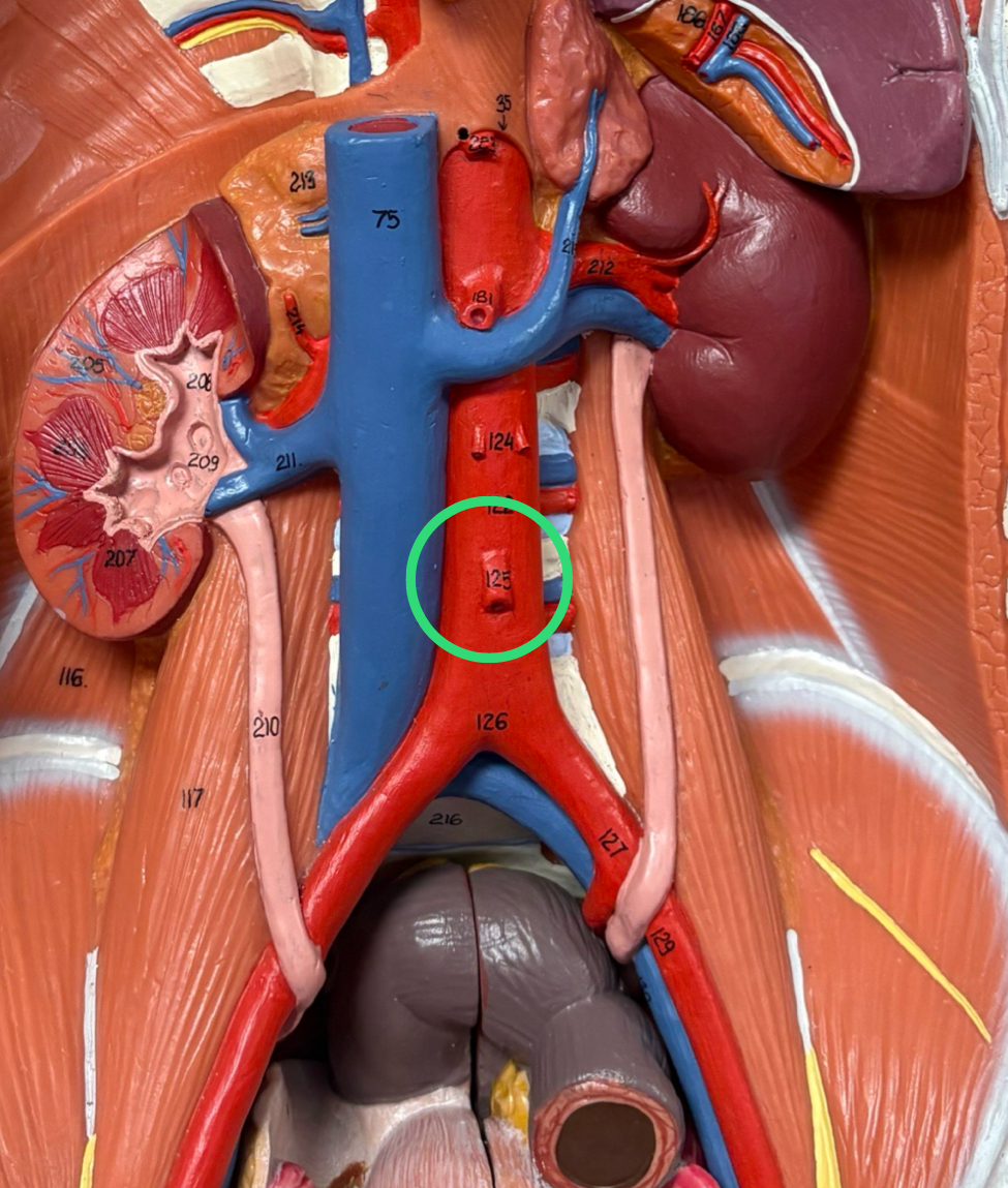
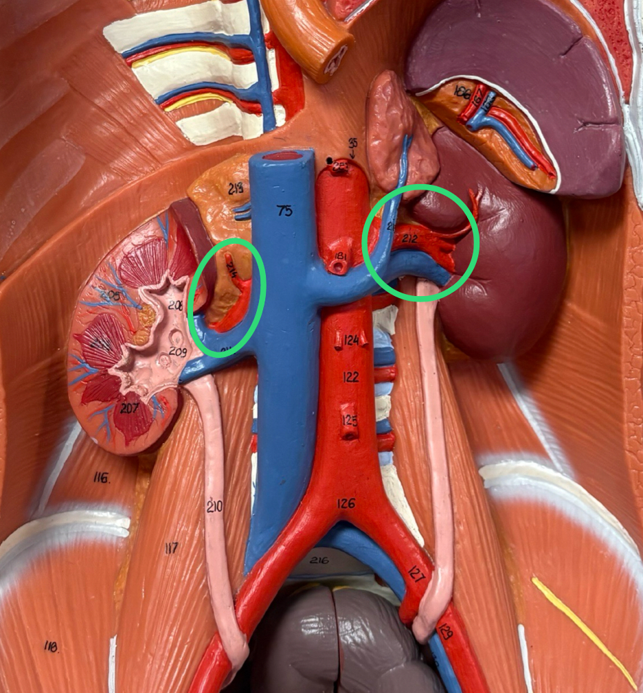
Renal Arteries (R & L)
2 short, wide arteries suppling each kidney
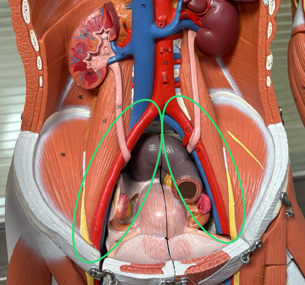
Common Iliac Arteries (R & L)
aorta splits into 2 common iliac arteries ~ L level
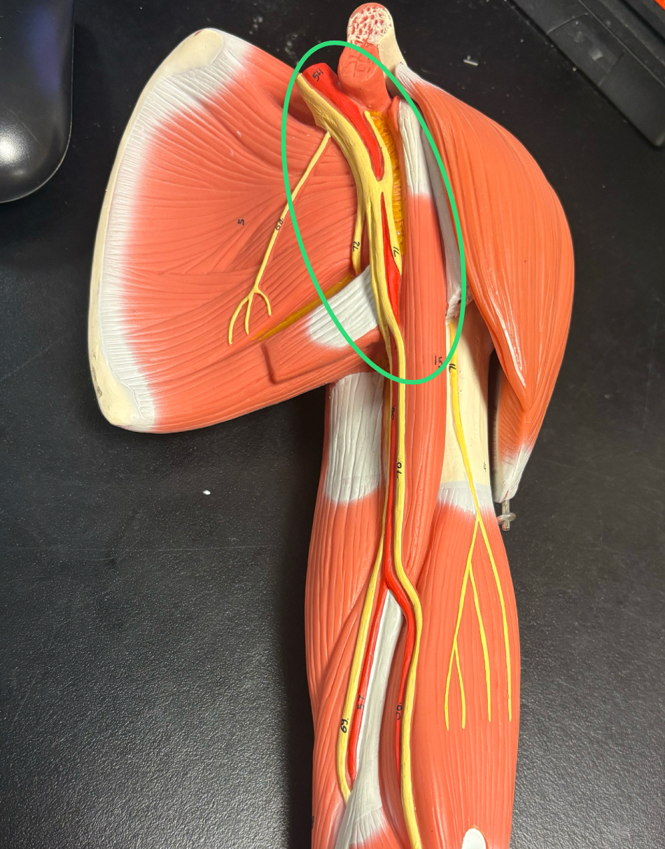
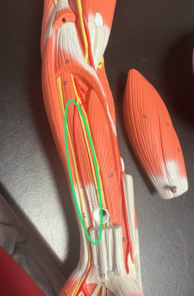
Axillary Artery
runs in armpit region
subclavian a. becomes this once it crosses lateral border of 1st rib
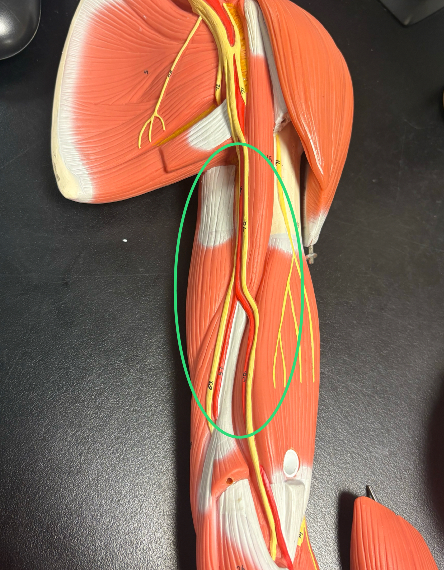
Brachial Artery
once axillary artery leaves axilla
Radial Artery
brachial artery splits into forearm forearm to thumb side artery
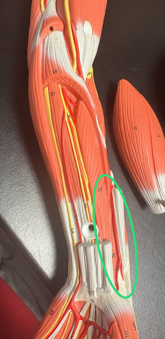
Ulnar Artery
runs down forearm towards pinky finger
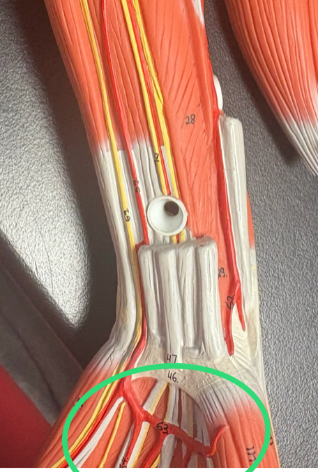
Superficial Palmar Arch
top arch that splits from radial to ulnar to combine in the palm
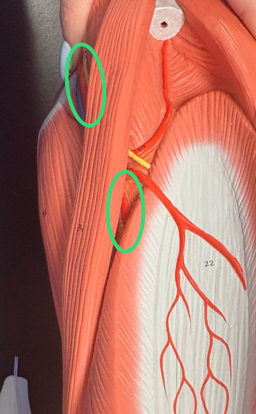
Femoral Artery
anteromedial thigh
main blood supply to thigh muscles & femur head
Popliteal Artery
once femoral artery runs posteriorly to back of knee
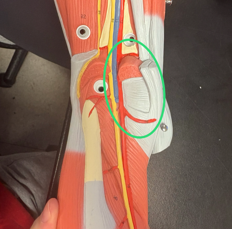
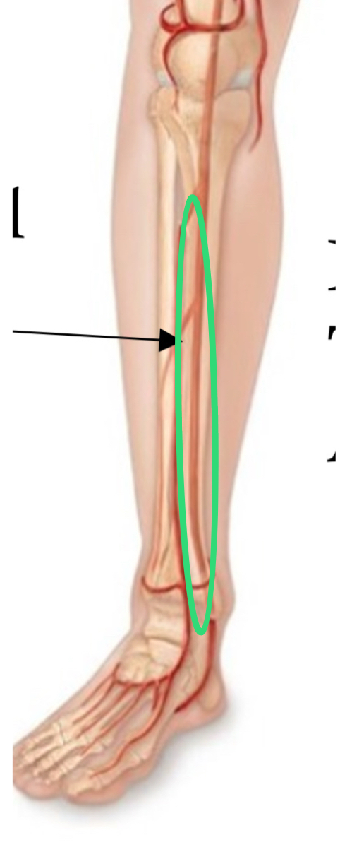
Posterior Tibial Artery
popliteal artery splits into forearm forearm posterior leg
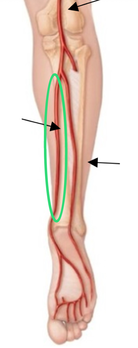
Anterior Tibial Artery
split off popliteal artery
supples anterior leg