MSK Unit 3 Special Tests
 Call Kai
 Learn
Learn Practice Test
 Spaced Repetition
 Match
Match Flashcards
 Knowt Play
1/30

There's no tags or description
Looks like no tags are added yet.
Name | Mastery | Learn | Test | Matching | Spaced | Call with Kai | Chat |
|---|
No analytics yet
Send a link to your students to track their progress
31 Terms
Valgus / Varus Stress Tests
Purpose
Valgus stress: medial structures/MCL
Varus stress: lateral structures/LCL
30° only = isolated collateral-ligament injury
0° & 30° = more global ligamentous instability
How to perform
pt supine with 0° knee extension
Apply a valgus force to assess the medial structures
Apply a varus force to assess the lateral structures
Repeat both tests w/ 30° knee flexion
Compare pain, gapping, and end feel with the opposite knee
Positive test
Pain and/or excessive joint-line gapping
Increased laxity or a softer end feel compared to non-test leg
Interpretation
Positive at 30° only: MCL or LCL only
Valgus = isolated MCL injury
Varus = isolated LCL injury
Positive at both 0° and 30°: global ligament injury
Collateral-ligament injury (MCL/LCL) plus involvement of additional stabilizers, such as the ACL, PCL, or joint capsule
MCL/LCL injury + ACL, PCL, and/or joint capsule involvement
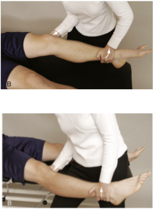
Lachman Test
LIGAMENTOUS STABILITY (anterior instability)
Purpose
ACL integrity (gold-standard ACL special test)
How to perform
pt supine w/ knee between full extension and <30° flexion
Stabilize femur
Grasp the proximal tibia, placing the thumb near the tibial plateau or joint line
Translate the tibia anteriorly while keeping the femur stable
Feel the amount of translation and the end feel
Optional:
If the leg is difficult to hold, support the thigh with a bolster or the clinician’s knee
Positive test
Excessive anterior tibial translation
Soft end feel
Clinical pearl: Keep the heel from digging into the table and make sure the hamstrings are relaxed. Hamstring contraction can produce a false negative
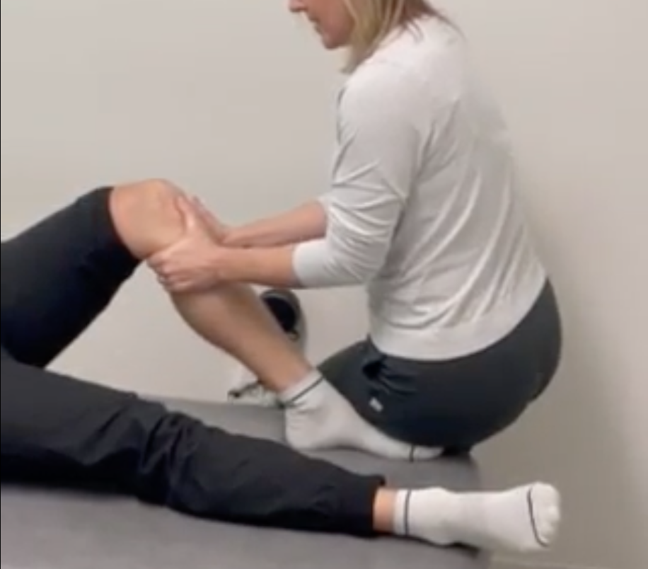
Anterior Drawer Test
LIGAMENTOUS STABILITY (anterior instability)
Purpose
ACL integrity
How to perform
pt supine w/ 90° knee flexion or slightly less
Stabilize the foot by sitting on it
Place both hands around the proximal tibia with the thumbs near the joint line
Translate the tibia anteriorly
Assess the amount of translation and the end feel
Positive test
Excessive anterior tibial translation
Soft end feel
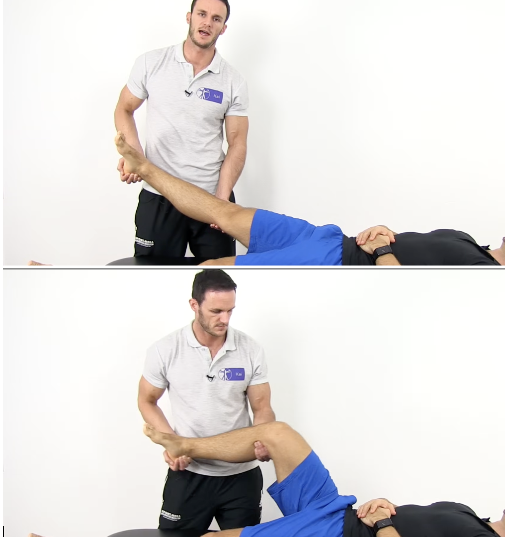
Pivot Shift Test - Needs Work
LIGAMENTOUS STABILITY (anterior instability)
Purpose
ACL tear
Anterolateral rotary instability
Assesses dynamic anterior subluxation and reduction of the tibia in an ACL-deficient knee - youtube
Assesses anterolateral rotatory instability/anterior translation of the tibia relative to the femur - CPG
How to perform - Youtube
pt supine w/ the hip flexed and abducted 30°
Grasp the calcaneus from the outside to create tibial IR (some authors add axial compression)
Apply a slight valgus force at the level of the fibula
Move the knee from extension into flexion while maintaining the forces
How to perform - CPG
pt supine w/ knee fully extended
IR the tibia holding at the ankle w/ hand 1
Place hand 2 on the lateral aspect of the proximal tibia
Apply a valgus stress while slowly flexing the knee from full extension
Look for a sudden reduction at approximately 20° of knee flexion = ACL tear
Positive test - Youtube
The tibia reduces or drops posteriorly at approximately 30–40° knee flexion as the IT band tightens
A palpable or audible clunk may occur
The patient may report the familiar giving-way sensation experienced during walking
Positive test - CPG
A sudden clunk or shift as the anteriorly subluxed lateral tibial plateau reduces at approximately 20° knee flexion = ACL disruption
Grading
Normal = equal/no shift
Grade 1 = glide
Grade 2 = clunk
Grade 3 = gross/severe shift
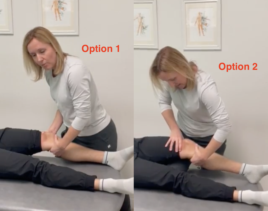
Posterior Drawer Test
LIGAMENTOUS STABILITY (posterior instability)
Purpose
Posterior instability
PCL tear
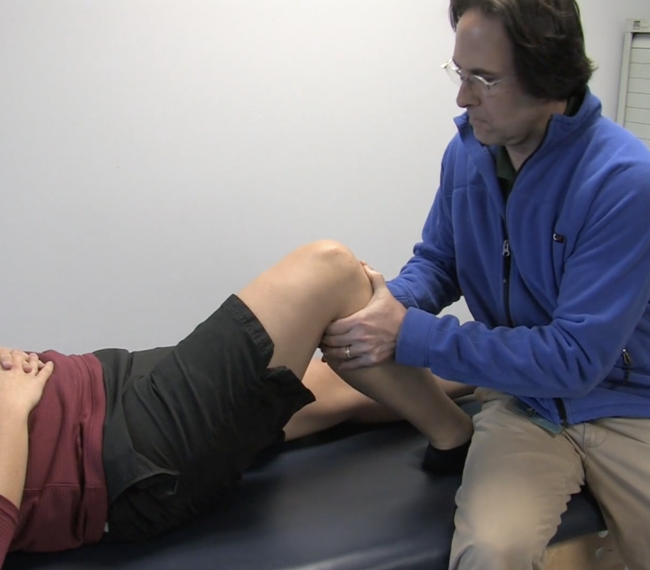
How to perform
pt supine w/ knee at 90° and the foot in neutral
Stabilize the foot by sitting on it
Place both hands on the proximal anterior tibia w/ thumbs on the medial and lateral joint lines
Translate the tibia posteriorly
Compare the amount of movement with the opposite side
Translation may be estimated or measured in millimeters
Positive test
Greater posterior tibial movement on the affected side than the unaffected side = PCL tear
Aka more posterior motion vs the unaffected side
Posterior Sag Test
Godfrey’s Test
LIGAMENTOUS STABILITY (posterior instability)
Purpose
Posterior tibial instability
PCL tear
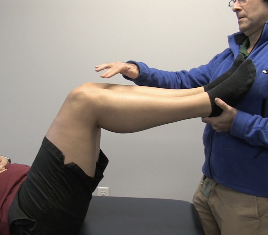
How to perform
pt supine with the hips and knees at 90°
Support both lower legs and heels
Compare & palpate tibial tuberosities
Optional: have pt contract hamstrings to increase posterior tibial translation
Positive test
The proximal tibia sags posteriorly on the affected side?? A torn PCL lets the tibia sag backward???
Affected tibial tubercle less visible than unaffected side = PCL tear
McMurray’s Test
LIGAMENTOUS STABILITY + MENISCUS
Purpose
Torn medial or lateral meniscus
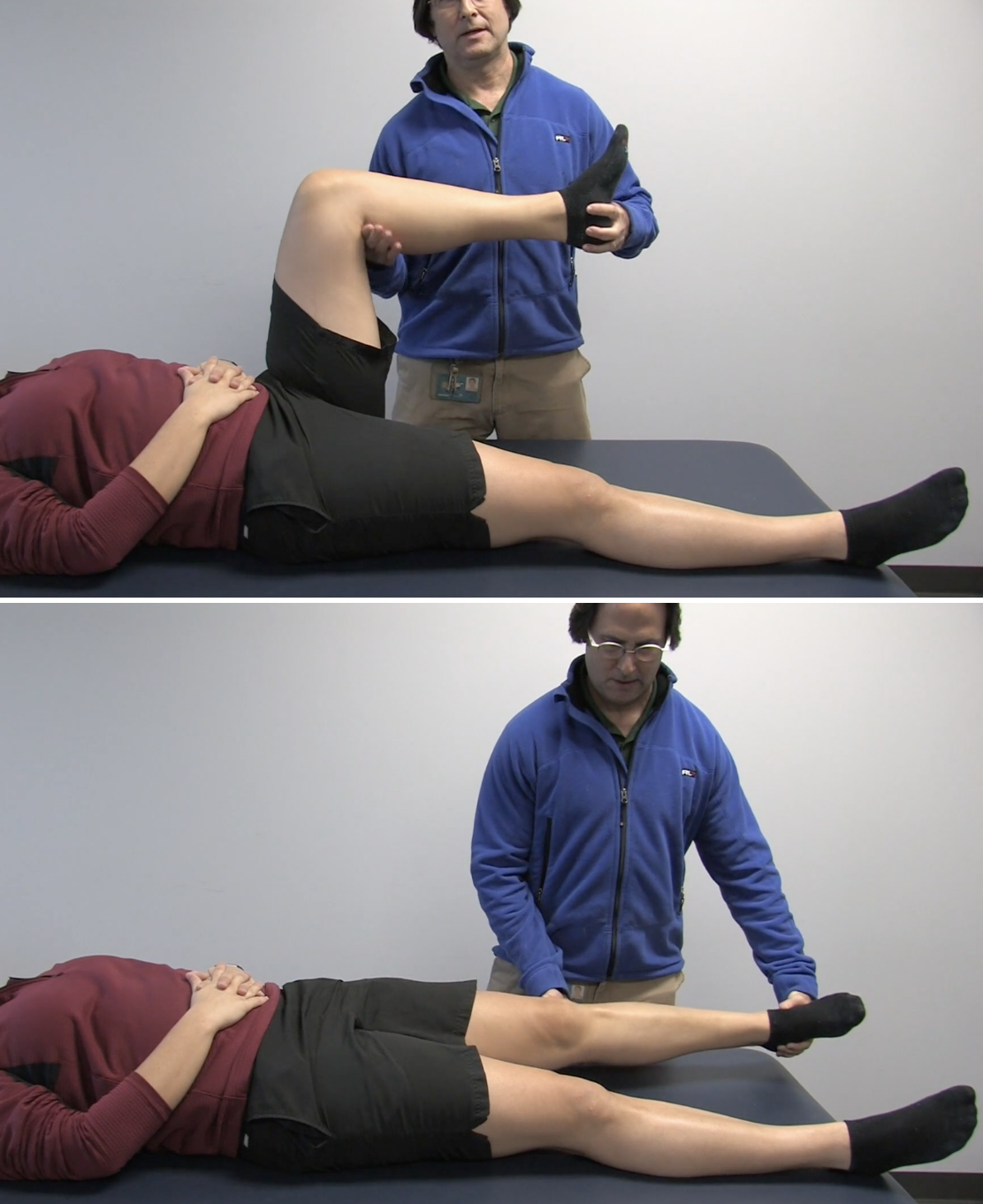
How to perform
pt supine
Grasp heel & flex hip and knee to approximately 90°
Palpate the medial and lateral tibiofemoral joint lines with the thumb and index finger of opposite hand
When the knee is flex, rotate the tibia
Medial meniscus → ER
Lateral meniscus → IR
Slowly extend the knee (some variations include simultaneous valgus force)
Positive test
Audible or palpable thud, pop, or click (, especially when accompanied by joint-line pain???
Pain reproduction
Apley’s Test
LIGAMENTOUS STABILITY + MENISCUS
Purpose
Rotation sprain of the soft tissue
Meniscal tear
How to perform
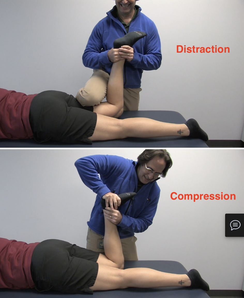
pt prone w/ knee at 90°
Stabilize the femur by half-kneeling over the pt’s hamstring
Grab pt’s foot w/ both hands
During the distraction phase, distract the tibia and rotate it
During the compression phase, compress through the foot and rotate the tibia
Compare the symptoms produced during compression and distraction.
Positive findings and interpretation
Worse pain during rotation = rotation sprain of the soft tissue
More pain with compression than distraction = meniscal tear
Bounce-Home Test
MENISCUS
Purpose
Meniscal pathology
How to perform
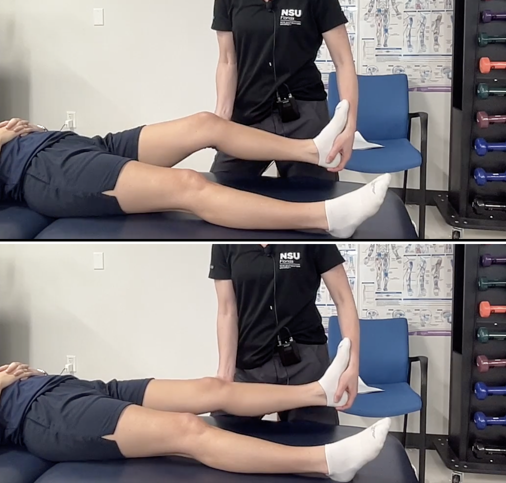
pt supine w/ slight knee flexion
Support underneath the knee as a guide
Allow the knee to move quickly and passively into extension
Positive test
Joint-line pain with an inability to reach full extension = meniscal pathology
Anterior pain over the infrapatellar fat pad may suggest a different source.
Clinical pearl: Bounce-home is not diagnostic by itself and should be interpreted as part of a cluster of findings
Thessaly Test
Disco Test
LIGAMENTOUS STABILITY + MENISCUS
Purpose
Meniscal tear or injury
Note: the only loaded meniscal test → better stats
How to perform
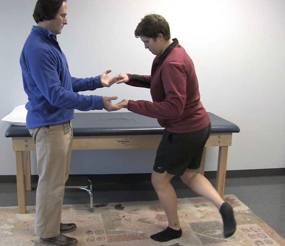
pt standing on involved leg at 20° knee flexion (provide support as needed)
Have pt IR & ER (femur on tibia) 3x in each direction
Positive test
Medial or lateral joint-line pain
Sense of catching/locking
Eccentric Step-Down Test
LIGAMENTOUS STABILITY + “Patellofemoral & Other”
Purpose
Patellofemoral dysfunction
May be used as a functional screen or knee-rehabilitation exercise
How to perform
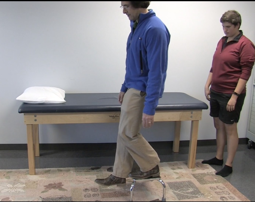
pt stands on a six-inch step or stool
Have the pt keep trunk straight as they lower one leg until the heel touches the floor
Test unaffected side before affected side (which side is affected??)
Assess anteriorly, posteriorly, and laterally
Palpate knee for crepitus if needed
Positive or abnormal findings
Any pain during the step down
Compensations: increased mvmt, trunk lean, loss of pelvic height symmetry (hip drop), increased femoral IR, increased knee mvmt in transverse plane
Zohler Sign
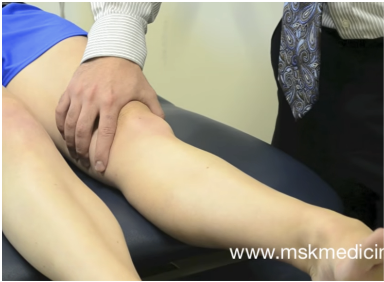
Clarke’s Sign
Purpose
Patellofemoral pain or chondromalacia
May identify irritation or roughening of the patellar articular surface
How to perform
pt supine w/ knees extended & quads relaxed
Glide the patella slightly inferiorly & apply gentle compression/resistance to superior patellar movement
Ask the pt to contract the quadriceps while the patella is held
Positive test
Reproduction of patellofemoral pain
Marked crepitus during quadriceps contraction
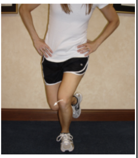
Unilateral Squat
Waldron Test
Purpose
Patellofemoral dysfunction during loaded knee flexion
How to perform
pt standing on test leg
Ask pt to perform a slow, partial single-leg squat
Observe patellar tracking, trunk/pelvic control, femoral rotation, knee alignment, and balance
Palpate around the patellofemoral joint may be used to identify pain or crepitus and the knee flexion angle at which sx occurs
Positive test
Patellofemoral pain or crepitus during the squat
Clinical pearl: Abnormal tracking or loss of lower-quarter control should be documented, but it is not tissue-specific by itself
Q-Angle Measurement
Purpose
Estimates the quadriceps’ line of pull & tendency for lateral patellar tracking
How to perform
pt supine
Identify the ASIS, center of the patella, and center of the tibial tubercle
Measure the angle between:
ASIS to patella center
patella center to tibial tubercle
Course values
Males: approximately 8–14°
Females: approximately 15–17°
Interpretation
Values above approximately 15–20° may be concerning.
The lecture notes describe an angle greater than 20° as abnormal and potentially associated with lateral patellar displacement.
Clinical pearl: Interpret the Q-angle with symptoms, patellar tracking, and dynamic movement rather than alone.
Medial Patellar Plica Test
Mediopatellar Plica Syndrome
LIGAMENTOUS STABILITY + “Patellofemoral & Other”
Purpose
Medial patellar plica syndrome
How to perform
pt supine
Glide the patella medially and hold it
Passively IR tibia & flex knee
Passively extend knee from the flexed position
Positive test
A palpable clunk under the patella as the knee extends = medial patellar plica syndrome
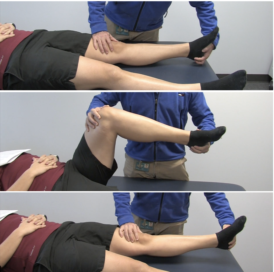
Hughston's Plica Test
Mediopatellar Plica Test
Noble’s Compression Test
LIGAMENTOUS STABILITY + “Patellofemoral & Other”
Purpose
Iliotibial band friction syndrome
How to perform
pt supine or side-lying w/ hip & knee at 90° (w/ bolster under knees on table - not stated)
Apply pressure over the lateral femoral condyle directly over the IT band
Maintain the compression while the patient actively extends the knee
Positive test
Pain before or at 30° knee flexion = iliotibial band friction syndrome
Pain at <30° is negative
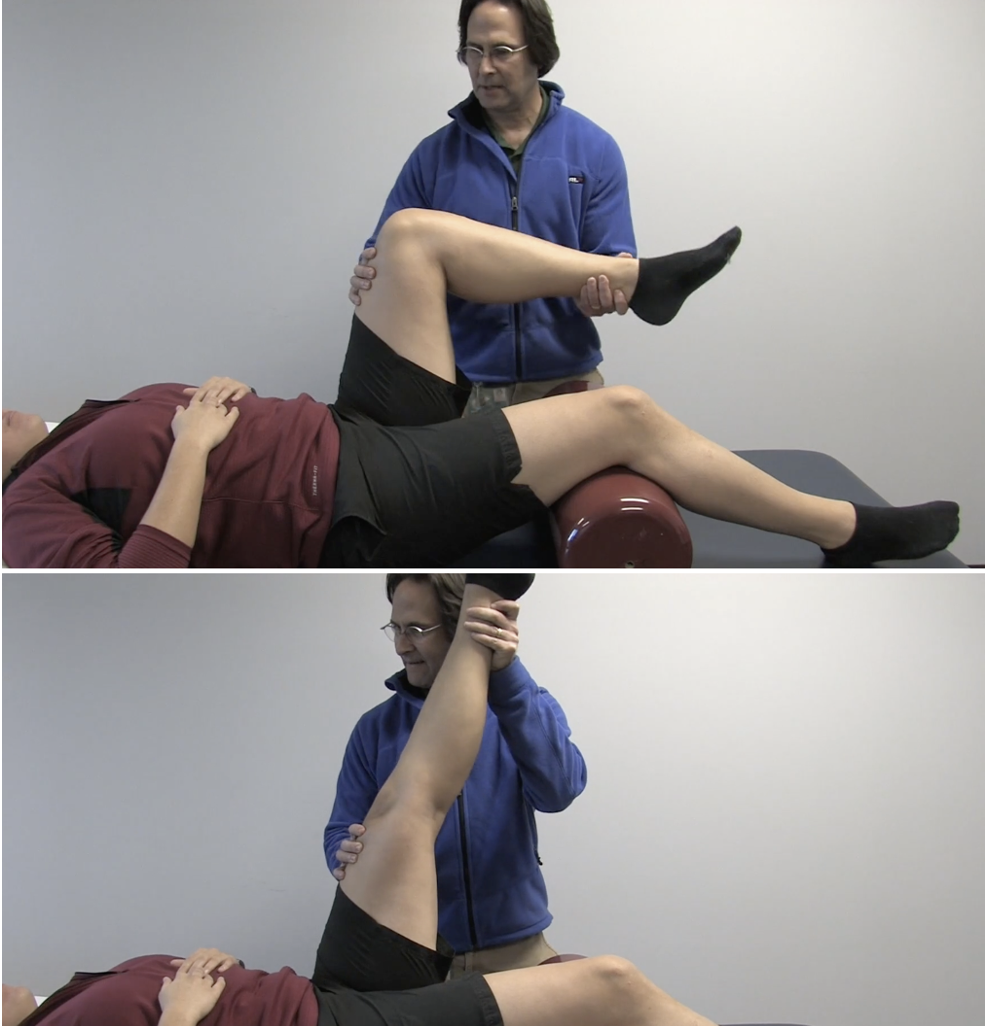
Stroke or Fluid-Displacement Test
Purpose
Knee-joint effusion
How to perform
pt supine
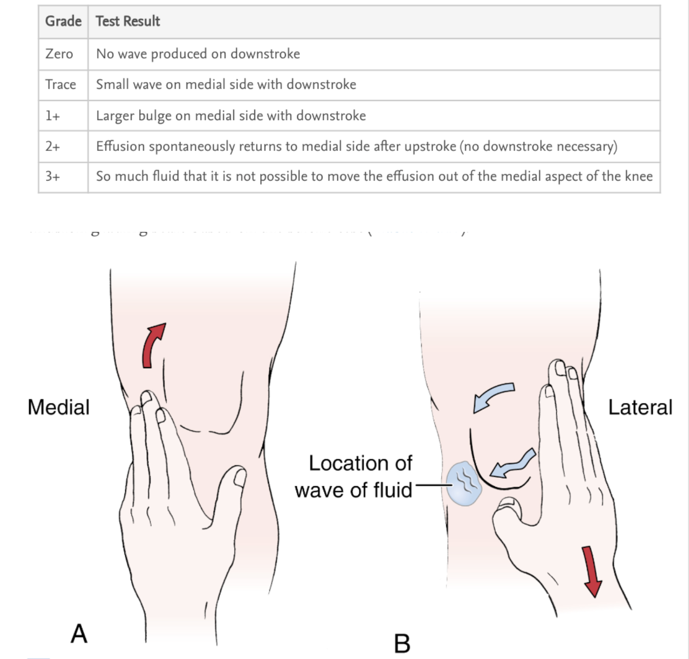
Using the palm w/ a flat hand, sweep fluid upward (“up and out”) along the medial side of the knee from distal to proximal 2-3x
Sweep downward along the lateral side toward the knee
Watch the medial side for refilling
Positive test
A visible return of fluid on the medial side following the medial upstroke and lateral downstroke = knee-joint effusion
Grading
Zero: no wave produced on downstroke
Trace: small wave on medial side with downstroke
1+: larger bulge on the medial side with downstroke
2+: effusion spontaneously returns to the medial side after the upstroke (no downstroke necessary)
3+: so much fluid that it is not possible to move the effusion out of the medial aspect of the knee
Navicular Drop Test
Purpose
Excessive medial longitudinal arch collapse
Abnormal pronation
How to Perform
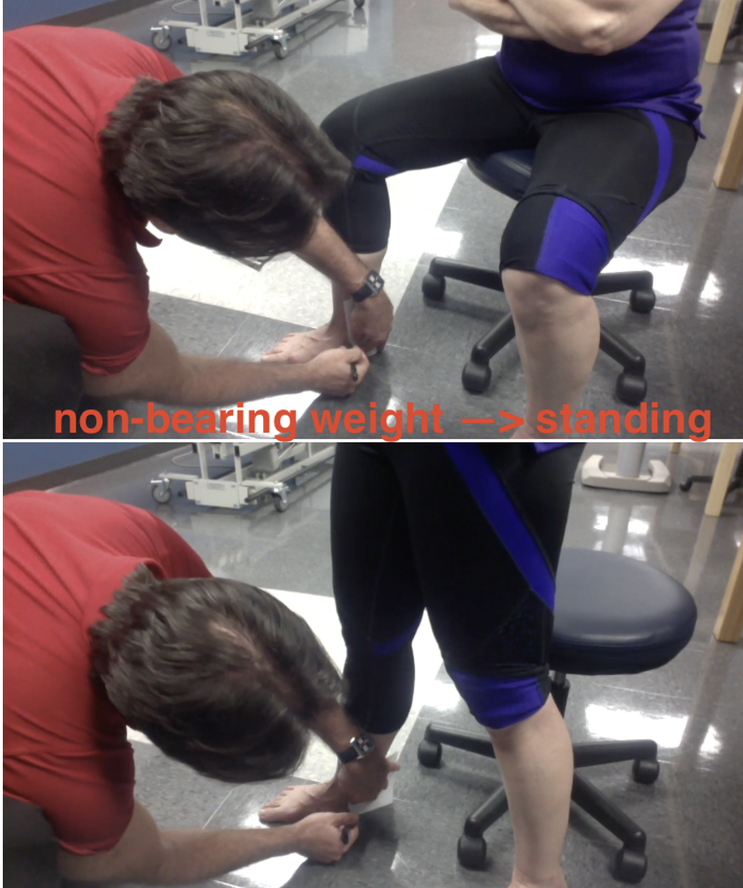
Pt seated
Place subtalar joint in neutral
Palpate and mark the navicular tubercle height from the floor using an index card
Have pt stand
Repeat measurement and calculate the difference
Positive Test
Navicular drop >10 mm
Too Many Toes Sign
Purpose
Abnormal pronation
Flat-foot posture
How to Perform
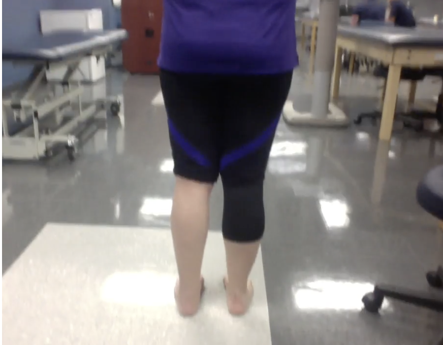
Pt standing
Observe the calcaneus and toes from directly behind the pt
Abnormal Test
Abnormal: more than the 5th toe and half of the 4th toe are visible lateral to the calcaneus
Normal: only the 5th toe and approximately half of the 4th toe are visible
Windlass Test
Purpose
Assess the windlass effect of the plantar fascia
Assess the response of the medial longitudinal arch
How to Perform
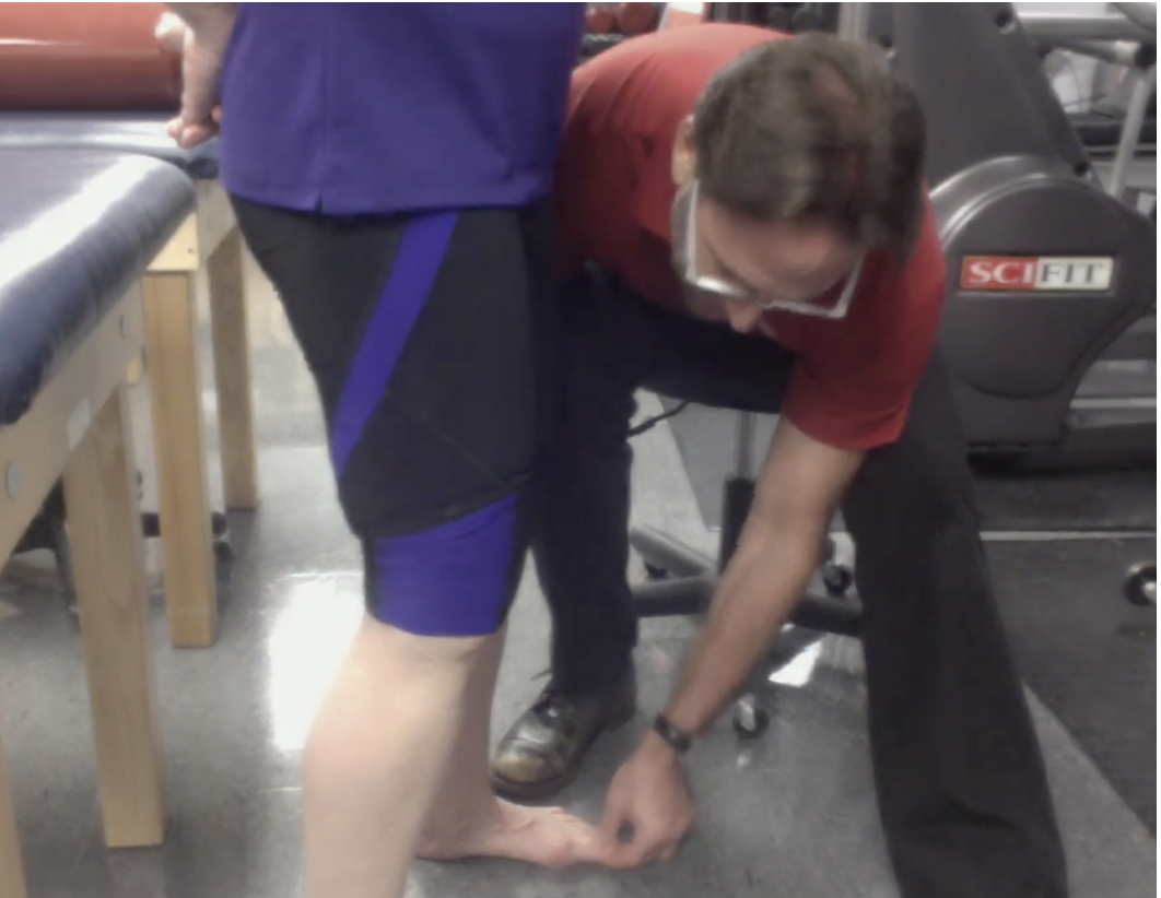
Pt stands completely relaxed in a natural base of support
Passively extend the hallux with one hand
Place the other hand around the calf to ensure the pt is not using muscle activity
Observe the medial longitudinal arch
Abnormal Test
Abnormal: medial longitudinal arch fails to rise with hallux extension
Normal: the arch rises as the great toe is extended
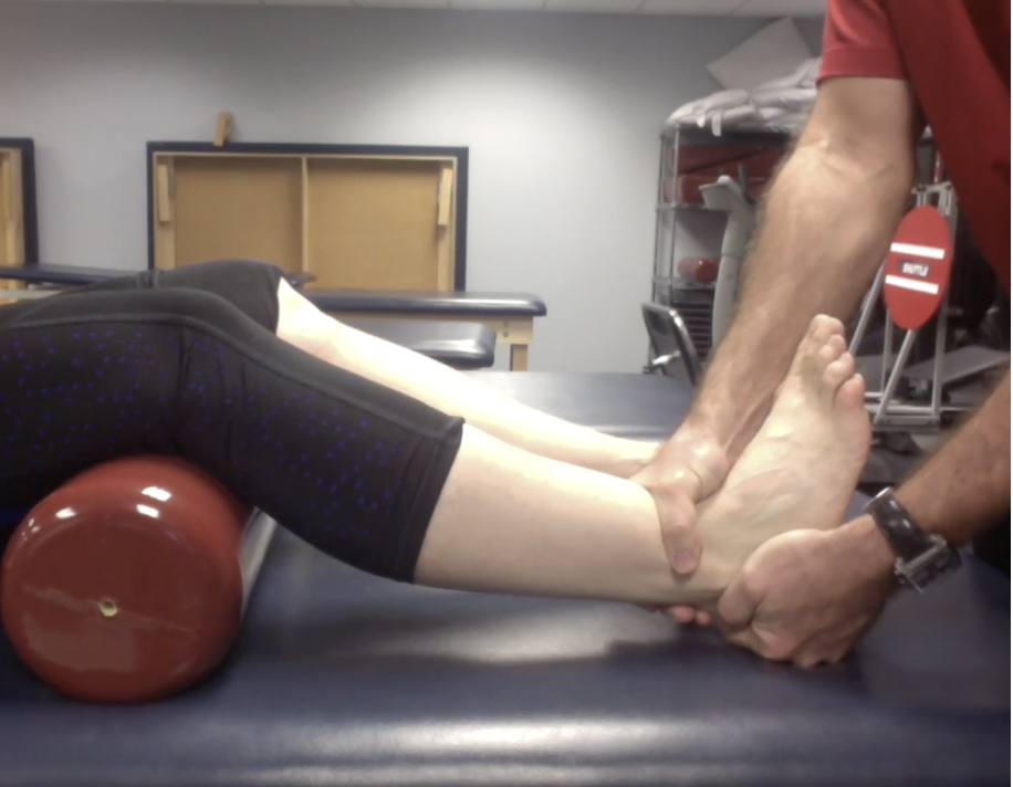
Anterior Drawer Test
Purpose
Assess Anterior TaloFibular Ligament (ATFL) stability
How to Perform
Pt seated or supine w/ bolster under knees w/ gastrocnemius and soleus relaxed
Stabilize the distal leg and grasp the heel
Position the ankle in 10–20° PF
Pull the heel anteriorly, or move the tibia posteriorly
Compare movement and end feel with the uninvolved side
Positive Test
Pain and/or increased movement compared to uninvolved side
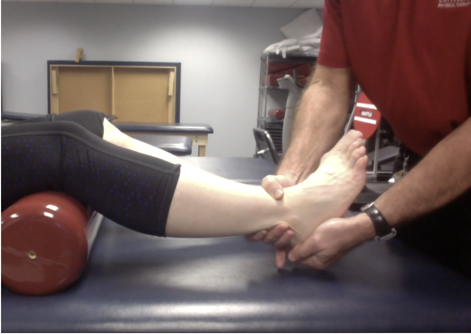
Talar Tilt Test
NEEDS CHECKING
Purpose
Assess the integrity of the ankle ligaments:
CFL
ATFL
Deltoid ligament
How to Perform
Pt supine with a bolster under the knees or side-lying with the knee flexed and foot relaxed
Stabilize the distal leg and grasp the calcaneus
With the ankle in neutral, apply inversion/adduction stress
Repeat in 10–20° PF with inversion/adduction stress
Apply eversion/abduction stress to assess the medial ankle
Compare pain, laxity, and end feel with the uninvolved side
Positive Test
Pain and/or increased laxity compared with the uninvolved side
Interpretation
Neutral + inversion/adduction → CFL
10–20° PF + inversion/adduction → ATFL
Eversion/abduction → deltoid ligament
Easy way to remember
Foot tilts inward → lateral ligaments
Foot tilts outward → medial deltoid ligament
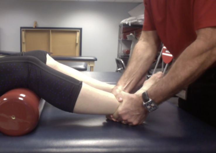
Squeeze Test
Purpose
Assess the distal tibiofibular syndesmosis for a high ankle sprain
How to Perform
Pt supine with a bolster under the knees or side-lying
Squeeze the syndesmosis (tibia and fibula) together above the malleoli
Positive Test
Pain in the distal 1/3 of the leg
Pain occurs distal to the area being squeezed
Rule Out
Tibia or fibula fracture, calf contusion, compartment syndrome
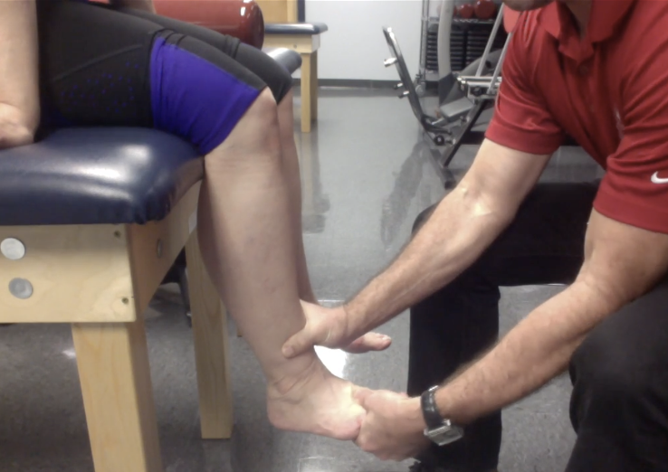
Kleiger Stress Test
ER Stress Test
Purpose
Assess the distal tibiofibular syndesmosis for a high ankle sprain
Assess for a deltoid ligament tear
How to Perform
Pt seated with the legs over the edge of the table and knee flexed to 90°
Stabilize the lower leg with one hand
ER the foot with the other hand
Positive Test
Reproduction of ankle pain
Anterolateral distal tibiofibular pain → high ankle sprain
Medial ankle pain → deltoid ligament tear
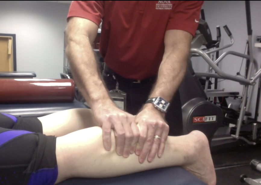
Thompson Test
Purpose
Assess for a complete Achilles tendon rupture
How to Perform
Pt prone with feet relaxed over the edge of the table
Squeeze the calf muscle
Observe the ankle for PF
Positive Test
Absence of PF when the calf is squeezed = complete Achilles tendon rupture
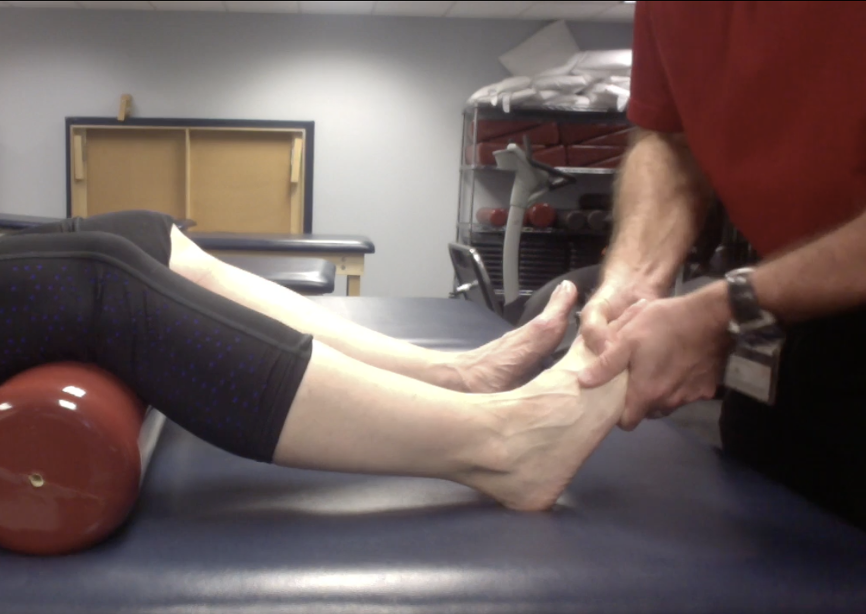
Morton’s Test
Purpose
Stress fracture
(Morton’s?) neuroma
How to Perform
Pt supine w/ bolster under knees
Squeeze the metatarsal heads together
Monitor the pt’s symptoms
Positive Test
Reproduction of forefoot pain = stress fracture or neuroma
Homan’s Test
Purpose
Deep Vein Thrombosis (DVT)
How to Perform
Pt supine with knees extended
Stabilize the thigh & passively DF the ankle
Monitor for calf symptoms
Positive Test
Calf pain with passive DF
Important Limitation
Poor sensitivity and specificity
Positive in fewer than 30% of pts with DVT
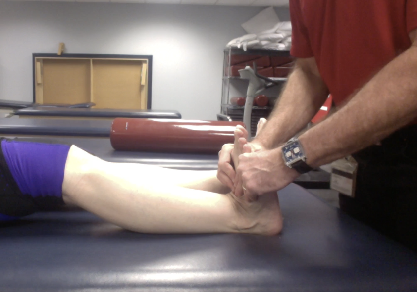
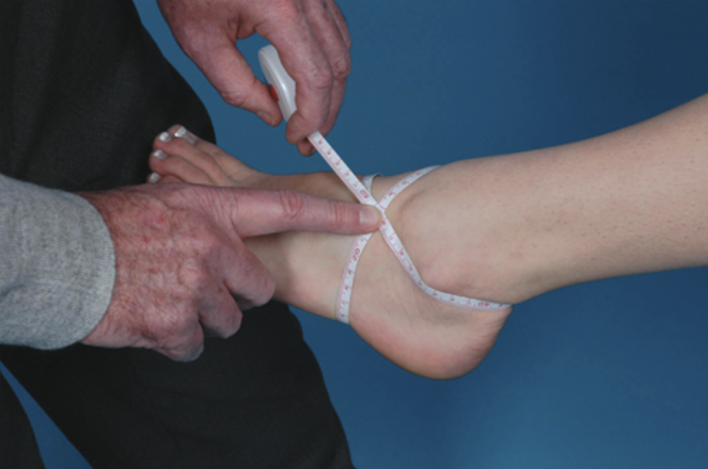
Figure-of-8 Measurement
Purpose
Ankle edema measurement and tracks change over time
How to Perform
Pt supine or seated with ankle in a neutral or comfortable position
Start midway between the tibialis anterior tendon and lateral malleolus
Pass the tape:
Just distal to the navicular tuberosity
Across the plantar arch toward the 5th metatarsal base
Across the tibialis anterior tendon
Just distal to the medial malleolus
Across the Achilles tendon
Back just distal to the lateral malleolus
Record the circumference using the same landmarks each time
Result
This is a measurement, not a positive/negative test
Larger value compared with the opposite side or a prior measurement → greater edema
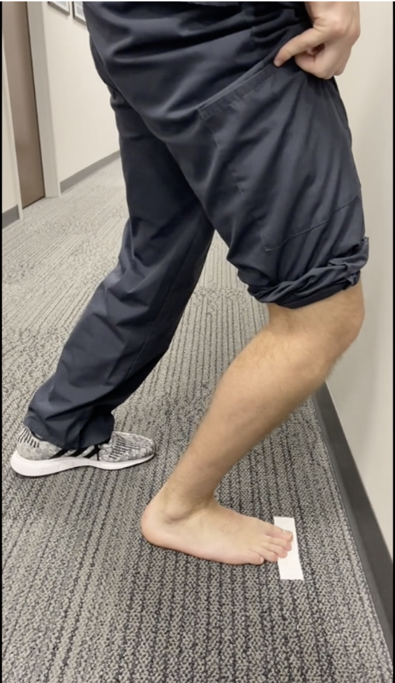
Weight-Bearing Lunge Test
Purpose
Assess weight-bearing ankle DF ROM
How to Perform
Mark 10 cm from a wall using tape
Align the foot so an imaginary line through the heel and great toe points toward the wall
Lunge forward and attempt to touch the knee to the wall while keeping the heel on the floor
Positive Test
Failure to touch the knee to the wall from 10 cm → restricted DF / ankle stiffness
May reflect decreased joint mobility or soleus tightness