ANS 123 Quiz 3
1/95
There's no tags or description
Looks like no tags are added yet.
Name | Mastery | Learn | Test | Matching | Spaced |
|---|
No study sessions yet.
96 Terms
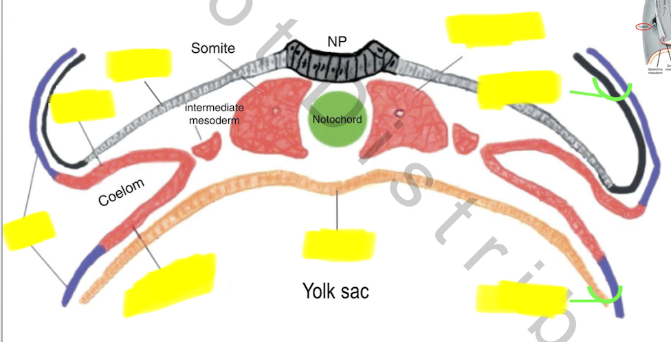
Mesoderm differentiates during neurulation
_______ ectoderm is still continuous w _______ ectoderm
_______ present (beginning of neurelation)
primitive streak separates ectoderm into ______ & ______ allowing _______ below to differentiate into notochord formation
Left & right sides of notochord = different plates of _______
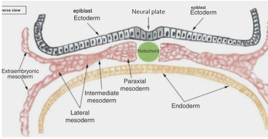
Epiblast , amnion
Notochord
left & right sides, mesoderm
mesoderm
Mesoderm differentiates during neurulation
_______ mesoderm
Portion of mesoderm closest to notochord
on both sides of notochord, “hugging” notochord
_______ mesoderm
Portion of mesoderm farthest from notochord
Intermediate mesoderm
Portion of mesoderm between ______&______ mesoderm
Paraxial
Lateral
paraxial & lateral
Mesoderm differentiates during neurulation
Lateral, Intermed, Paraxial
Further along in development, these sections of mesoderm _______ into _______ regions
Also important to note that the _______ plate mesoderm splits
Once separated, these regions ________ further
separate, distinct regions
lateral
differentiate
Mesoderm differentiates during neurulation
Once separated, these regions differentiate further
_______ mesoderm
→ head
→ somite
_______ mesoderm
→ kidney
→ gonads
Lateral plate mesoderm
→ __________ (dorsal, closest to eCTOderm)
→ __________ (ventral, closest to eNDOderm)
Paraxial
—
Intermediate
—
somatic mesoderm
splanchnic mesoderm
Somites
differentiated from _________ mesoderm
the 1st ________ structure in embryo
Influences _________ of body (key to correct body layout) & formation of _________ structures (ribs, vert, cranial/spinal nerves, muscles, etc.)
May influence/trigger neural crest cell migration
paraxial
segmented
regionalization, segmented
neural crest cell
Somitogenesis
Formation of somites
occurs during primitive streak ________, & ________
Begins in _______ portion of embryo
regression, neurulation
anterior
Somitogenesis
________ mesoderm ”pinches‐ off” on each side of neural ______ & ________
→→ Paired blocks of _________
(1st separated from intermed mesoderm, then paraxial mesoderm itself separates into chunks that ea become somite)
Somites form in ________ on either side of neural _______ & _______
1. entire _______ mesoderm separates from ___________ mesoderm as 1 separated column / piece of ________ mesoderm
2. once you have a whole thing of _______ mesoderm, then you start to chunk up the ________ mesoderm as you're gradually moving _________
3. this chunking up creates the somites
Paraxial, neural tube, notochord
paraxial mesoderm
(first separated from intermed mesoderm, then paraxial mesoderm itself separates into chunks that ea become somite)
columns, neural tube, notochord
1. paraxial, lateral plate, paraxial
2. paraxial, paraxial, posteriorly
Somitogenesis
Stages:
1st visible somite pair , 4 somite pairs, 7 somite pairs, 10 somite pairs, etc…
as ____erior somites form, ____erior somites already doing job
that's why it's very difficult during devel to get an image w full range of intact somites anterior→posterior
wave of maturation anterior→posterior
posterior
anterior
Somites
Each somite is very ________
They then differentiate into 3 diff cell lineages:
__________
__________
__________

multipotent

Each somite has 3 sections
Dermatome
_______ most portion (closest to _____derm)
Myotome
__________
Sclerotome
_______ most portion (closest to _____derm)
Each section differentiates into _______ arranged mesodermal derivatives
Dorsal, ectoderm
Middle
Ventral most portion (closest to endoderm)
segmentally (segment)
Each somite has 3 sections (dermatome, myotome, sclerotome)
Each section differentiates into segmentally arranged mesodermal derivatives:
Dermatome
→ ______ of ______
Myotome
→ ________
Sclerotome
→ Axial ________ structures (______)
(becomes _______ of ________)
Dermis of skin
Muscles
skeletal (bones)
bones of vertebral column
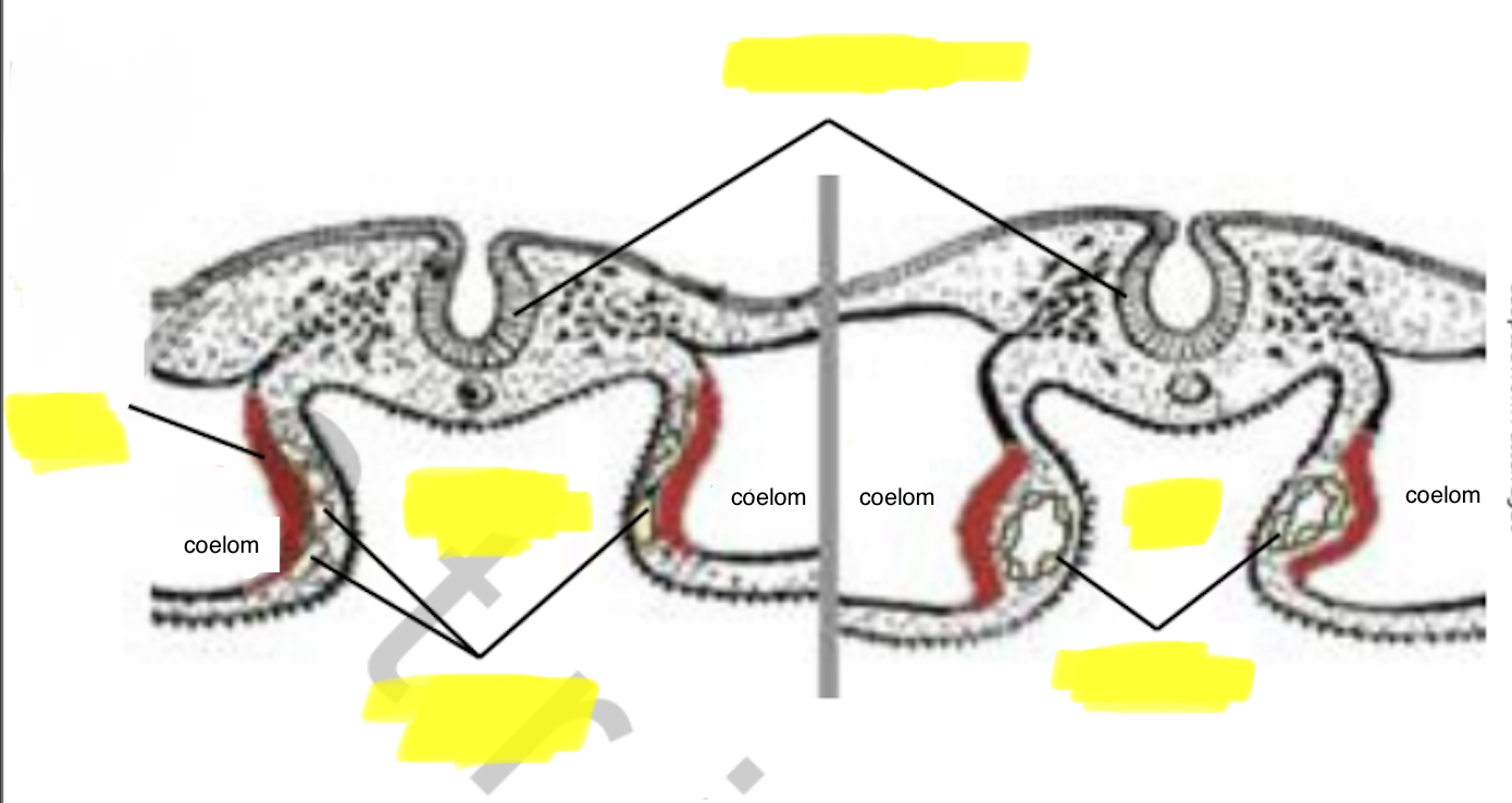
Somites
Transverse v. Dorsal view

Left pic = wedge shaped ________ on either side of notochord, can assume image is around time of ________
Right pic = somites get numbered from ________ to ________ (anterior = more _______)
somites, neuralation
anterior → posterior ; mature
________ = How the embryo becomes 3D ( 2d → 3d )
body folding
Body folds
Before folding:
Flat stanley / pancake
Flat ectoderm, flat mesoderm, flat endoderm
Endoderm continuous w _______
After folding:
close sides of 2D structure to create lateral __________ & enclose the ______/______
________ (intraembryonic cavity) forms when ______ & ______ mesoderm separate (required for body folding)
YSE
—
body walls, gut tube / organs
Coelom, somatic & splanchnic
Starting point of body folding
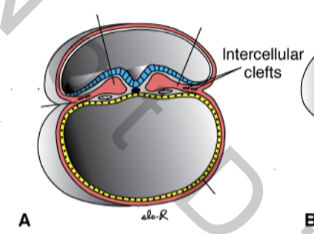

Body folds
A) For body folding to occur, need to pinch definitive ________ away from ________
pinching & folding begins to occur on the side
effectively, pushing in on ______ of embryo

definitive endoderm, YSE
sides
Body folds
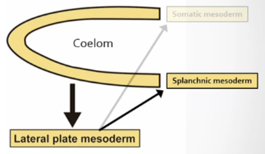
B) Now, _______ mesoderm has split into 2 branches:
_______ mesoderm (track up around amnion)
_______ mesoderm (adhere to endoderm)
Creates gap as pinches even further; this larger gap eventually becomes _________ body cavity (_______)
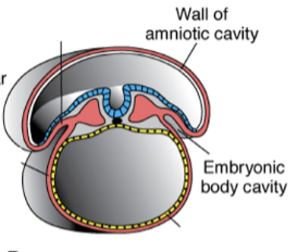
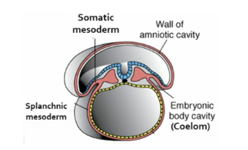
lateral plate
somatic
splanchnic
intraembryonic, coelom
Body folds
C) Further along, ______ getting bigger, beginning to pinch off _________
D) Further folding occurring; ______ getting pinched away from ______
E) Completely folded; ______ no longer present (got pinched off); fully formed ______ (lined w cells that used to b definitive endoderm)
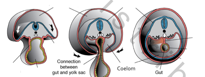
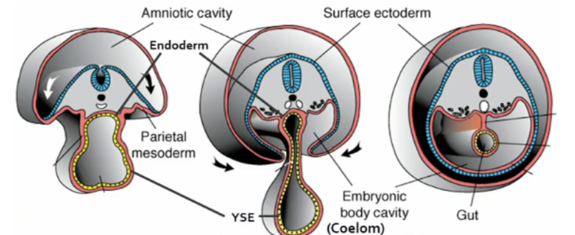
gap, definitive endoderm
endoderm, YSE
YSE, gut tube (definitive endoderm)
Body Folding
process of “pinching off” embryo from _____/_____
In order:
1. ________ fold (head fold)
2. ________ body folds
3. ________ fold (tail fold)
As embryo’s body pinches off, _____&_____ fuse ventrally at midline
Incomplete _______ = hair lip, cleft palate
Head region = more _______ than lower
yolk/YSE
—
1. Subcephalic (head)
2. Lateral
3. Caudal (tail)
—
right & left
fusing
mature
Body Folding
Results:
3D embryo “______” fused at ______
Marks off ________ from ________ region
Forms sides & ________ surface of embryo
________ of embryo “tucked in”
tube, midline
embryo proper, extra‐embryonic
ventral
Germ layers
Heart Formation
Heart formation = form of ___________
In chicken, 1st heartbeats ~33‐38 hrs ___________
_________ = well‐established by 51‐56 hrs
Heart is formed along the way as _________ occurs
organogenesis
post‐fertilization
Circulation
body folding
Heart formation
Prior to folding: ______ fills w _______ cells & _______ forms
A) ________ still in process; ________ has fallen into ________; __________ is shoving hypoblast out of the way
B) _________ cells proliferate & fill up ________ w ________ cells; _______ ______ splits creating new cavity called _______
blastocoel, mesoderm cells, coelom
primitive streak - mesoderm , blastocele - definitive endoderm
B) mesoderm, blastocele, mesoderm cells - lateral plate mesoderm, coelom

Heart formation
During regression:
pre‐_______ ________ mesodermal cells get situated
1. Migrate & merge _______ to _______ process
2. _____________ cells undergo ________ & proliferate
pre‐cardiac splanchnic
anterior, head process
Cardiac crescent cells, determination
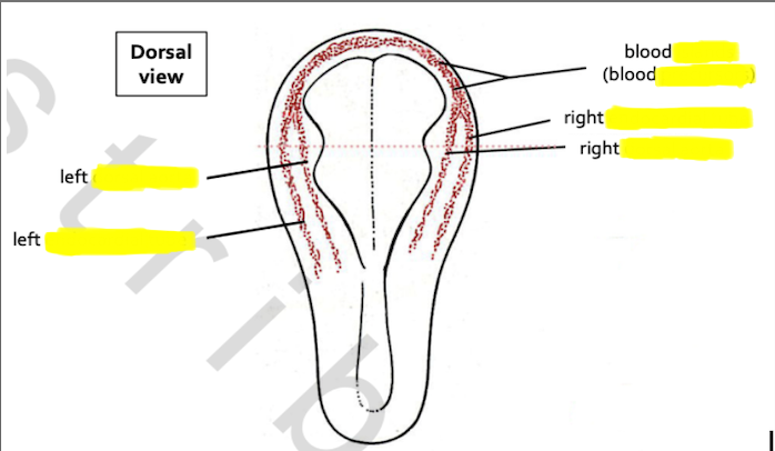
Heart formation
During regression:
pre‐cardiac splanchnic mesodermal cells get situated
1. Migrate & merge anterior to head process
have _____ & _____, proliferate/merge to create crescent
Cardiac crescent = 1o ___________ (HFR) = ________ area
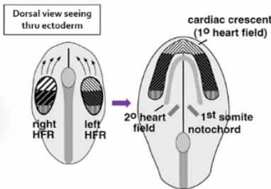
2. Cardiac crescent cells undergo determination & proliferate
right & left
Heart field region , Cardiogenic
As embryo begins folding: early stages of formation of heart, foregut, coelom, & ventral body wall = interrelated
1. On each side of embryo, ________ mesoderm migrates ________
Coelom = space/cavity created when _____&_____ layers separate
2. Foregut forms as _______ migrates
________ cells differentiate into _____cardial (heart) & ______cardial (vessels) precursor cells
3. Endocardial precursors form endocardial ______
4. Foregut closes → _______
(_______ precursors pulled ventrally)
5. ________ tubes & _______ precursors fuse @ midline
(fusing caused by apoptosis of touching walls)
Bringing these to midline forms ______ cardiac tube
within tube, differentiation of myocardial precursors into ______ fibers begins
All this is possible because of body folding
splanchnic, ventrally
splanchnic & somatic mesoderm
splanchnic mesoderm
Cardiac crescent cells, myocardial , endocardial
tubes
gut tube, Myocardial
Endocardial, myocardial
simple
cardiac
All this is possible because of body folding
Cardiac crescent cells differentiate into myocardial (heart) & endocardial (vessels) precursor cells

Endocardial precursors form endocardial tubes

Simple cardiac tube anatomy
Heart starts as simple _______
To become complex organ w separated compartments, tube must contort (left & right atria fold ________; looping)

tube
upwards
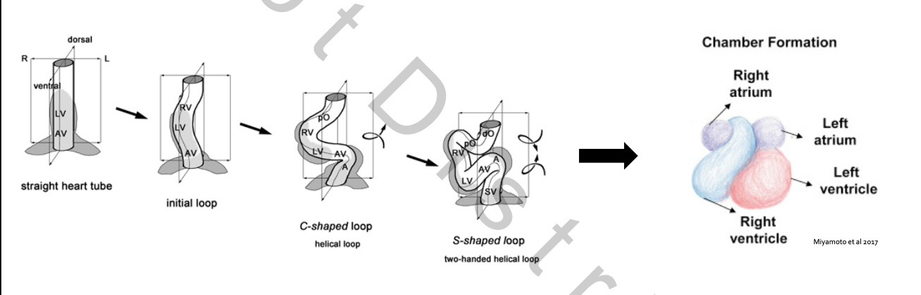
Heart Looping
Heart tube continues to grow & bend
“Squashes” down on itself
Twists to move _____&_____ to correct positions
Septation occurs as heart loops (______ begin to form)
while heart is looping, massive ________ also occurring → muscle _______ beginning to form
left & right ventricles
walls
proliferation, walls
Heart Looping
.
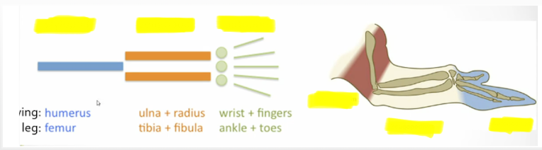
Heart formation
Lateral plate mesoderm → ________ mesoderm
Limb Development
Lateral plate mesoderm → ________ mesoderm
splanchnic
somatic
Limb fields (limb devel)
________ from ________ mesoderm undergoes determination to become Limb field (either wing or leg)
Undifferentiated site of ________
Morphogen involved: possibly _______ family products
(morphogen = protein/molecule that serves as the signal during induction)
Mesenchyme =
_______ organized mesoderm cells
Derived from ________ mesoderm
Gives rise to _____&_____ tissue
bilateral at this point; right & left somatic mesoderm begin to form diff limb fields
Mesenchyme, somatic
future limb
Hox gene family products
Mesenchyme =
Loosely
somatic
connective & muscle
Limb bud
1. Myotome from ______ enter limb fields
2. _____+______ contact & push overlying surface ectoderm outward
3. Limb bud = Combo of ________+_______+________
somites
Mesenchyme + myotome
mesenchyme + myotome + surface ectoderm
Limb bud
1. Myotome from somites enter limb fields
Somites ~15‐20 form ______; ~23‐32 form ______
Future muscles
Morphogens: ______&_______
Only specific somites respond to these
2. Mesenchyme + myotome contact & push overlying surface ectoderm outward
mesenchymal cells from __________ joined by mesenchymal cells from _______ of _______
once in respective limb field area, now going to begin differentiation process (rapid division/ proliferation)
Buldges result from rapid division / proliferation, pushing against overlying ectoderm
3. Limb bud = Combination of mesenchyme + myotome + surface ectoderm
Made of rapidly ________ cells; need to stay in contact for limb devel
wing, leg
FGF‐8 & FGF‐10
—
somatic mesoderm, myotome of somites
dividing
Limb Bud formation
the most _______ ectoderm cells differentiate & are induced to become ___________ (____)
Morphogen: FGF____
key activator of ______
From here on out, _______ drives bud formation
(thumb always anterior, pinky posterior)
AER induces adjacent mesenchyme to differentiate into ________ zone
______ form via cell line interactions
distal, Apical Ectodermal Ridge (AER)
FGF‐10
AER
AER
(thumb always anterior, pinky posterior)
Progress
Axes
Limb Bud formation
AER induces adjacent mesenchyme to differentiate into Progress zone
sends morphogens back towards _________ region. Morphogen: FGF____
Overall: communication process where diff morphogens get shoved at a tissue, tissue receives morphogen causing differentiation, then uses morphogen (same or diff) to signal back
Concentration gradient
cells of limb field (mesoderm) closest to ______ undergo most _______ division rates
ie cells closest to ________ hear message loudest to start rapid division
limb field region. FGF‐8
AER, rapid
AER
Limb Bud formation
important morphogens = ______ & ______
—> allow establishment of AER & ZPA signaling ceters

FGF8, FGf10
Limb Development
Axes via cell line interactions
3 induction intrxns in limb bud generate 3 limb axes
informs cells which part of the limb it needs to be
1. P_____-D_____ axis
2. D_____-V_____ axis
3. A_____-P_____ axis
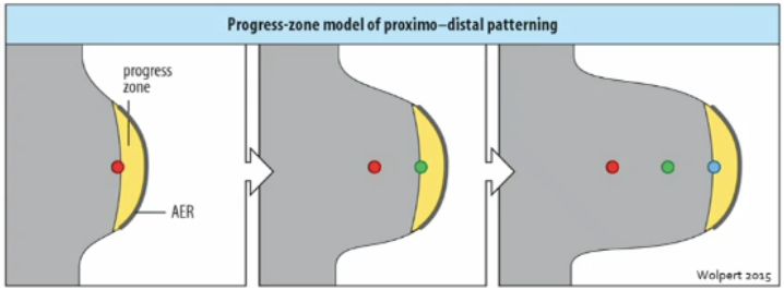
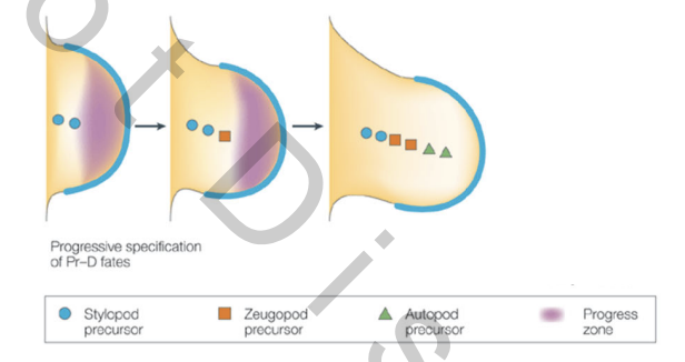
1. Proximal‐distal axis
2. Dorsal‐ventral axis
3. Anterior‐posterior axis
Limb Development
1. Proximal‐distal axis
_______ + _______ zone
2. Dorsal‐ventral axis
_______ overlying sides of developing limb bud + _______ cells in body of limb bud
3. Anterior‐posterior axis
_______ cells @ _______ (posterior margin in base of limb bud) + _______ zone
AER + Progress zone
Ectoderm + Mesenchyme
Mesenchyme ZPA + Progress zone
Limb Devel
1. Proximal‐distal axis (AER + Progress zone)
________ = area of mesoderm closest to AER that hears message loudest
AER induces _______ cells to proliferate
Using morphogens: ______&______
PZ cells in close contact w AER continue to proliferate
Progress zone
progress zone
FGF8 & 10
Limb Devel
1. Proximal‐distal axis (AER + Progress zone)
PZ cells left behind experience ________ concentration fx:
Some undergo apoptosis (_______ concentrations = low‐to‐none)
Some just slow prolif & come under induction influence from other inducers
Effects of other inducers = ______ & ______ expand distally
morphogen
morphogen
AER & progress zone
Limb Devel
1. Proximal‐distal axis (AER + Progress zone)
Effects of other inducers = AER & progress zone expand distally
Cells “_______” develop into limb regions & structures
1st ones left behind become _______
Mechanism still uncertain

left behind
proximal
Proximal‐distal axis (limb)
Cells “left behind” develop into limb regions & structures
SZA
SZA

Limb Devel
1. Proximal‐distal axis (AER + Progress zone)
Disruption/destruction/removal of ______ results in truncation of limb
if you shave off layer of ______, limb development comes to grinding halt at that point in development
AER
ectoderm
Limb Devel
2. Dorsal‐ventral axis
(______ overlying sides of developing limb bud + _______ in body of limb bud)
Dorsal & ventral patterns give rise to different structures
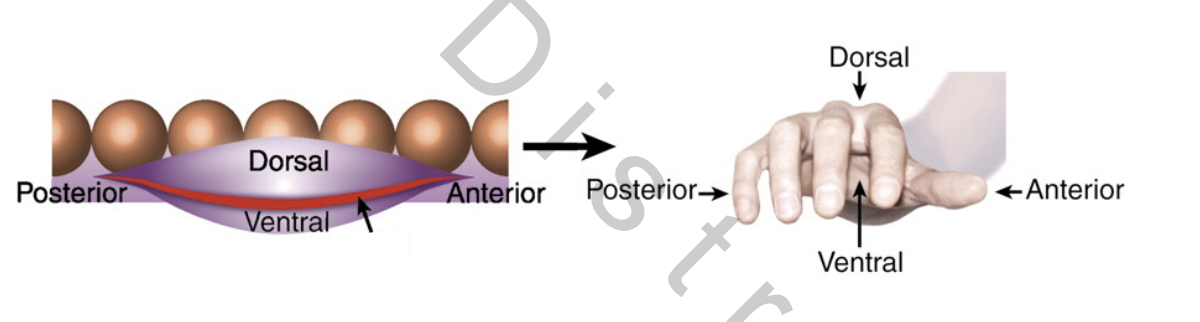

(Ectoderm overlying sides of developing limb bud + Mesenchyme cells in body of limb bud)
Limb Devel
2. Dorsal‐ventral axis
Remember: AER & progress zone expand distally → Cells “left behind” develop into limb regions & structures
Along the way…
_______ ectoderm sends message “______”
Morphogen: WNT7a
_______ ectoderm sends message “______”
Morphogens: BMPs & EN1
(bone morphogenic proteins, engrailed1)
Dorsal “dorsal”
Ventral “ventral”
2. Dorsal‐ventral axis
_______ Morphogen: WNT7a
_______ Morphogens: BMPs & EN1

dorsal
ventral
Limb Devel
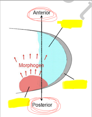
3. Anterior‐posterior axis
(Mesenchyme cells @ ZPA + Progress zone)
Prior to outgrowth, edge of ______ contacts small group of ________ cells (from _______ mesoderm) in ________ region of early limb bud
AER activates these cells, using Morphogen FGF___, to become _______
AER, mesenchyme, somatic mesoderm, posterior
FGF‐8, ZPA
Limb Devel
3. Anterior‐posterior axis
ZPA characterized by ________ gene activity
Releases Morphogen: _______(___)
shh ____________ (posterior → anterior) determines digit formation
higher shh concentrations induce ________ structures

sonic hedgehog
Sonic hedgehog (shh)
concentration gradient
posterior
Anterior‐posterior axis (limb)
bank: ZPA, PZ, AER

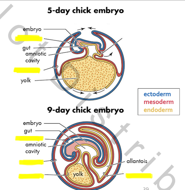
Extraembryonic membranes (EEM)
Membranes that develop outside of _______
Support embryonic ________
Develops from _______ & _______ cell lineages
Temporarily _______ with embryo
Body folds establish boundary btwn _______ & _______ regions
4 EEM form during _______ develop

embryo
development
embryonic & extraembryonic
continuous
embryo & extraembryonic regions
vertebrate
Extraembryonic membranes (EEM)
4 EEM form during vertebrate develop
1. A______
2. C______
3. A______
4. Y______
Present to some degree in ALL _______
_____&_____ fuse after formation to become chorioallantoic membrane (CAM)
1. Amnion
2. Chorion
3. Allantois
4. Yolk sac
—
vertebrates
Chorion & Allantois
EEM Formation
Derived from ____ germ layers (mesoderm/endoderm/ectoderm), extends out of _______
Somatopleure
fusion of __________ + ______derm
Splanchnopleure
fusion of __________ + ______derm
3, embryo
Somatic mesoderm + Ectoderm
Splanchnic mesoderm + Endoderm

EEM Formation

EEM Formation – MAMMALS
Mammals have same 4 EEM
1)A______ 2)C______ 3)A______ 4)Y______
_____&_____ 1st fuse to become _______ (CAM)
Then, (in mammals) fuse w maternal endometrium to become ________

1)Amnion 2)Chorion 3)Allantois 4)Yolk sac
Chorion & Allantois 1st fuse to become chorioallantoic membrane (CAM)
placenta
EEM Summary
Amnion & Chorion originate from _________
Allantois & Yolk sac originate from _________
—
_______ = hydration; protection
_______ = no function until fusion with chorion
_______ = nourishment
_______ = respiration
Somatopleure
Splanchnopleure
—
amnion
allantois
yolk sac
chorion
EEM Summary
Fused Chorion + Allantois
Birds (chorioallantoic membrane – CAM)
Origin: _____+_____
Function: _____+_____ management
Mammals
________ forms from splanchnic mesoderm + trophectoderm
Fuses w ______, then maternal _______, to become placenta
Splanchnopleure + Somatopleure
respiration + waste management
Chorion, allantois, endometrium
Growth that occurs when cells increase in size = __________
Growth that occurs when cells increase in number = __________
(Can occur in combination or separately)
Hypertrophy
Hyperplasia
Neural progenitor = Produces ______ AND _______ cells; _____‐potent
________ = generic term for ANY neuronal process
________ = bundles of axons from a ganglion
________ = Terminally post‐mitotic cell of nervous system capable of receiving & transmitting information via electric impulses
________ = group of neuronal soma in the ____NS
Sensory or Autonomic
Basal lamina/Basement membrane
Extracellular matrix that attaches, anchors, & surrounds almost all epithelial tissues & structures in the body
neurons & glial cells; multipotent
Neurite
Nerve
Neuron
Ganglion, PNS
Basal lamina/Basement membrane
Extracellular matrix that attaches, anchors, and surrounds almost all epithelial tissues and structures in the body
Nervous tissue growth & devel
Review: Brain development
Neural tube constricts & _______ via _______
⇒ Regional _____& _____
Progresses from 3‐part brain → 5‐part brain
End of neurulation = Fully formed _______
thickens, cell division
differentiation & proliferation
neural tube
End of neurulation = Fully formed neural tube
Cells produced proteins that coated outer surface of neural tube
= ________
Neural tube consists of _______ layer of multipotent ___polar neural ________
___polar – 1 process extending from each end of soma
1 process extends towards ______ (future _______)
Other process extends away from cavity toward ______ of neural tube (_______)
______ MIGRATES back & forth between ventricular & outer surfaces
Basal lamina
SINGLE, bipolar, progenitors
Bipolar
cavity (ventricle)
outer surface (basal lamina)
Soma

neural tube wall
epiblast ectoderm
part of neural tube up against ventricle
ventricular zone
basal lamina
Neurogenesis = making neurons
While migrating, neural progenitors _________
Division occurs only when soma are near _______ surface (_______ zone)
(divison = physical separation / cytokinesis / splitting & production of 2 daught cells)
Near _______ surface, cells enter rest phase of mitosis (_______ zone)

proliferate
ventricular surface, ventricular zone
outer surface, Marginal zone
Neurogenesis
Due to combination of ______ soma and massive _______, neural tube cells take on _______ appearance
BUT still are only a single layer (_______)

migrating, proliferation, stratified
Pseudostratified
Neurogenesis - 2 phases
1. ________
Symmetric division only
2. ________
Symmetric + Asymmetric division
1. Expansion
2. Neurogenic
Neurogenesis
1. Expansion
Symmetric division only (2 identical daught)
Prelude to true _______
Neural tube ________ (remember brain region formation?)

neurogenesis
thickening
Neurogenesis
2. Neurogenic
Symmetric + Asymmetric division
aka __________
Asymmetric division ⇉ 1 _______ + 1 _______ neuron
(_____ neuron = neuron PRECURSOR); can still prod daught cells

True neurogenesis
1 progenitor + 1 transiently amplifying (TA) neuron

Neurogenesis
Division outcomes as progenitor’s DNA ages:
Expansion
Symmetric division → 2 identical ________
______s can return to the beginning to continue cycle
Neurogenic
Asymmetric division → progenitor cell splits into 1 progenitor cell + 1 ________ (TA / Terminally post‐mitotic)
_______ can keep dividing to continue cycle
neural progenitors
NPs
—
TA neuron
Progenitor
Neurogenesis
Neurogenic phase
Progenitor can keep dividing to continue cycle
symmetric divis → 2 identical _______
Asymmetric divis → 1 _______ + 1 _______ cell
Symmetric divis (last stage) → 2 _______ cells
(______ first; then astrocytes)
2 TA neurons
TA neuron + glial cell
2 glial cells
(Oligodendrocytes first; then astrocytes)
Neurogenesis
Neurogenic phase
Asymmetric division
1 daughter cell not _____polar
Wraps around ______ zone process of ________ cell & “climb” toward _______ zone


bipolar
marginal zone, progenitor, marginal
Neurogenic phase (neurogenesis)
Neural tube changes
Along tube, regional difference in _______ rates & types of _______
Produce distinct regions of __NS in specific anterior‐posterior pattern
_______ begin to differentiate & become post‐______ as leave _______ zone (lose ability to divide)
As things get more crowded, newer cohorts of _______ must migrate further out
Forms ______ of cortex (no longer _________)
most recently produced ones at _______ layer
cell division rates, daughter cells
CNS
Neurons, post‐mitotic, intermediate zone
neurons
layers of cortex, pseudo-stratified
outermost
Post‐natal neurogenesis
Rare in ENDOtherms (warm blooded)
General mammalian pattern:
______ evidence of ______ beyond sexual maturity
In adults, typically restricted to _____campus, sub______ zone of lateral ventricle w migration to olfactory ______
Exceptions….
Rare
General mammalian pattern:
Little, neurogenesis
hippocampus, subventricular, bulb
Post‐natal neurogenesis
Exceptions (to general mammalian pattern):
_______ that engage in seasonal, dramatic changes in behavior
Neurogenesis in ________ related to:
Song acquisition & performance
Migration
Caching (storing & successfully retrieving stored food items)
______ during olfactory memory formation
Neurogenesis in forebrain w migration to _______ (late as 4 wks)
Neurogenesis in ________ possible throughout life
Birds
hippocampus
—
rodents
olfactory bulb
hippocampus
Most post‐natal _________ growth = result of “growth” of neurites, especially ________
Nervous tissue growth postnatally occurs largely thru ______ (incrs size)
nervous tissue, axons
hypertrophy
Axon initiation
______ of ______ neuron adjacent to basal lamina = signaled to become _______
As layers form in neural tube, _______ signal _______ to exit neural tube

Neurite, post‐mitotic neuron, axon
morphogens, axon

Axon growth cone = how processes grow
Tip of axon differentiates into _______ growth cone (free from soma)
Cytoplasmic “suitcase” – _______ loaded w everything needed for _______ synthesis
Cytoplasmic proteins (ribosomes, mRNA)
Actin molecules
Microtubules
Enables ________ survival, elongation, & ________ proteins
Emancipates growth cone from having to wait for ______ to travel from ______
_______ of growth constantly pull & cause axon to elongate (fingers reaching out for target)
autonomous
growth cone, protein
axon, navigation
proteins, from soma
Filopodia
Axon growth cone behavior = dependent on which signals the ______ encounter
______/adhesion molecs → “_______”
Ex. Cell Adhesion Molecs (CAMs)
________ molecs → “______”
Physical/mechanical ________ → “_______”
Stiff cells (eg. w collagen)
filopodia
Attractant/adhesion → “Advance”
Repellent → “Retreat”
barriers → “Go around”
Pioneer axons
1st axons to exit _______ / grow into a ________
have _______ growth cones; very _______ acting
Follow _______ signals & forge _______ to correct _______
Produce ______‐specific _______ & incorporate into ________ along way
Attract & guide _______ axons
Ex. In PNS, from neurons in the same ganglia as the pioneer axon
Cause ______ axons to ______
neural tube, new region
Hyperactive, fast
regional, pathway, target tissue
region‐specific CAMs, axon membrane
follower
follower, adhere
Growing axon behavior
1. Following ______ signals, growing axon advances to _______ target
Once shared _____ detected, growth cone pulls axon in that direction
2. Reach _______ target
3. Signals that attracted to _______ target become _____/non‐______ after leaving _______ target
depending on where axon is relative to _______ target, function of same molecule can switch between ______/______
4. At _______ target: _______ branches & each branch differentiates into ________
attractant, intermediate
CAM
intermediate
intermediate, repulsive/non‐attracting, intermediate
intermed target, attractant / repulsive
Final target, Growth cone, synapse
Where do PNS neurons come from?
Remember neural tube formation:
Primordial ______ comes from _____derm
Formation of tube via invagination
1. Notochord induces epiblast _______ ↠ _______ cells
2. Bending of neural ______ into neural _____
3. Closure/pinching off of neural ______ into neural tube

loose ends = Neural crest cells
CNS, ectoderm
Formation via invagination
ectoderm , neural plate cells
plate, groove
groove
PNS neurons come from Neural crest cells (NCCs)
Epiblast cells at _______ of closing ________ undergo determination → Future ________ cells
Become ______‐like, _____potent cells that ______ thruout body in well‐defined patterns
Capable of differentiating into either _______ or _______
Not fully pluripotent bc they cant differentiate into _______
margins, neural tube, neural crest
pluripotent‐like, multipotent, migrate
mesoderm or ectoderm
endoderm
Neural crest cells (NCCs)
Capable of differentiating into either mesoderm or ectoderm
Mesodermal cells → ________ (cells of mesoderm) differentiate into _____&______ tissues
Smooth muscle, Osteoblasts /clasts, Adipocytes, chondrocytes
Ectodermal cells → cells of _______
Melanocytes
Schwann cells
Neurons
mesenchyme, connective & muscle
PNS
Growth of PNS
Just like neurons of CNS, PNS neurons rely on ________ activity
Start w ________
Ea ______ axon then produces ______‐specific ______ that attract, guide, & adhere subsequent ________
Single ______ → form ______ bundles → which form ______ → which form ______
growth cone
pioneer axons
follower, region‐specific CAMs, follower axons
Single axons → Axon bundle → Fascicle → Nerve
PNS ________ = held together by layers of connective tissue
endo /peri /epi neurium
axon bundles
PNS axon bundles = held together by layers of connective tissue
ENDOneurium
Wraps around a single neuron’s ______, its associated ______ cells & _______
PERIneurium
Wraps around bundles of _______-wrapped _______, ________, & ________ tissue
Forms a ______
EPIneurium
Wraps around bundles of _______, ________, & ______ tissue
Forms a ______
axon, glial cells, capillaries
axons (endoneurium wrapped), blood vessels, adipose tiss
fascicle
fascicles, blood vessels, adipose tissue
nerve
PNS axon bundles – held together by layers of connective tissue
Endoneurium, Perineurium, Epineurium


Once axons synapsed:
Functional verification = _______
An axon will _______ its connections
Send out new ________ w/in target area
Allow existing ______ to _______
Response to activity between _____&_____
“Use it or lose it.”
“Use it a lot, recruit more.”
Response to _______ of organism
As organism grows, _______ to _______ will change
Axon ________ as body grows
Axon growth shifts to ________ (growth between 2 landmarks)
No longer ________ driven (not growing from ________ anymore; axon gets ________ as it’s lengthened)
Axon grwth b4 synapsing: growth cone
After synapsing: interstitial growth
Fine tuning
shift
new branches
branches, regress
axon & target
growth of organism
physical distance, ganglia
lengthens
interstitial
growth cone (end of axon; stretched out)
Axon grwth b4 synapsing: growth cone
After synapsing: interstitial growth
Dendrites
Less is known, ______ are more murky
Dendrites have their own _______
Appear as mini _______
Dendrite _______ behavior can be variable
Sometimes responds opposite to ______ growth cone
Sometimes responds ______ (when synapsing w axon is required)
dendrites grow throughout life
morphogens
growth cones, growth cones
growth cone
axon, identically
Dendrites
Complexity of _______ = dependent on amount of incoming ________
Multi‐dendrite vs simple dendrite
Innervation activity levels affect dendritic complexity
Dendrites = VERY dynamic
Ex. In mice, some dendrites in visual system can appear & disappear within minutes when exposed to stimuli

branching, innervation
Myelin
Both _______ & _______ produce myelin sheaths around some axons
1 ______ makes multiple sheaths, 1 ______ makes 1 sheath
_______ (CNS); _________ (PNS)
myelin appears as white matter of _______
______ matter = Not myelinated (e.g., soma, dendrites, etc)
Enables fast _______ conductance along _______
is an electrical _______
Accelerates conductance by localizing ______ channels at ________ (short gaps btwn adjacent myelin sheaths)
Myelination = dynamic throughout life
______ myelin plasticity different from _______ myelin plasticity
oligodendrocytes & schwann cells
oligo, schwann
Oligo = CNS, Schwann = PNS
CNS; Gray
electrical, axons
insulator
Na+, Nodes of Ranvier
CNS, PNS
CNS: Oligogenesis
In rodents: ____________ cells (OPCs) can…
...undergo ______ to produce more _______
AND/OR
...differentiate into a post‐______ pre‐_______ oligodendrocyte (PO)
~80% PO attrition rate (fail to ______)
Remaining ~20% of POs migrate to ______ & become _______, producing myelin
CNS myelin plasticity
_______ occurs throughout life
In human adults, ______ still active
Numbers peak during _______ age
Oligodendrocyte precursor
division, oligodendrocytes
post‐mitotic pre‐myelinating oligodendrocyte
survive
axon, oligodendrocytes
Oligogenesis
OPCs
middle age
PNS: Schwann cell (SC) classes
Myelinating SC
Produce ______ & wrap ______ in _______
Nourish axons
Remak SC
__________
Nourish axons
myelin sheaths, axons, myelin sheaths
—
Non‐myelinating
PNS: Schwann cell development
________ ↠ Schwann cell precursors (SCP)
SCPs _______ & bounded by associated _______
Once associated w ______, SCPs become dependent on _____ for _______
SCPs stop migrating:
now classified as _______ (___)
_______ still growing
figure out which axons to myelinate via _______ sorting
Based on _______ of axon
Big diameter (thick) = ______
Small diameter (thin) = ______
Become “self‐supporting” & mature into myelinating Schwann cells
NCCs
migrate, axons
axon, axon, survival
Immature Schwann cells (ISC)
Axon
Radial sorting
diameter
Big = Myelin
Small = No
Become “self‐supporting” & mature into myelinating Schwann cells
PNS myelin plasticity: Response to injury
_______ nerves can undergo _______
PNS ________ possible through _______ plasticity (specifically _______ SCs)
Caveats:
1. Effective only on _________
Regeneration rate too ______ to completely heal traumatic nerve damage
2. SCs competence ______ with _______
Peripheral, regeneration
regeneration, Schwann cell (remak SCs)
minor injuries
slow
diminishes w/ age
SC-mediated PNS regeneration
1. Peripheral nerve injury
2. Both ______&______ SCs undergo _________
_________ de‐myelinate
& both convert into _______ SCs
3. Newly programmed _______ SCs & ________ break down _______ part of axon
= Wallerian degeneration*
* occurs to lesser extent & mechanism less understood in CNS
4. _______ SCs clear out ________ tissue
Produce _______ factors to ________ re‐growing axon
5. ______ SCs form scaffold to guide re‐growing _______ back to ______
6. _______ proliferate; reprogram to turn on ________ genes (promote axonal growth)
7. Newly re‐programmed ________ SCs then re‐myelinate axon
Myelinating & Remak, reprogramming
Myelinating SCs
Repair SCs
Repair, macrophages, damaged
= Wallerian degeneration*
Repair, damaged
trophic factors, nourish
Repair, axon, target
Repair SCs, myelinating
myelinating