Anatomy Lab Exam 1
 Call Kai
 Learn
Learn Practice Test
 Spaced Repetition
 Match
Match Flashcards
 Knowt Play
1/125

There's no tags or description
Looks like no tags are added yet.
Name | Mastery | Learn | Test | Matching | Spaced | Call with Kai | Chat |
|---|
No analytics yet
Send a link to your students to track their progress
126 Terms
Anatomical Plane- frontal plane
divides body into anterior and posterior

Anatomical Plane- sagittal plane
divides body down the middle, into right and left

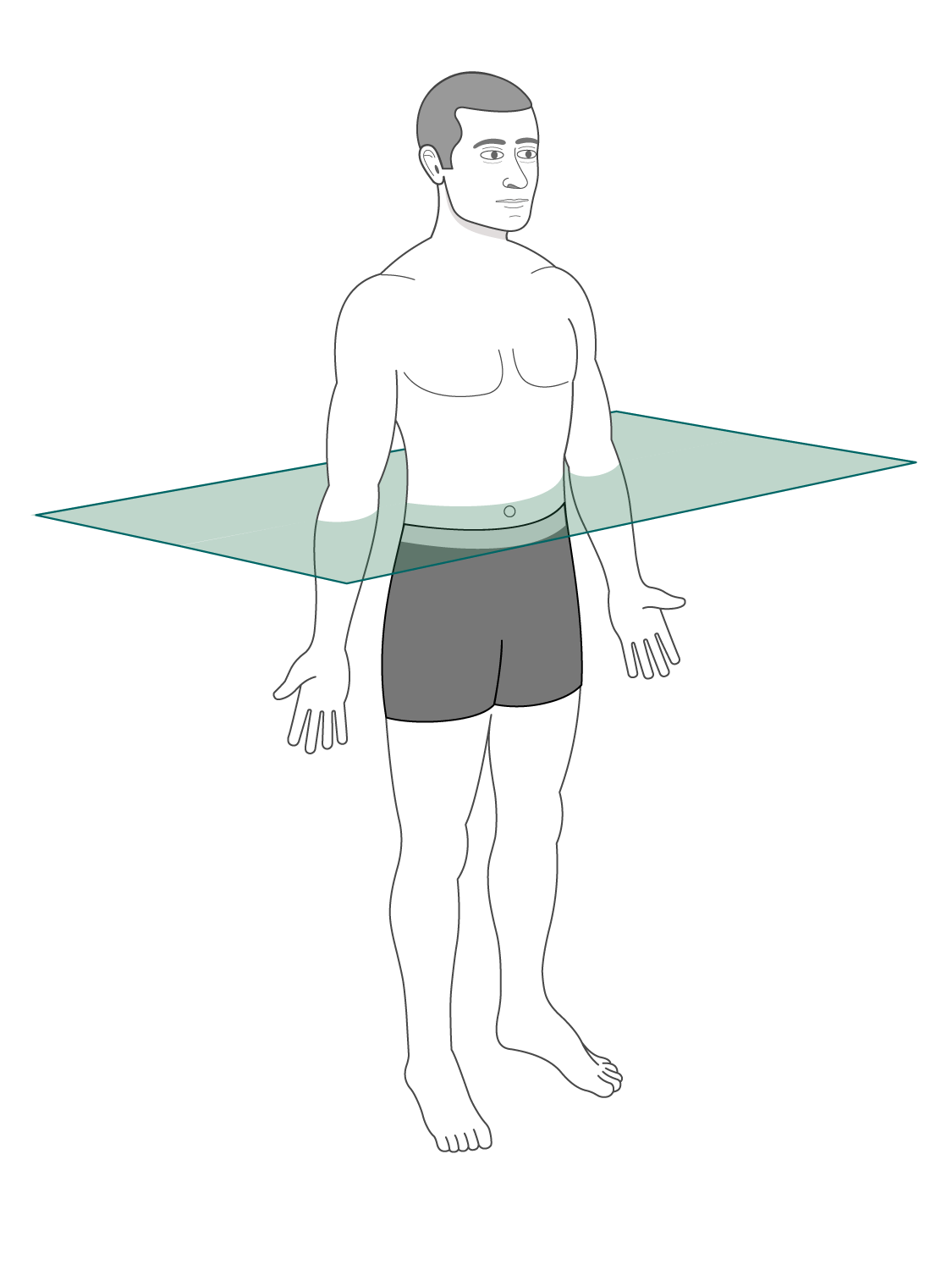
Anatomical Plane- transverse plane
divides body (near hips) into superior and inferior (upper and lower)
two main body cavities
dorsal cavity
ventral cavity
dorsal cavity, what it houses and its subdivisions
houses the brain and spinal cord
Subdivides into the cranial cavity (brain) and vertebral cavity (spinal cord)

ventral cavity subdivisons
abdominopelvic cavity - the abdominal and pelvic
thoracic cavity
abdominal cavity carries the…
many organs for digestion

pelvic cavity carries the…
bladder, reproductive organ, and the digestive tract
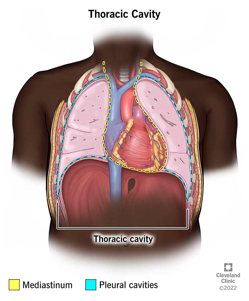
the thoracic cavity carries the..
heart and lungs
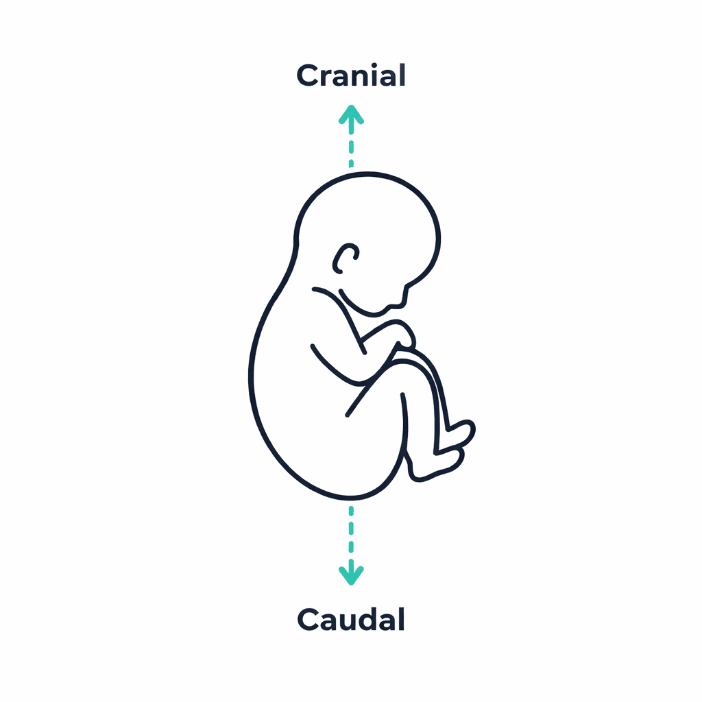
cranial
toward the head
caudal
toward the tail
posterior/dorsal —- same in humans
toward the back
anterior/ventral —- same in humans
toward the front
superior
toward the head/ top
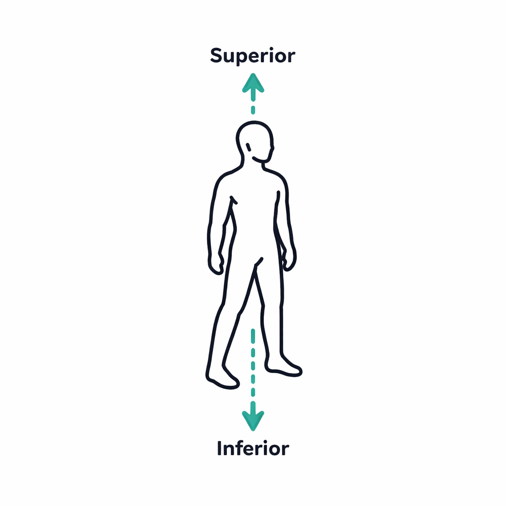
inferior
toward the feet/down

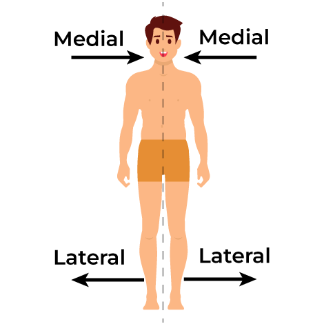
medial
toward the midline

lateral
away from the midline
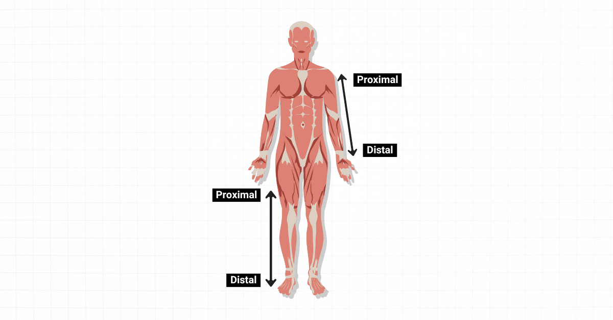
proximal
closer to the point of attachment

distal
further (more DIStant) from the place of attachment
superficial
toward the surface (shallow)
deep
toward the core
parietal
toward the body wall
visceral
toward the organs
epithelial tissues
tight cells that line all external and internal body surfaces
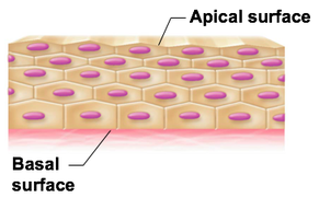
apical surface of epithelial tissue
environment side

basal surface of epithelial tissue
contact with neighboring tissue
Epithelial tissues are avascular, meaning…
there are no blood vessels within the tissue
simple squamous epithelial tissue
flat cells, flat nuclei
simple— single layer
helps rapid diffusion/filtration because it is flat
found in either in alveoli of lung OR glomerular capsule in kidney

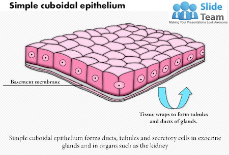
simple cuboidal epithelial tissue
cells are cubed, nuclei is sphere
simple—single layer
it absorbs and secretes because of the extra internal space from the shape
found in the kidney tubules

simple squamous epithelial tissue in the lung
(look at squished nuclei )

simple squamous epithelial tissue in kidney
(look at squished nuclei )

simple cuboidal epithelial tissue in kidney
(look at sphere nuclei )
simple columnar epithelial tissue
Column-shaped cells, oval nuclei
simple— single layer
shape gives better surface area to help for secretion and absorption
found in the digestive tract, the duodenum and jejunum

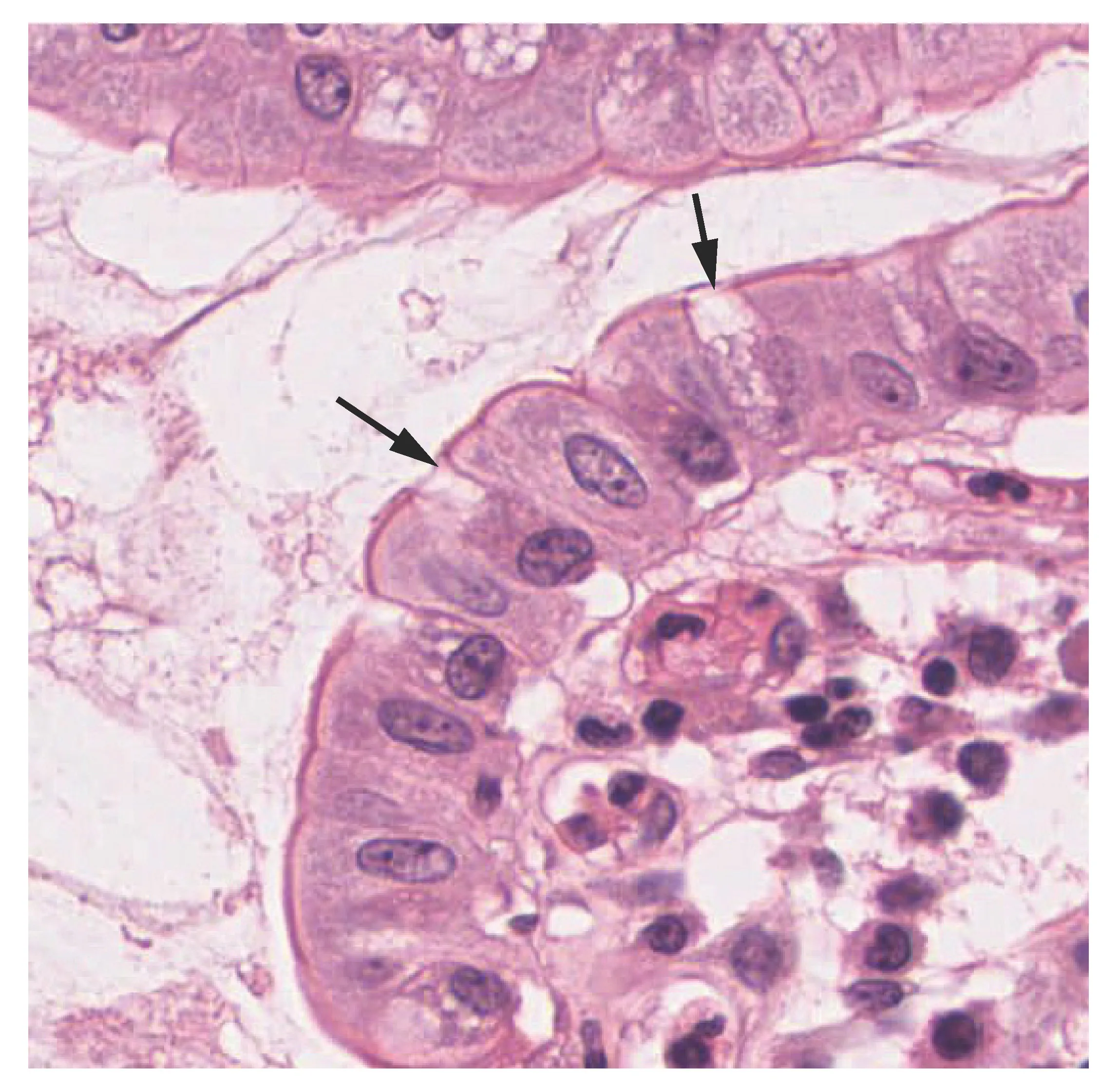
simple columnar epithelial tissue in digestive tract
pseudostratified columnar epithelial tissue
column shaped cell, oval nuclei
looks like many layers, but is only one
functions in absorption and secretion
found in the lining of the trachea and upper resp tract


pseudostratified columnar epithelial tissue
(nuclei is oval and appears to be overlapping/messy)
stratified squamous epithelial tissue
surface cells have squamous shape, where deeper cells have column/cube shape
stratified— many layers
protect underlying body structures from constant friction
can be found in the upper layer of skin, the epidermis (also found in esophagus)
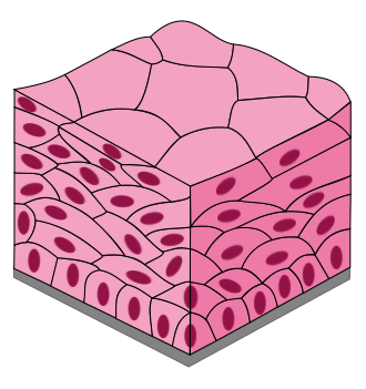
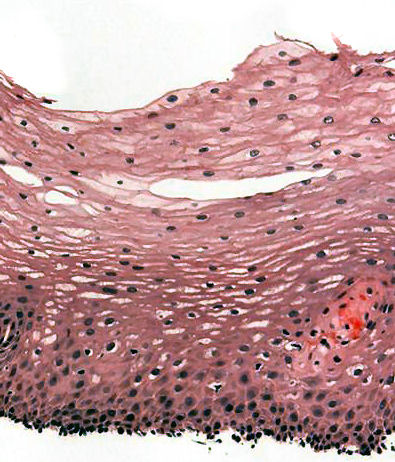
stratified squamous epithelial tissue in the skin
(it starts squashed, then starts to cube out)
extracellular matrix part in the connective tissue
it supports to help bear weight and withstand large amounts of tension and pressure
ground substance part in the connective tissue
stuff that fills the space between cells and fibers. Its made up of interstitial fluid, cell adhesion proteins, and proteoglycans
collagen fibers part in the connective tissue
the proteins that provide strength and support
elastic fibers part in the connective tissue
allowing organs and structures to return to their original shape after being deformed
All connective tissues share three characteristics..
origin from the embryonic tissue, the mesenchyme
degrees of vascularity, some are rich in blood vessels, some are avascular
mostly extracellular matrix (composed of ground substance and fibers)
Loose connective tissue
main cell- fibroblasts
containing ground subst, collagen and elastic fibers
the most widely distributed connective tissue
binds body parts together yet still allows them to move freely
found in most epithelial tissues, providing network of blood vessels to support the avascular epithelium

dense regular connective tissue
main cell- fibroblasts
tightly packed collagen fibers, with little ground substance, and poorly vascularized
flexible structure with great resistance to tension from ONE direction (slight “give” but once straight there is no more)
found in tendons, ligaments, and aponeuroses
** this tissue has a system/order

dense irregular connective tissue
main cell- fibroblasts
many collagen fibers that are thick and in random order, with little ground substance
these deal with tension exerted from MANY directions
found in the in the reticular layer of the dermis of the skin, as well as the fibrous coverings of specific organs such as kidneys, bones, cartilage, and muscle.

adipose connective tissue
main cell-- adipocytes
no visible fibers in this tissue
nucleus is pushed to one side, causing a fish-net look, then has more room to store fat
found throughout the body, but a thick layer is seen the creating the hypodermis.

fibroblast ( cell in CT)
Makes fibers like collagen
Adipocyte (cell in CT)
Stores fat for energy, insulation, and cushioning
epidermis
outermost layer of the skin
mostly- stratified squamous epithelium

dermis splits into two different layers
papillary layer AND the reticular layer

papillary layer of dermis
composed of loose CT just under the epidermis
this helps anchor the epidermis and dermis

reticular layer of dermis
much thicker of a layer
composed of dense irregular connective tissue
this layer helps hold glands and hair follicles in place

hypodermis
works to connect the skin to the underlying tissues
Composed of adipose tissue (insulation) and loose CT (anchors skin)

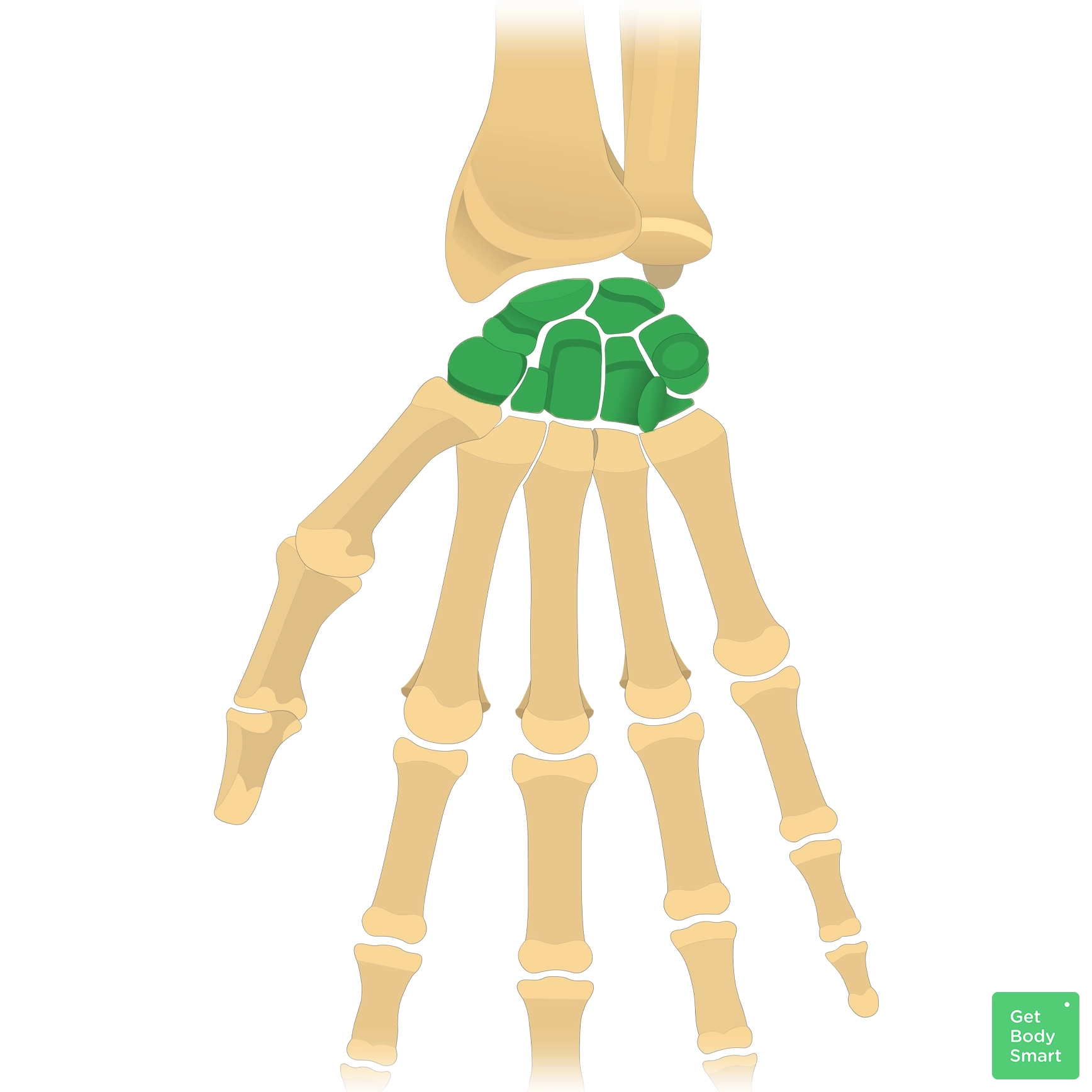
carpals (8 total)

metacarpals

proximal phalanges

middle phalanges
(note, not on thumb)

distal phalanges

clavicle

conoid tubercle
on clavicle
(think where it concaves; it's the conoid)

acromial end of clavicle
(flatter side)

sternal end of clavicle
(makes sense, bc it connects near sternum)

scapula
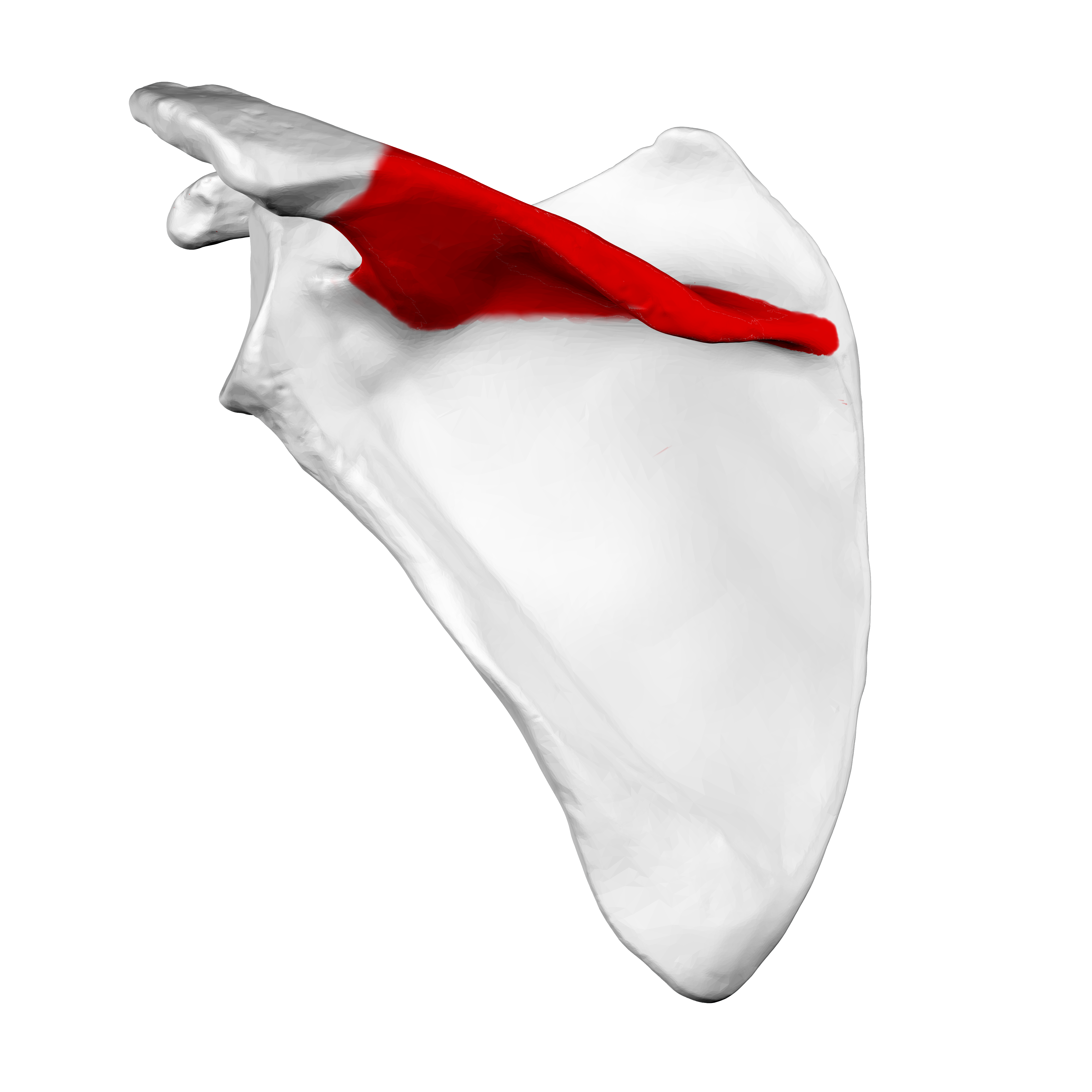
spine on scapula
(posterior side)
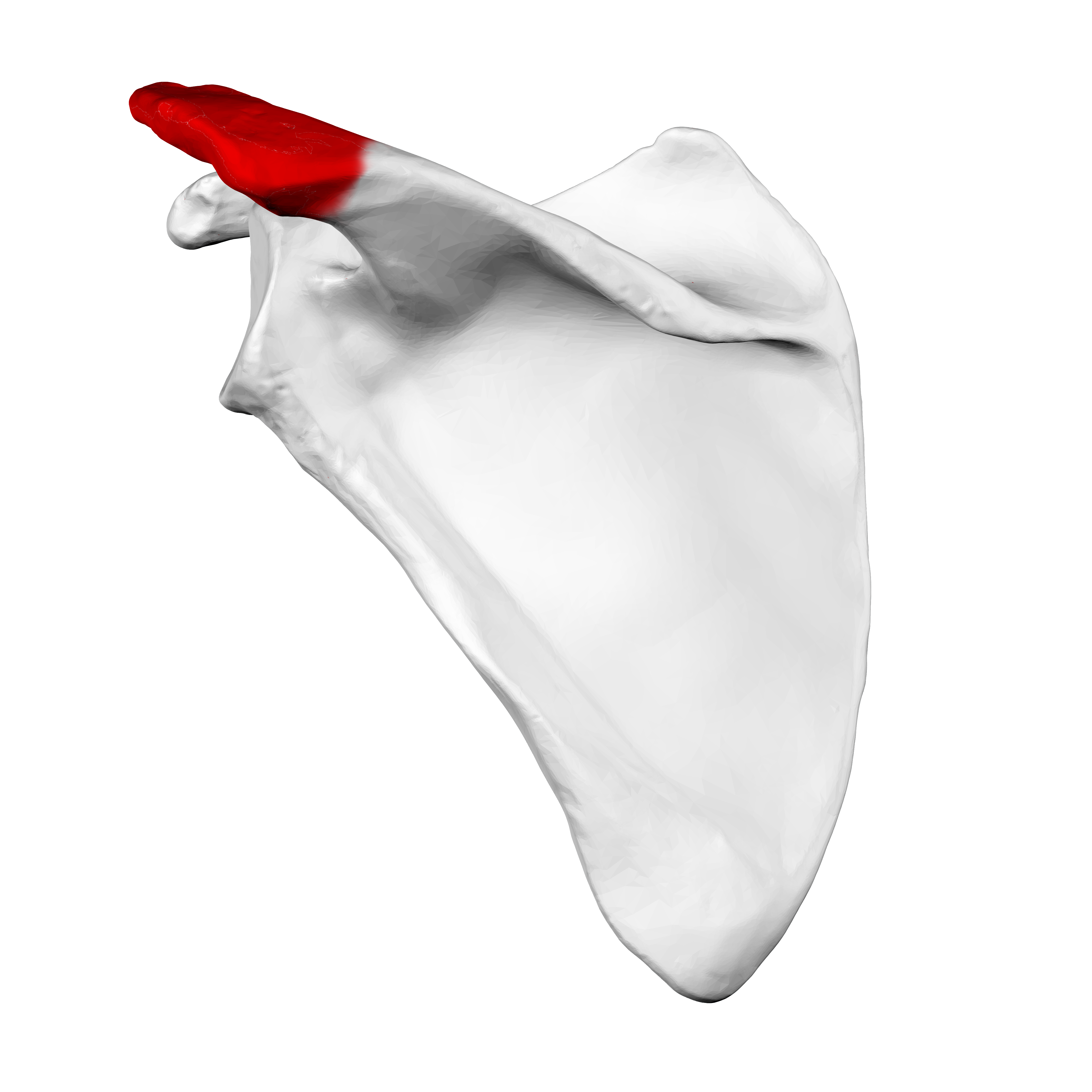
acromion on scapula

coracoid process on scapula
(looks like crow beak)

supraspinous fossa on scapula
(posterior side)

infraspinous fossa on scapula
(posterior side)

subscapular fossa on scapula
(anterior side)
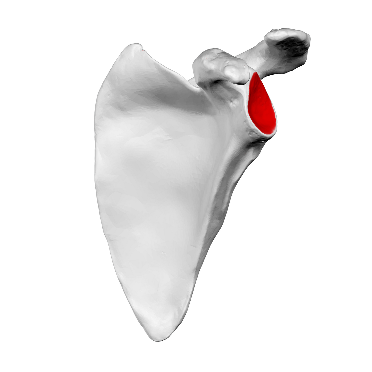
glenoid fossa on scapula
(anterior side)

humerus

humeral head of humerus

greater tubercle on humerus
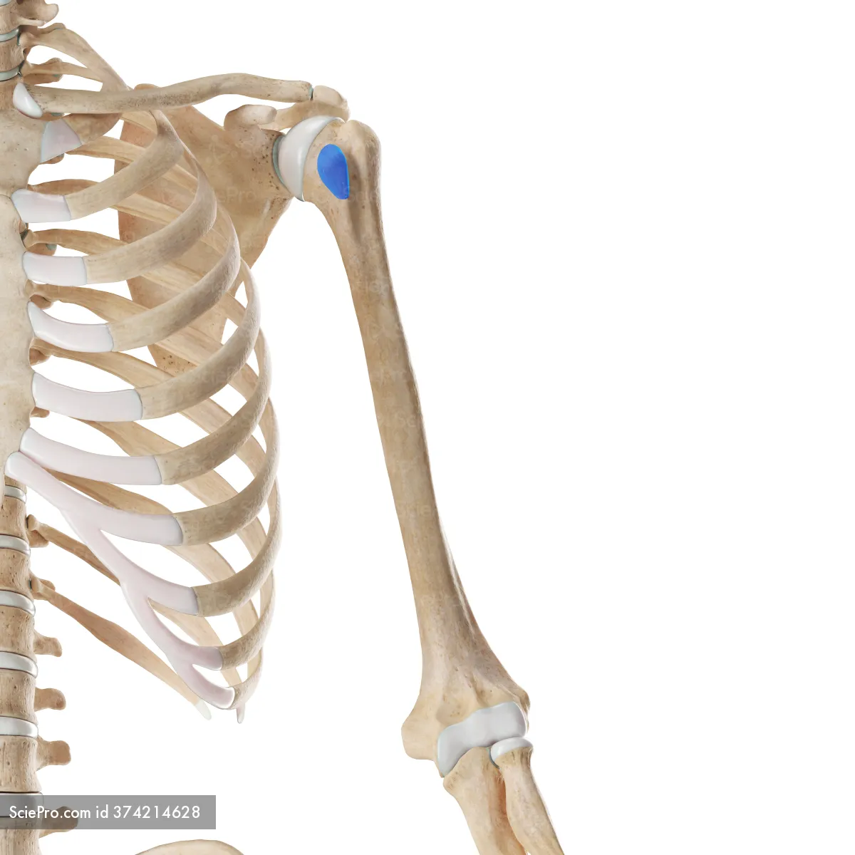
lesser tubercle on humerus
(anterior side)

intertubercular groove on humerus
(anterior side)

deltoid tuberosity on humerus
(anterior side)

medial epicondyle on humerus
(posterior side)

lateral epicondyle on humerus
(anterior side)

coronoid fossa on humerus
(anterior side)

capitulum on humerus
(sticks out the most, think captain)
(anterior side)
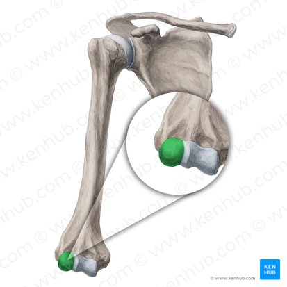
NOT GREEN PART, whats light blue part
trochlea on humerus
(think it looks like two toes, TROchlea)
(anterior side)

what is the name referencing to both of these parts on the humerus?
humeral condyles
(anterior side)

what is the divot on the back of the humerus
olecranon fossa
(posterior side)
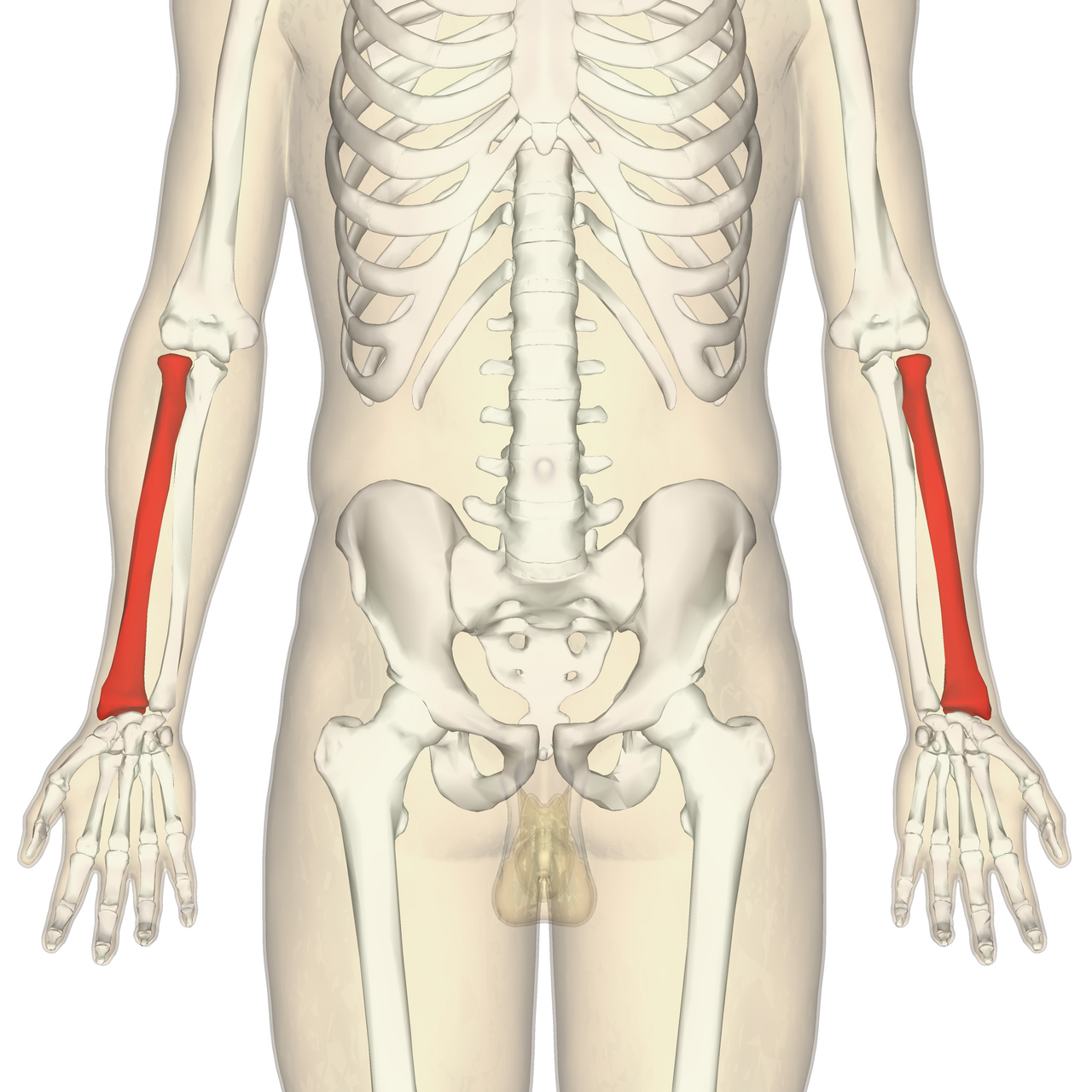
radius
(little bit shorter than ulna)
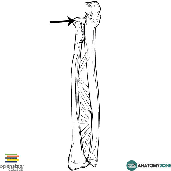
head of radius
right arm, anterior side
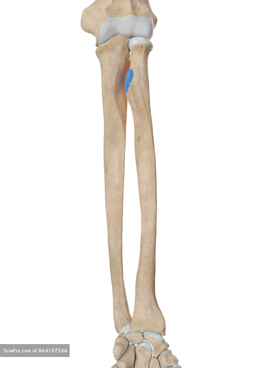
radial tuberosity
left arm, anterior view

radial styloid process
right arm, anterior view
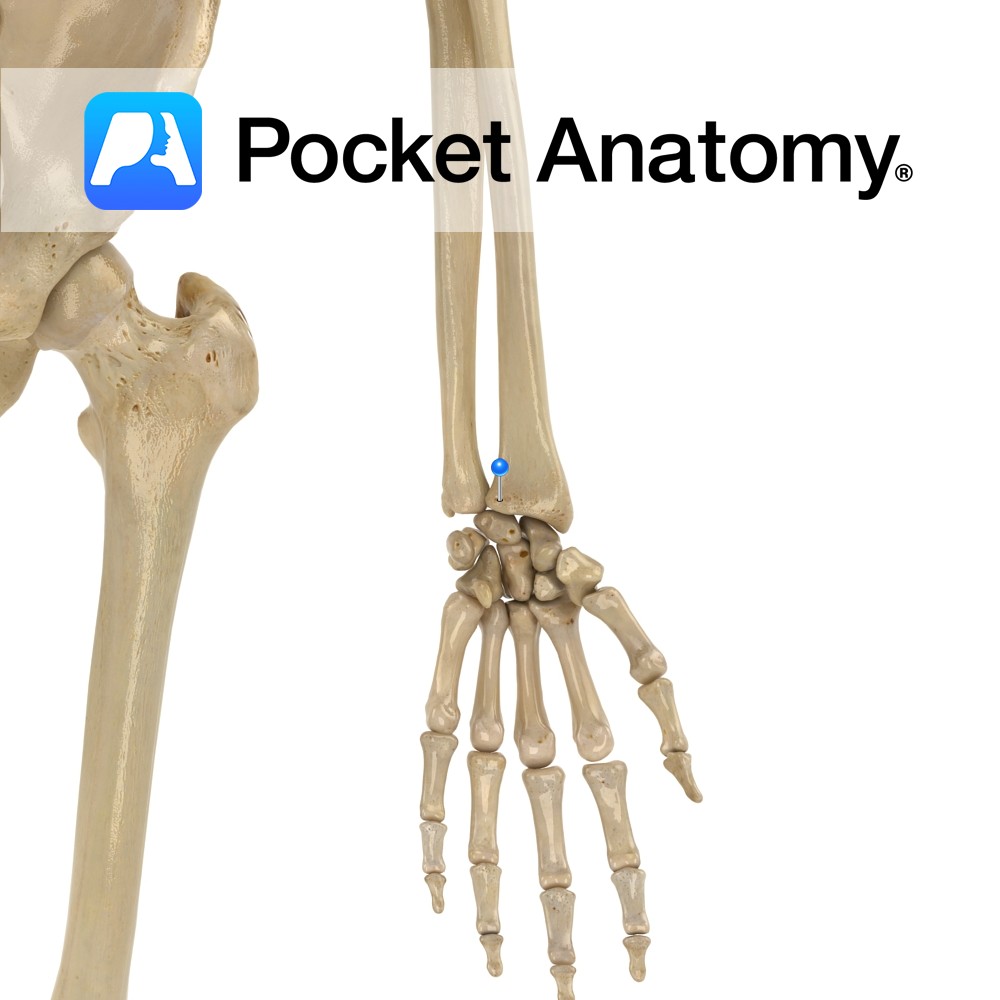
ulnar notch on radial
left arm, anterior view
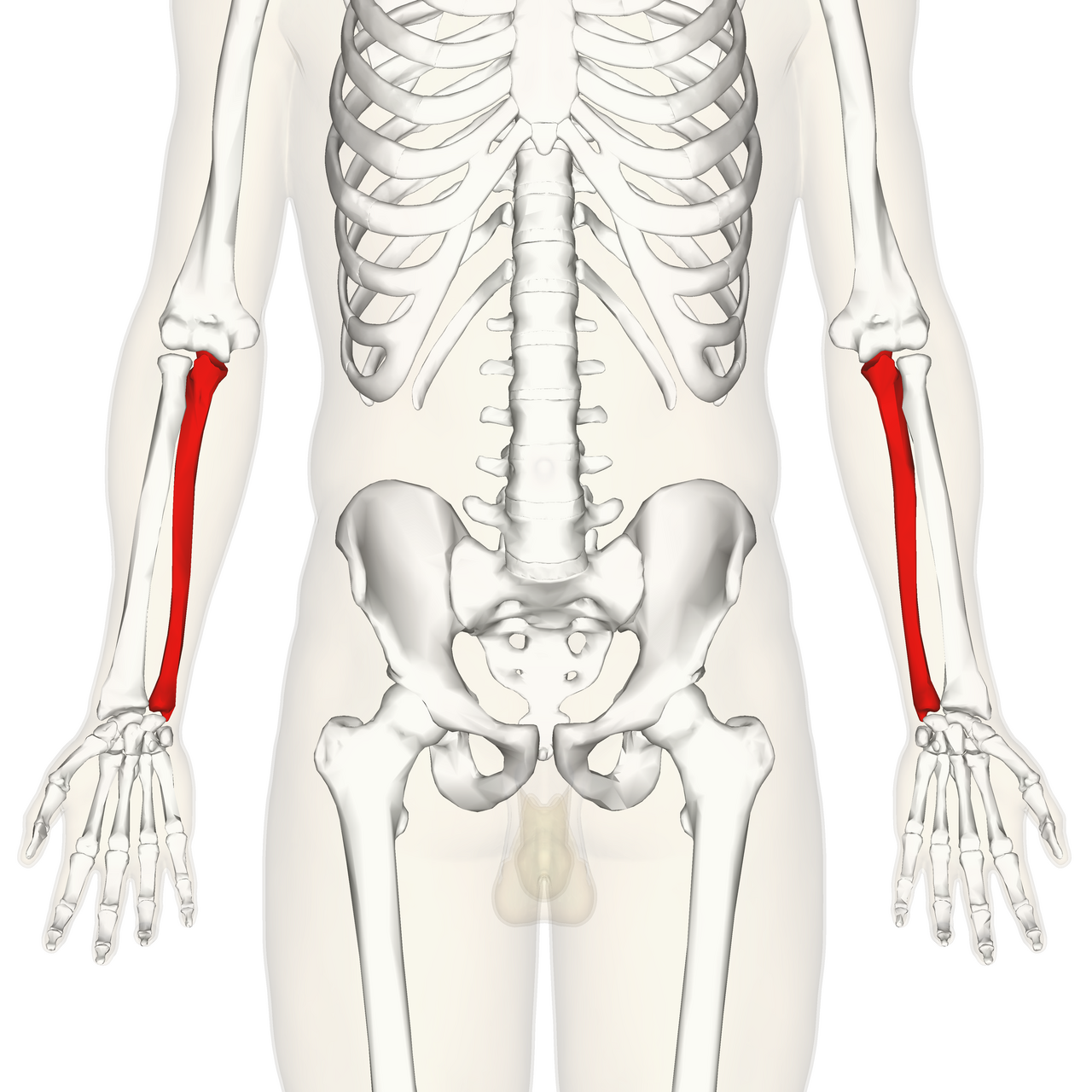
ulna
(little bit taller than radius)

trochlea notch on ulna
(anterior view)
right arm
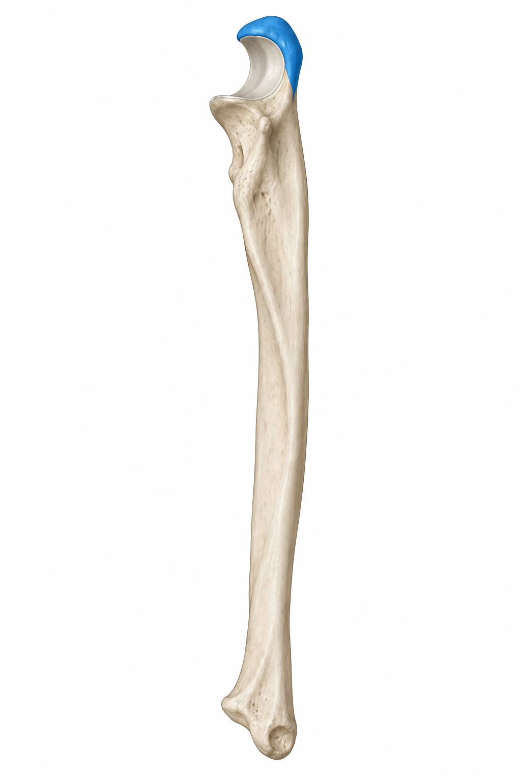
olecranon process of ulna
(anterior view)
right arm
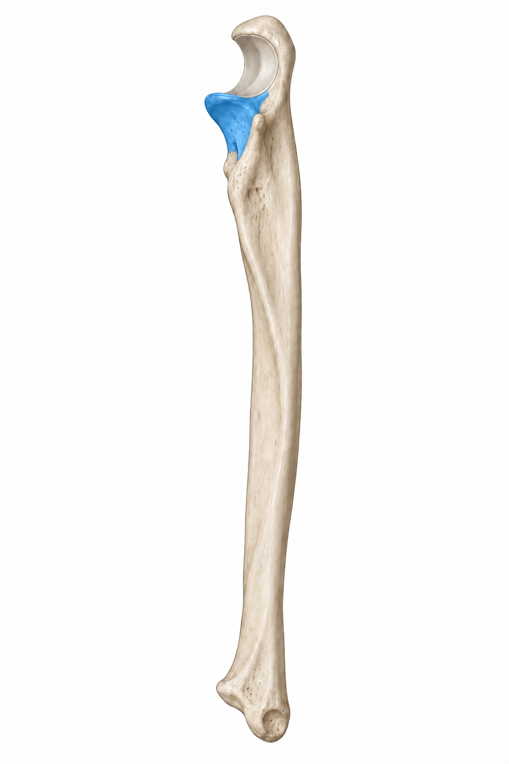
coronoid process of ulna
right arm, (anterior view)

radial notch on the ulna
posterior side

ulnar styloid process

what joint is this
shoulder joint
landmarks of the bones for the shoulder joint
humerus head and glenoid fossa of scapula connect together for this joint
structural classification of shoulder joint
synovial joint
synovial type of shoulder joint
ball and socket
where is the glenoid labrum, joint capsle, and tendon of the long head of the biceps brachii in the shoulder joint
glenoid labrum- wrapped around glenoid fossa
joint capsle- wraps around whole joint
