18. Metabolic supportr of photoreceptors
1/42
There's no tags or description
Looks like no tags are added yet.
Name | Mastery | Learn | Test | Matching | Spaced |
|---|
No study sessions yet.
43 Terms
Muller cells do what?
Supportive functions in retina:
Regulates and maintains retinal vasculature
metabolism and transport of glucose derivatives
neurotransmitter metabolism
Helps protect retina from oxidative stress (glutathione production)
Ionic/pH homeostasis
Light conduit
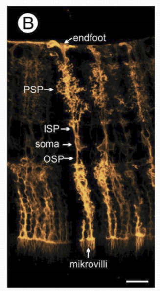
How do Muller cells regulat and maintain retinal vasculature?
Muller cells ensheath retinal capillaries.
What do mullers cells secrete?
Anti-angiognetic gactors: inhibit endothelial cell proliferation.
Where does neovasularization occur in diabetic retinopathy?
Towards the vitreous and not within retina.
What types of metabolism does the retina rely on for function?
Both glycolysis and aerobic metabolism.
What is most of the glucose taken up by the retina converted into?
Lactate
What type of glial cell takes up large quantities of glucose in the retina?
Müller cells.
What is retinal capillary ensheathment and which cells perform it?
It is the surrounding of capillaries by Müller cells, allowing efficient glucose uptake.
Where is glycogen exclusively produced in the retina?
In Müller glial cells.
What happens to glucose in muller cells?
It is converted to lactate via lactate dehydrogenase and exported out to be utilized in retinal cells.
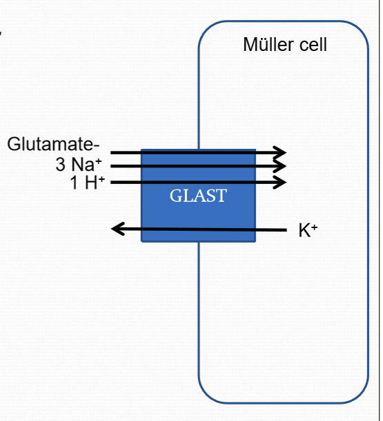
What do muller cells take up?
Extracelluloar glutamate to prevetn lateral spread of glutamate signal to neighboring synapses, modifies the synaptic response, and prevent glutamate induced neurotoxicitgy.
How does excess glutamate cause neurotoxicity?
Too much glutamate can cause cell apoptosis by triggering the release of calcium ion storage.
Where does the glutamate in the extracelluloar space come from?
They are derived from the inner plexiform synapses.
What channel/protein do muller cells use to uptake glutamate?
GLAST transporter (Glutamate Aspartate Transporter) using the sodium gradient for glutamate transport.
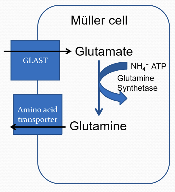
What do the muller cells covert glutamate to?
Glutamine using glutamine synthetase (requires ATP and Ammonia/NH4). Glutamine is then transported back to the retinal neurons via neutral amino acid transporters.
Where does ammonia come from?
It is a byproduct of glutamate synthesis in retinal neurons.
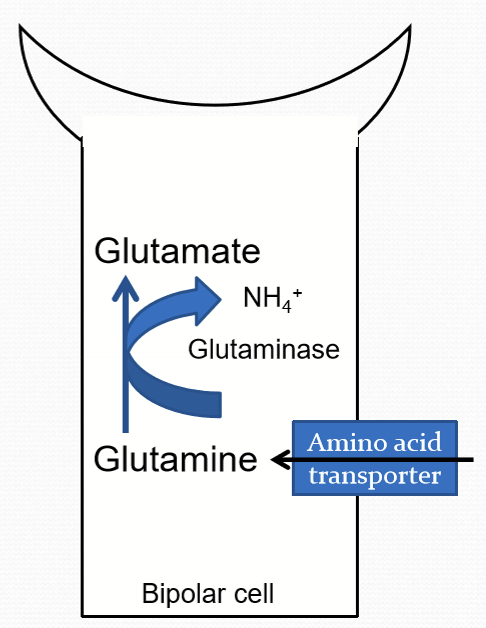
What cells use glutamine from muller cells?
Bipolar and inner retinal neurons.
What percentage of glutamine from muller cells do photoreceptors use?
only a small percentage
What is GABA?
The major inhibitory neurotransmitter in the vertebrate retina.
What is GABA’s function?
To process signal to retinal ganglion cells.
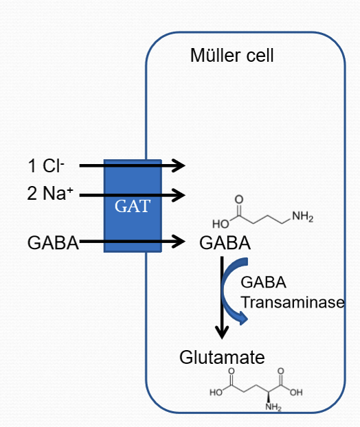
How do muller cells uptake GABA and why?
They uptake GABA via sodium/chloride ion dependent co-transport (GAT). Uptake of GABA terminates the synaptic action of GABA. GABA is then converted to glutamate via GABA transaminase.
Why does the retinal have an inner blood-retinal barrier?
To maintain the stable ionic environment to ensure proper function of the specialized environment of the retina.
How is the inner blood-retinal barrier maintained?
TJ between endothelial cells
no fenestrations
GLUT1 to transport glucose transcellularly
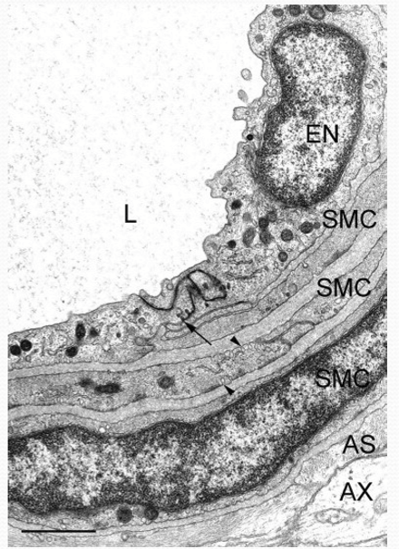
What is the structure of the distinct retinal artery structure that makes up the inner blood-retinal barrier?
Endothelium (EN)
3 layers of smooth muscle cells (SMC)
Astrocytes (AS)/muller cells ensheath superficial vessels
Muller cells ensheath outer plexuses
What makes up the outer blood-retinal barrier?
Non-leaky tight junctions between RPE cells. THey restrict paracellular transport.
What is the function of the outer blood-retinal barrier?
Maintain a stable ionic environment to ensure proper function of the specialized environment of the retina.
What is unique about the blood vessels of the choriocapillaris?
They are fenestrated and allow glucose and other nutrients to diffuse more freely, but need to pass transcellularly through RPE.
What tissue has the highest oxygen consumption rate?
Retina, most glucose (90%) used for glycolysis and converted to lactate. In dark, utilizes all available O2 for aerobic metabolism.
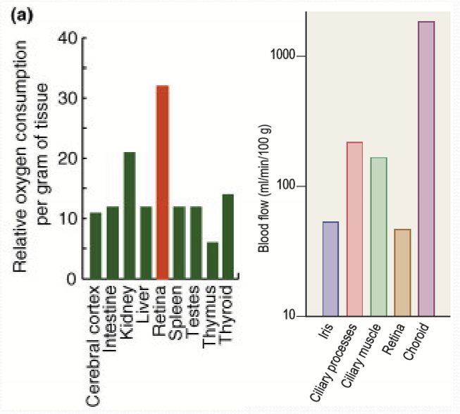
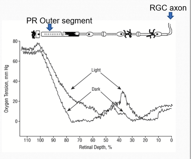
What are the sources of oxygen for the retina?
Retinal and choroidal vasculature. With highest O2 near choroidal vasculature.
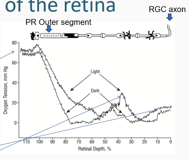
What is the O2 levels near the photoreceptors?
Near zero due to high energy demands of photoreceptor, especially in the dark.
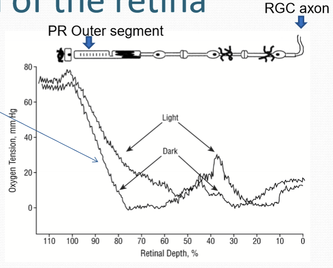
When is aerobic metabolism higher in the outer retina?
When in the dark vs light. Less O2 used in light.
What drives the metabolic demand for energy?
NKA and Generation of GTP for cGMP production.
The energy demands are altered based on illumination with:
light decreasing the need for NKA and increasing need for guanylyl cyclase for the recovery phase.
darkness increases need for NKA and decreases need for guanylyl cyclase.
What has a higher energy requirement? Illuminated or non-illuminated PR?
Non-illuminated PR outweighs illuminated PR energy requirements.
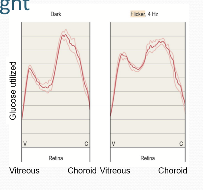
What causes the retina to maximize its energy use?
A flickering light. It causes an increase in demand of energy for RGCs and triggers vascular response in retinal vasculature.
Do retinal vessels and optic nerve head vessels have innervation?
No, they only rely on local vascular control mechanisms to match metabolic demand.
What cells in the retina respond to signaling pathways to help regulate blood flow?
Vascular endothelial cells
What role do glial cells (astrocytes/Müller cells) play in retinal blood flow?
They can mediate vasomotor response
What does the vasomotor system respond to in order to maintain constant blood flow in the retina?
Perfusion pressure.
What causes vasodilation?
Nitric oxide/NO
Prostaglandins/PGs
Lactate: stimulates release of NO and PGs
What causes vasoconstriction?
Endothelin-1 (ET-1) & pericytes express endothelin receptors and respond to ET1 to contract retinal arterioles.
What does sympathetic stimulation do to choroidal blood flow?
It reduces choroidal blood flow by vasoconstrictive alpha-receptors in smooth muscle cells being stimulated with neurtransmitter neuropeptide Y (NPY).
What does parasympatehtic stimulation do to choroidal blood flow?
It increases blood flow by acetylcholine, vasoactive intestinal peptide, PACAP, and NO.
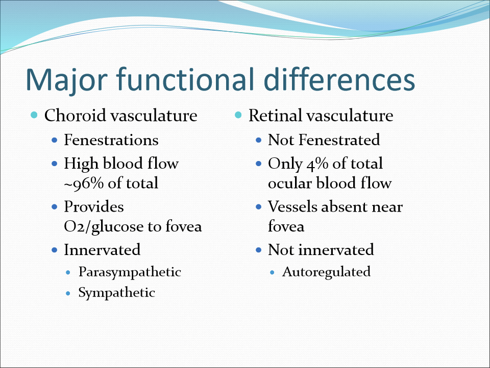
What are the major differences between choroid vasculature and retinal vasculature?
Choroid has fenestrations, high blood flow, provides O2 and glucose to fovea, and is innervated by para and sympathetic.