Skeletal System
1/69
There's no tags or description
Looks like no tags are added yet.
Name | Mastery | Learn | Test | Matching | Spaced |
|---|
No study sessions yet.
70 Terms
Metabolic Functions
Processes that deal with the buildup or breakdown of living cells for the purposes of providing energy and facilitating growth. One important metabolic function of bones is storing calcium and phosphorus (found in food)
Platelets
(Thrombocytes) Small, colorless cell fragments in blood that form clots and stop bleeding.
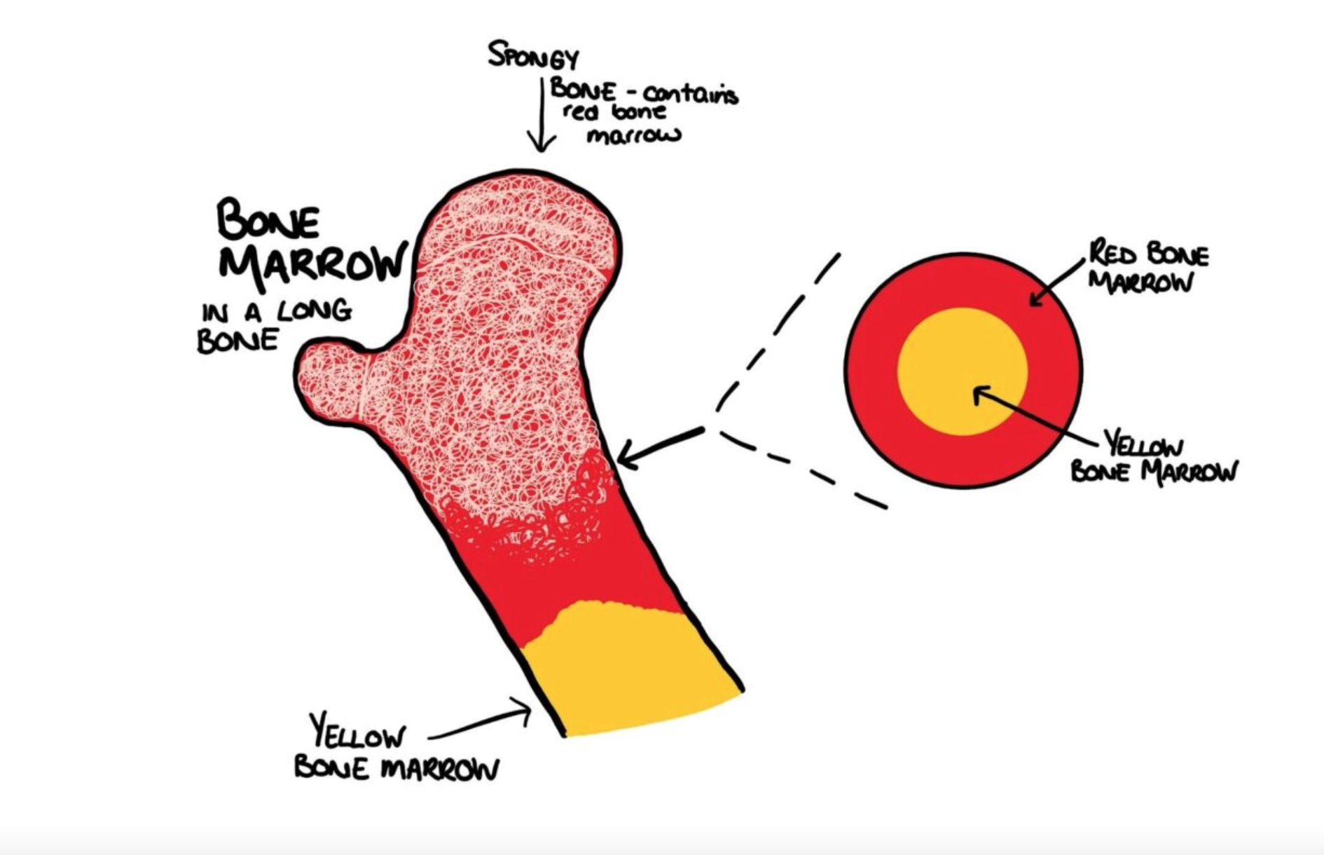
Bone Marrow
A soft tissue inside the bones that produces blood cells. The amount of bone marrow available to reproduce blood cells declines with age.
Peripheral Blood
The blood circulating throughout the body outside of the heart and the major blood vessels in the lungs. Readily accessible source for blood cells used for diagnostic testing.
Long Bones
Bones of the arms and legs - bones
Flat Bones
Bones of the ribs, shoulder blades, pelvis and skull - thin, flat and usually curved
2 Forms of Bone Structure
Cancellous Bone. Compact Bone
Cancellous Bone
(Spongy Bone, Trabecular Bone) Type of bone tissue characterized by its porous, honeycomb-like structure composed of interconnected rods and plates called TRABECULAE. It is found at the ends of long bones, in the pelvis, ribs, vertebrae and skull and houses red bone marrow
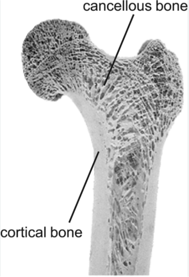
Compact Bone
(Cortical Bone) Dense outer layer of bone tissue. Provides strength and support to the skeleton. It is composed of functional units called OSTEONS (Haversian Systems) which are aligned parallel to the long axis of the bone and contain blood vessels and nerves

Haversian Canal
(Osteonic Canal , Central Canal) A microscopic channel within compact bone that houses blood vessels, nerves and lymphatic vessels providing essential nutrients and support to bone cells. Key component of the Haversian system (Osteon), which is the basic structural unit of compact bone.
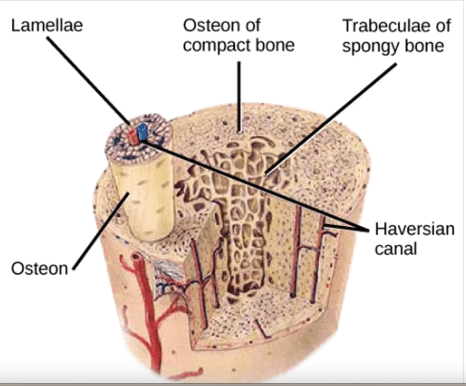
Osteon
(Haversian System) Fundamental structural and functional unit of compact bone. Appears as a cylindrical unit containing a central Haversian Canal surrounded by concentric layers of bone matrix called LAMELLAE where OSTEOCYTES reside in lacunae (bone cavities) connected by tiny canals (CANALICULI) that facilitate nutrient and waste exchange.
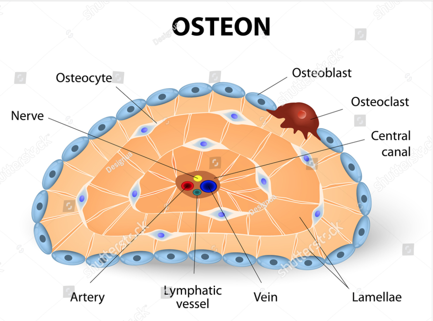
3 Types of Cells Present In Bone
Osteoblasts, Osteocytes, Osteoclasts
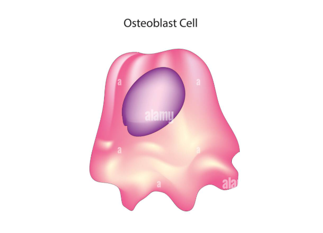
Osteoblasts
Specialized cells responsible for bone formation. They synthesize the organic matrix of bone (Osteoid) and then facilitate its mineralization ( the process where calcium and phosphate are deposited to create hardened bone. These cells play a crucial role both in the initial development of bone and the ongoing process of bone remodeling. As they age, they become osteocytes.
Osteocytes
Mature bone cells. The most abundant type of bone cell crucial for maintaining bone health. Derived from osteoblasts and reside within the mineralized bone matrix. They actively regulate bone remodeling and mineral homeostasis by sensing mechanical stimuli and communicating with other bone cells. Can revert back to osteoblasts
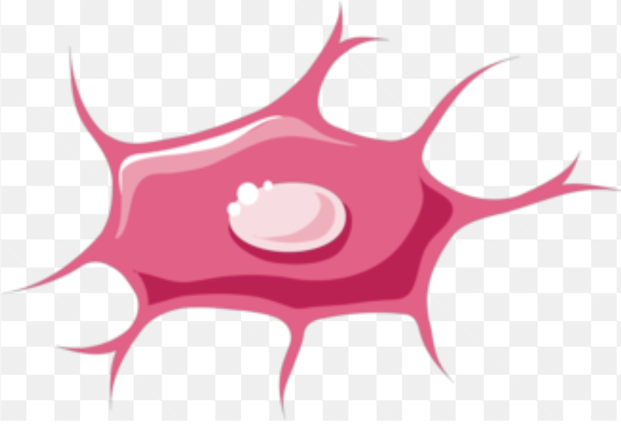
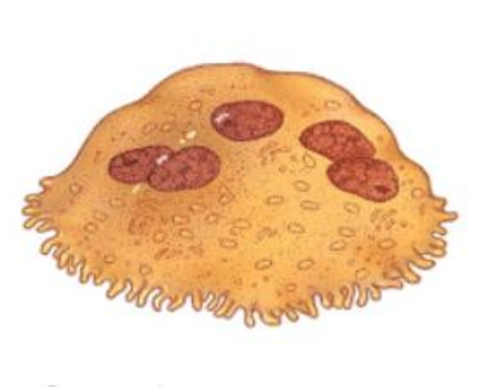
Osteoclasts
Specialized, large, multinucleated cells of hematopoietic origin that are primarily responsible for bone resorption (a crucial process in bone remodeling and maintenance of mineral levels). They originate from mononuclear cells in the hematopoietic lineage and possess a distinct ruffled border where they secrete enzymes and acids to break down bone tissue.
Hematopoietic
Refers to the process of blood cell formation (hematopoiesis) This process involves hematopoietic stem cells (HSCs) that reside primarily in the bone marrow and give rise to all mature blood cells, including red blood cells, white blood cells and platelets. Essential for maintaining a healthy circulatory system and immune function.
Periosteum
A dense, fibrous membrane covering the outer surface of bones except at the articular surfaces (parts with a joint space). Serves as an attachment point for muscles, tendons and ligaments. Inner layer contains osteoblasts Involved in the initial stages of bone fracture healing, contributing to the formation of new bone tissue
Stem Cells
Cells with the potential to develop into many different types of cells in the body. Able to renew themselves for long periods of time. Serve as a repair system for the body. Two main types of stem cells - Embryonic and Adult. Used for treating musculoskeletal injuries, mostly in horses and dogs.
Red Marrow
Primary site of blood cell production. Vital for diagnosing and understanding various conditions including anemias, leukemias, and certain infections and/or malignancies. The process of examining red marrow usually involves obtaining samples by aspiration or core biopsy which is then analyzed to assess cell types, abnormalities and overall marrow health.

Reticulin
Main intracellular material within bone. Reticulin fibers are composed of type III collagen secreted by reticular cells. They are mainly composed of reticulin protein and form a network or mesh. Reticular fibers crosslink to form a fine meshwork (reticulin) in which stem cells are interspersed. This network acts as a supporting mesh in soft tissues such as liver, bone marrow and the tissues and organs of the lymphatic system
Sinusoids
Vascular spaces (slightly larger than capillaries) within the bone marrow where blood cells enter as they mature. Sinusoids empty into veins that put newly formed blood cells into circulation.
Adipocytes
(Fat Cells) Crucial components of adipose tissue serving as energy storage and insulation and playing a significant role in endocrine functions and musculoskeletal repair through adipose derived stem cells. Adipose tissue, composed primarily of adipocytes, is recognized as a major endocrine organ secreting hormones like leptin, estrogen, resistin and TNF-a ( or TNF - Tumor necrosis factor). They replace stem cells as the body ages.
Resistin
Hormone studied for its roles in metabolism and inflammation.
Leptin
Hormone produced primarily by adipose tissue that plays a crucial role in regulating energy balance by influencing appetite and metabolism.
Adipose Tissue
(Body Fat) A specialized loose connective tissue composed of fat-storing cells called adipocytes. It serves multiple crucial functions, including energy storage, insulation, and cushioning for organs, and it also acts as an endocrine organ, secreting hormones and other bioactive molecules.
Yellow Marrow
Primarily known for its adipose storage and energy production. Large presence in older animals. It can convert back to red marrow under certain circumstances but its main function is not blood cell production. Found in the medullary cavities of long bones and the spaces of spongy bone.
White Bone Marrow
The active cellular portion of bone marrow responsible for producing white blood cells (leukocytes) which are crucial for the immune system.
Two Major Categories of Skeletal Bones
Axial Skeleton, Appendicular Skeleton
Axial Skeleton
Body’s central framework - skull, spine, ribs, sternum
Appendicular Skeleton
Bones of the limbs and limb girdles that are attached to the axial skeleton - legs shoulders and pelvis
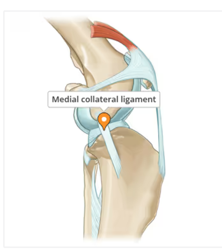
Ligaments
Strong, fibrous connective tissues that connect bone to bone providing stability and eliminating excessive movement in joints. Cranial Cruciate Ligament (CCL) rupture is a frequent orthopedic injury in dogs usually a degradation over time rather than a traumatic injury.
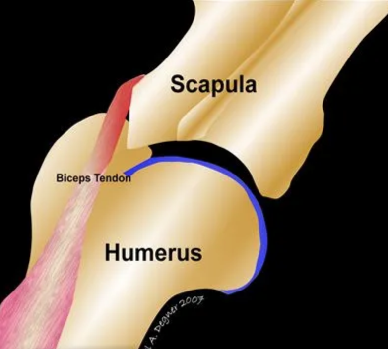
Tendons
Crucial, fibrous connective tissues that connect muscles to bones facilitating movement and absorbing forces. Composed primarily of collagen fibers and designed to transmit muscle forces to bones enabling locomotion and providing some protection to muscles against trauma
2 Areas of the Skull
Cranial Cavity. Facial Bones (nose, jaw, eye sockets)
Components of the Jaw
Maxilla (upper jaw) Mandible (lower jaw)
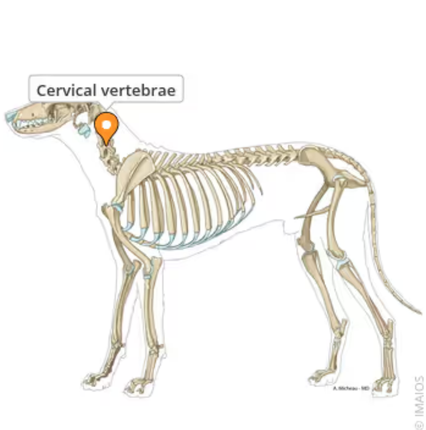
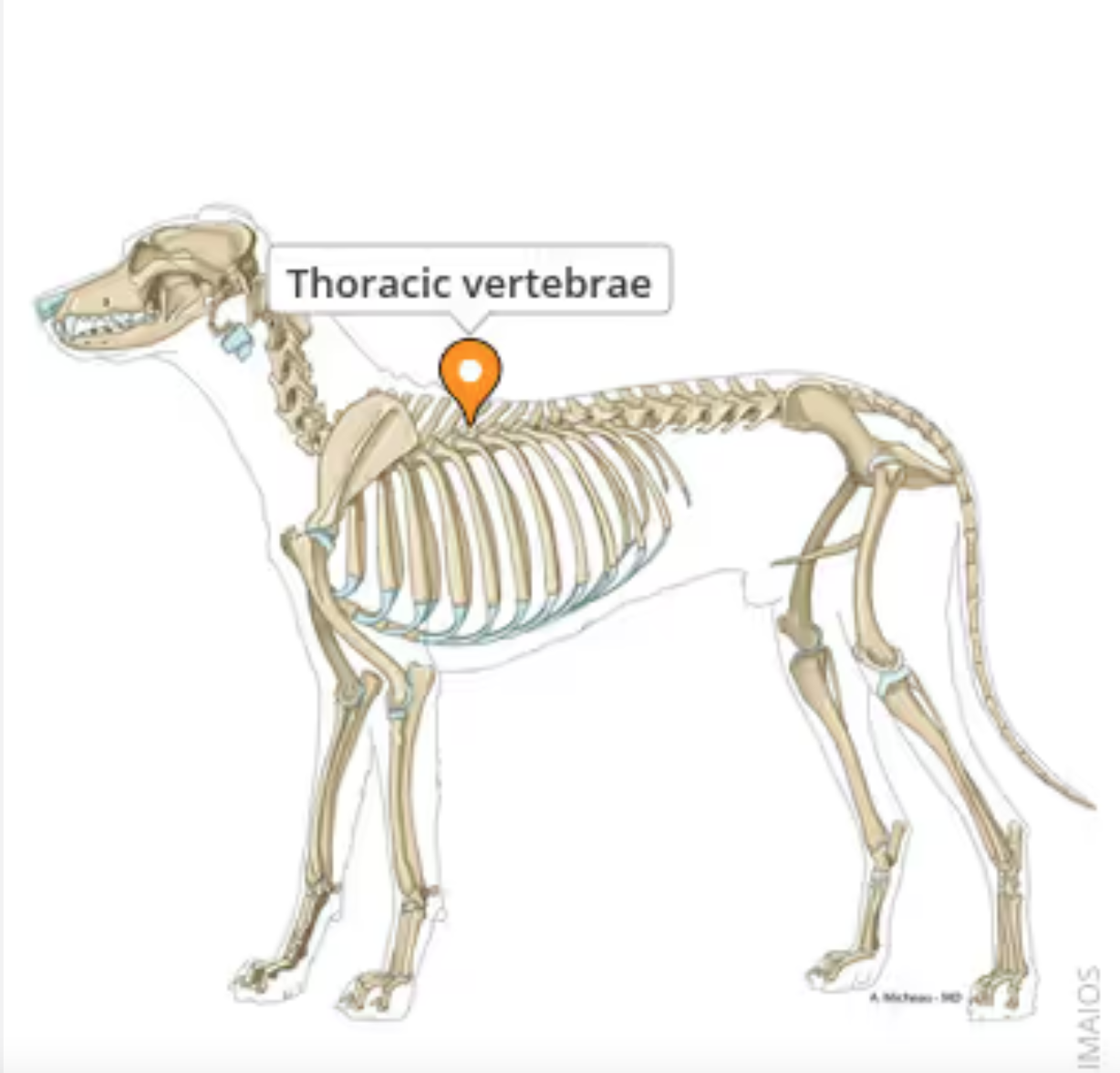
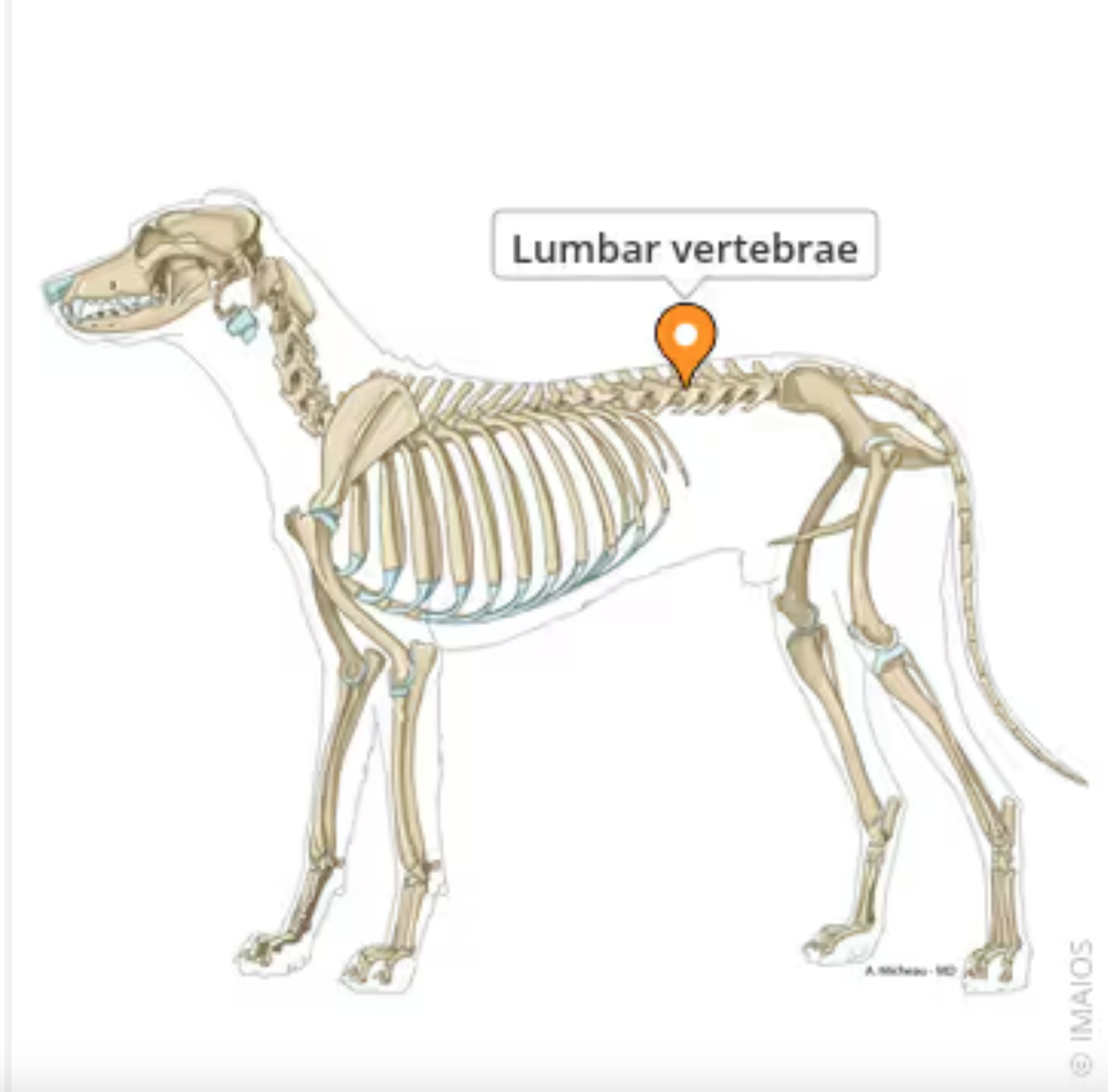
5 Areas of the Spine
Cervical (neck vertebrae), Thoracic (chest vertebrae), Lumbar (lower back vertebrae), Sacral (pelvic vertebrae), Coccygeal / Caudal (tail vertebrae)
Cervical Vertebrae
C1-C7 (all mammals)
Thoracic Vertebrae
Forms thoracic spine and articulates with the ribs. T1-T13 (carnivores, cows, sheep) , T1-T18 (horses), T1-T14 or T15 (pigs)
Lumbar Vertebrae
Bones that form the lower back region of the vertebral column. Distinct from the thoracic and sacral vertebrae due to their size, shape and lack of rib articulation.
L1-L6 ( horses, cows, some sheep, some pigs) , L1-L7 (dogs, cats, goats, other sheep, other pigs)
Sacral Vertebrae
Vertebrae that are fused together to form the sacrum, a single strong bone that articulates with the pelvic girdle and supports the weight of the body
S1-S5 (cows and horses), S1-S4 (pigs and sheep), S1-S3 (cats and dogs)
Coccygeal ( Caudal) Vertebrae
The bones that form the tail. Cd. Vary in number depending upon the size of the animal. In dogs and cats, the number of coccygeal vertebrae is variable with dogs typically having 20-23 and cats having fewer. These vertebrae gradually decrease in size toward the tip of the tail losing arches and processes and
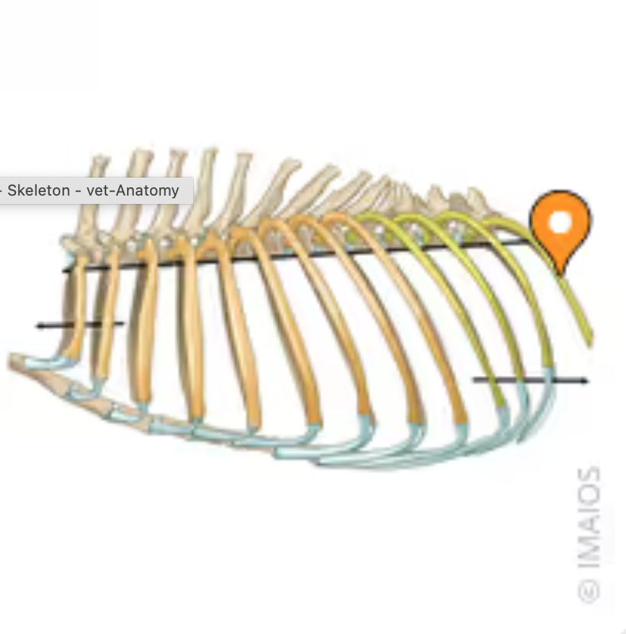
Ribs
(Costae) 12 pairs of long, curved, flat bones with a head that articulates with the caudal body of one thoracic vertebrae and the cranial body of the adjacent vertebrae. form a cage around the heart and lungs and assist in respiration
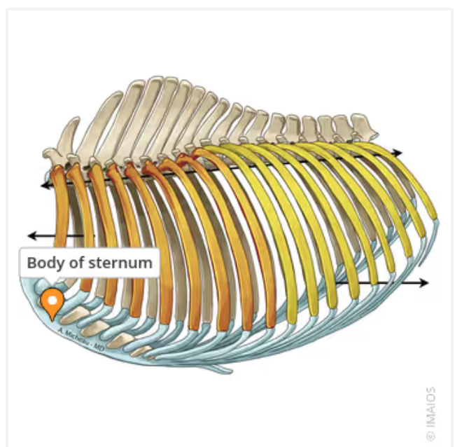
Sternum
Flat, dagger shaped bone located in the center of the chest. Helps protect the contents of the chest and assist in the breathing process.
Floating Ribs
The last two pairs of ribs. They do not attach to the sternum
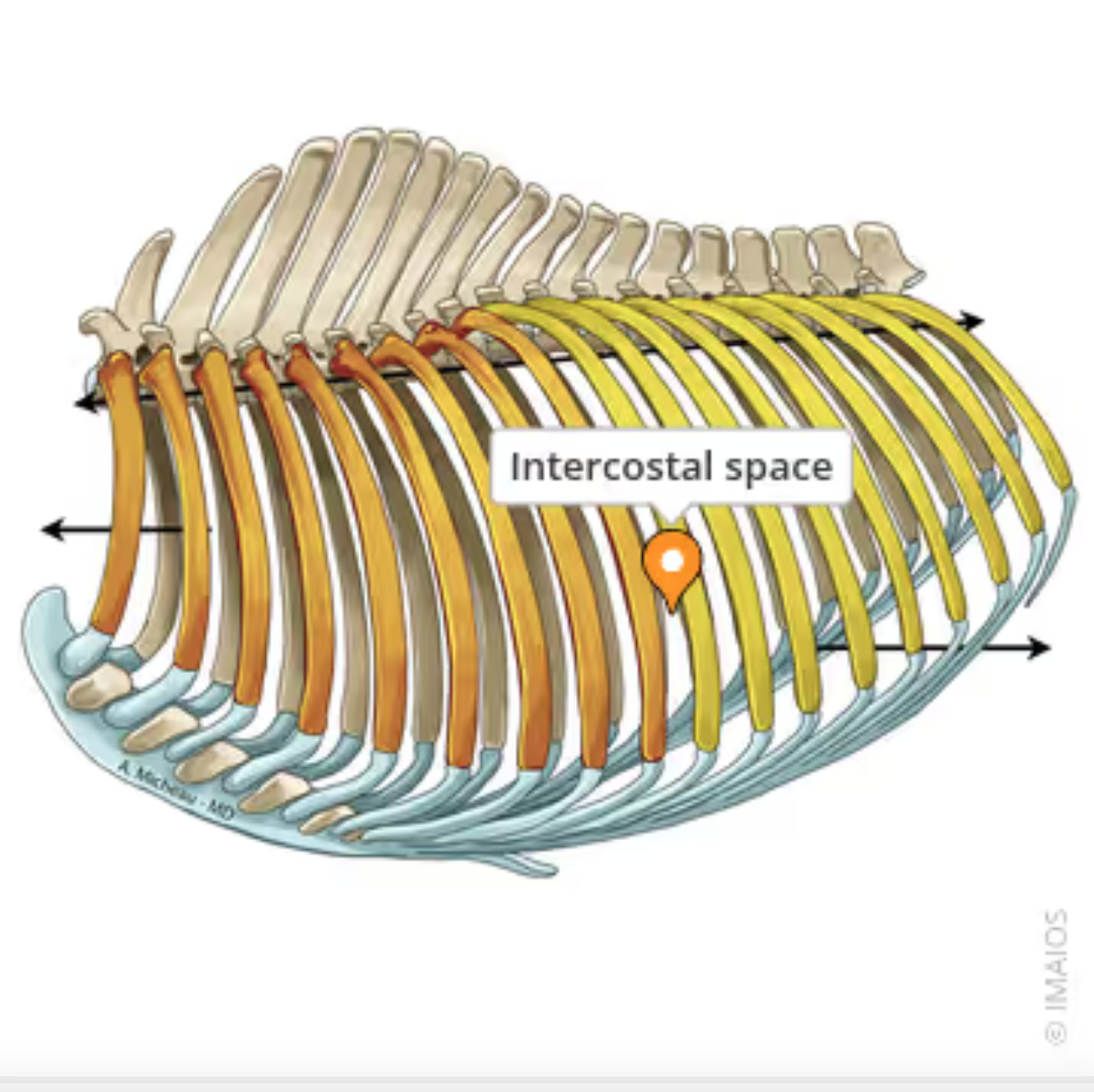
Intercostal Space
The anatomical area between two adjacent ribs housing intercostal muscles, blood vessels and nerves. Crucial for respiration and surgical access. Utilized for various veterinary procedures including exploration of the thoracic cavity, managing thoracic diseases, performing thoracic surgeries like intercostal thoracotomy and in diagnostic imaging such as ultrasound examinations.
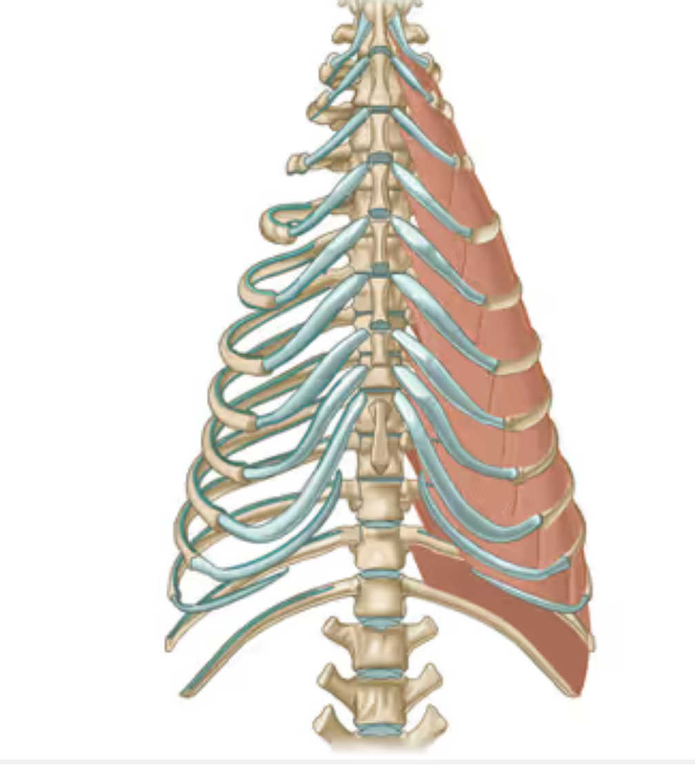
Intercostal Muscles
Muscles between the ribs crucial to respiration, comprised of internal and external layers. External layer assists in inhalation by elevating and expanding the ribs, which expands the thoracic cavity and reduces intrathoracic pressure and drawing air into the lungs. The internal layer assists in exhalation by pulling the ribs downward and compressing the thoracic cavity
Thoracic Limbs
Forelimbs
Pelvic Limbs
Back legs
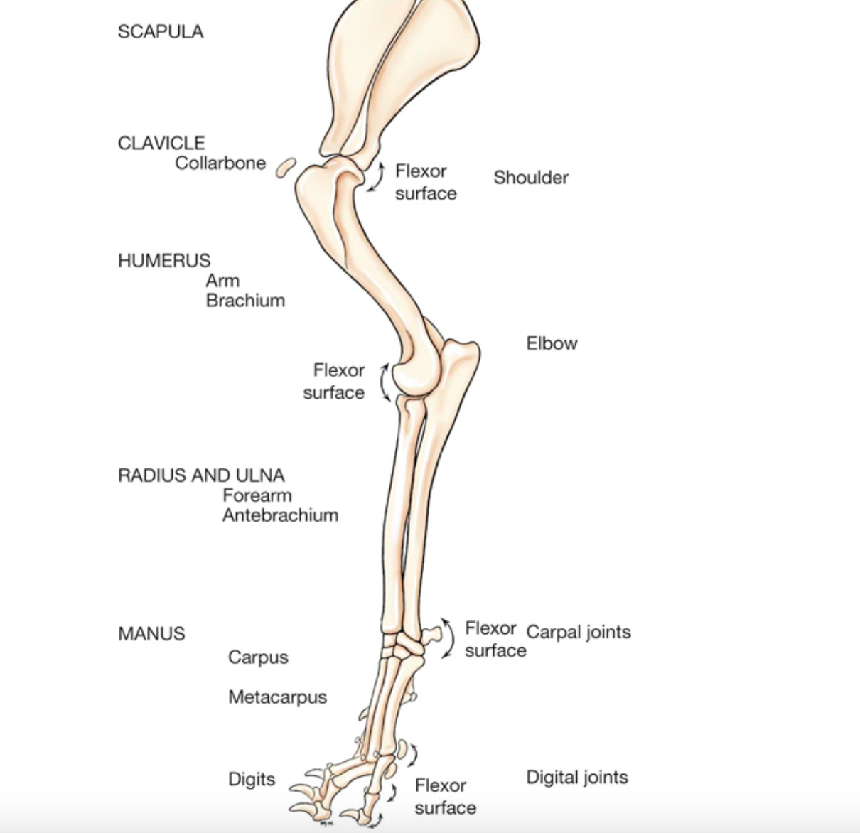
Bones of the thoracic limb
Scapula, humerus, radius, ulna, carpals, metacarpals, phalanges (digits)

Scapula
Shoulder blade. Attaches thoracic limb to the body’s dorsal surface. Articulates with the humerus at the shoulder joint. Medial side rests against the rib cage. Lateral side has a ridge (spine of the scapula) which serves as point of attachment for numerous muscles involved in shoulder and forelimb movement.
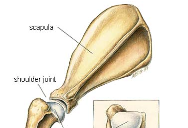
Humerus
The ‘single’ bone of the arm connecting the scapula at the shoulder joint and the radius and ulna at the elbow joint.
Radius and Ulna
Two ‘long bones’ of the forearm. Radius is the primary, weight bearing bone. Ulna supports less weight but its olecranon process (protruding part of the elbow) is crucial for the elbow joint.distinct bones in some animals (humans, dogs, cats, pigs) fused in others (ruminants, horses). distinct in carnivores to rotate the forearm (climbing, grasping). Fractures of these bones are common, especially in dogs and cats often resulting from trauma like falls or being hit by a car and typically require surgical stabilization.
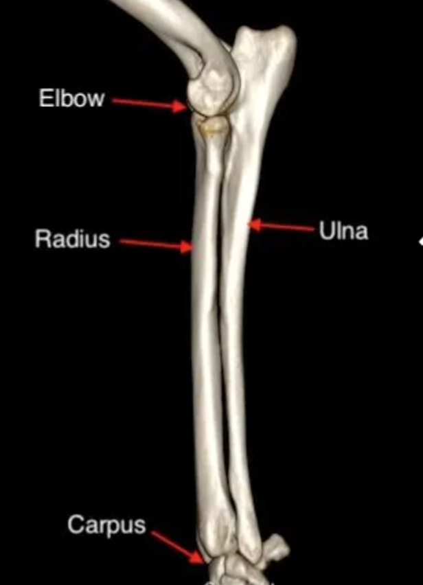
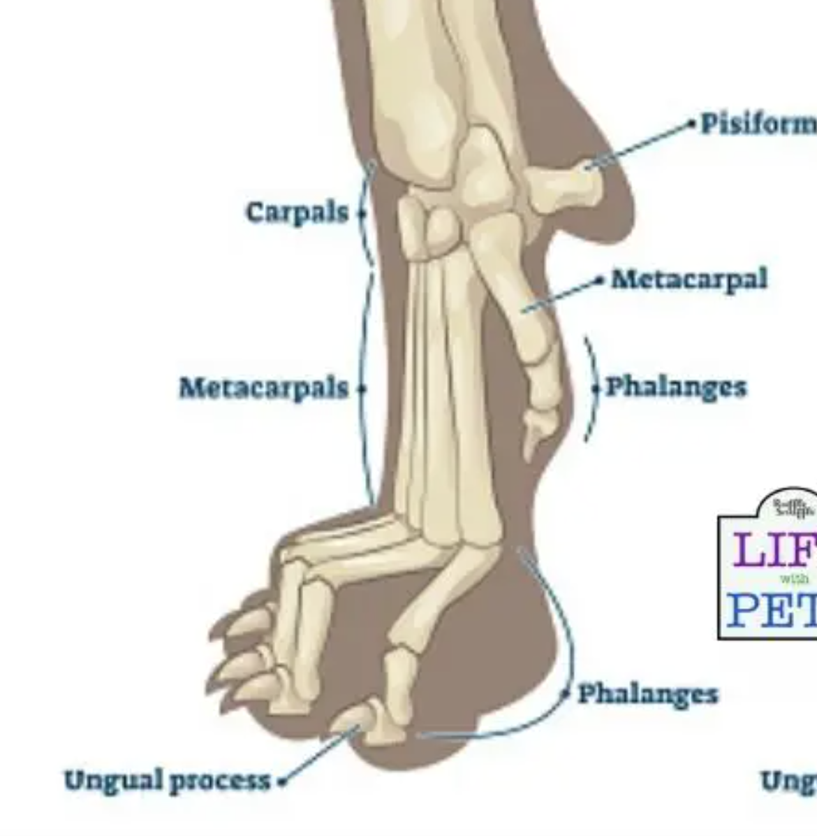
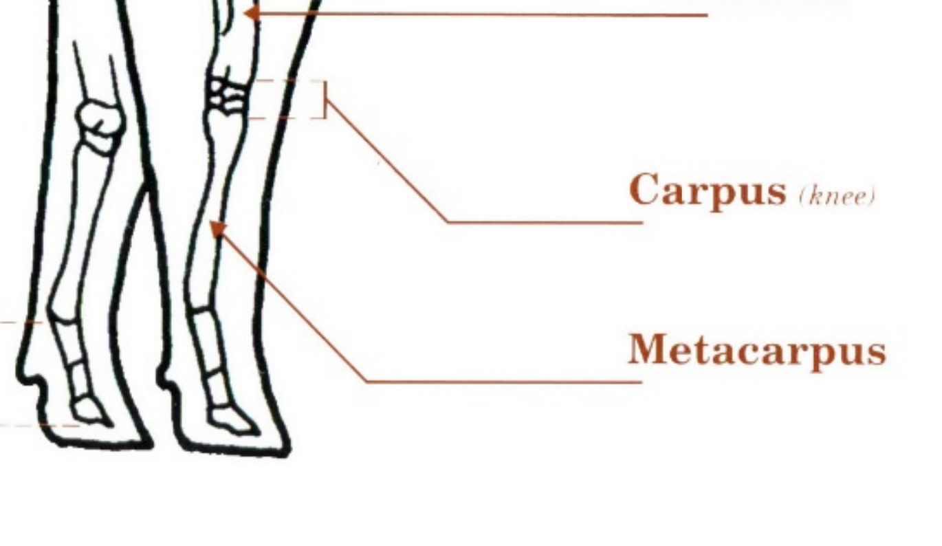
Carpals
the complex joint in the forelimb of animals, commonly known as the wrist or "knee" in some species, which is crucial for weight-bearing, flexibility, and locomotion. This anatomical region is composed of multiple small carpal bones arranged in proximal and distal rows, along with associated ligaments, tendons, and synovial joints, and is susceptible to injuries like fractures, sprains, and hyperextension due to trauma or overuse. Usually 8 bones (2 rows of 4). Some animals have one fused bone. Some have fewer
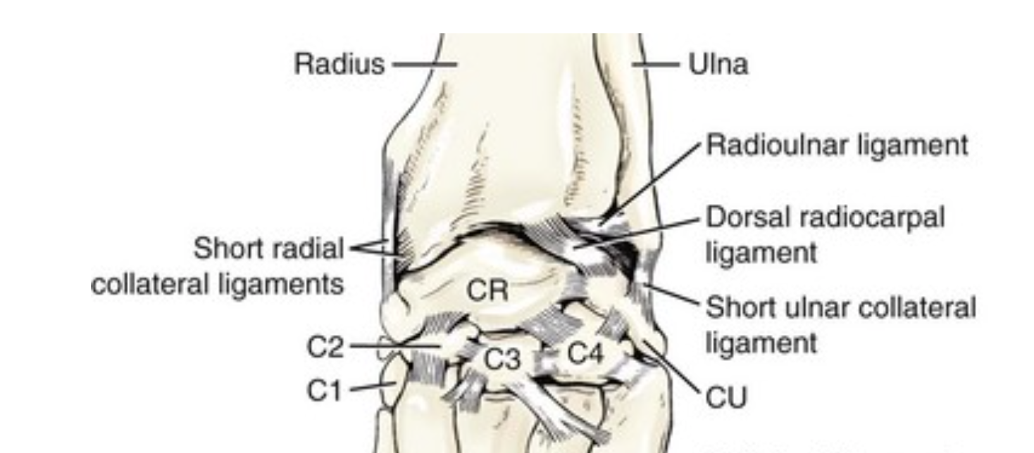
Metacarpals
Long bones that connect carpals to phalanges. Up to 5 in a row numbered 1 (most medial) to 5 (most lateral), Some are fused in some animals and may number less than 5.
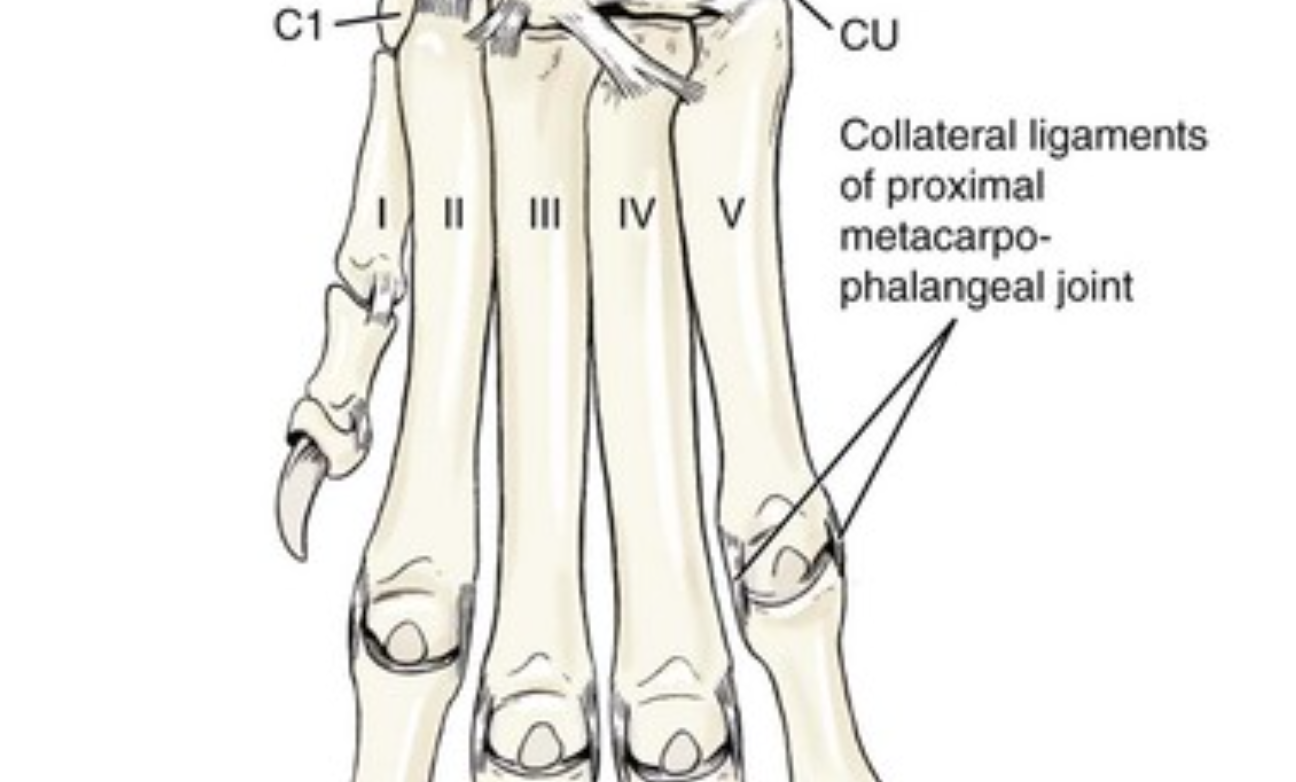
Dog and Cat Metacarpals
Dogs and Cats have 5 metacarpal bones
Digitigrade
Walking on toes and not touching the ground with heels
Pig Metacarpals
Pigs lack the first metacarpal bone. The third and fourth (two middle ones) are longer than the outermost (second and fifth) so that only the two middle toes are supporting the pig’s weight
Cow and Sheep Metacarpals
Cows and sheep lack the first and second metacarpal bones. The third and fourth are fused into one bone (the cannon bone) and the fifth metacarpal bone is very small
Horse Metacarpals
Horses lack the first and fifth metacarpal bones. The second and fourth (the splint bones) are smaller than the third (cannon bone) The third metacarpal is very large and supports all of the leg’s weight
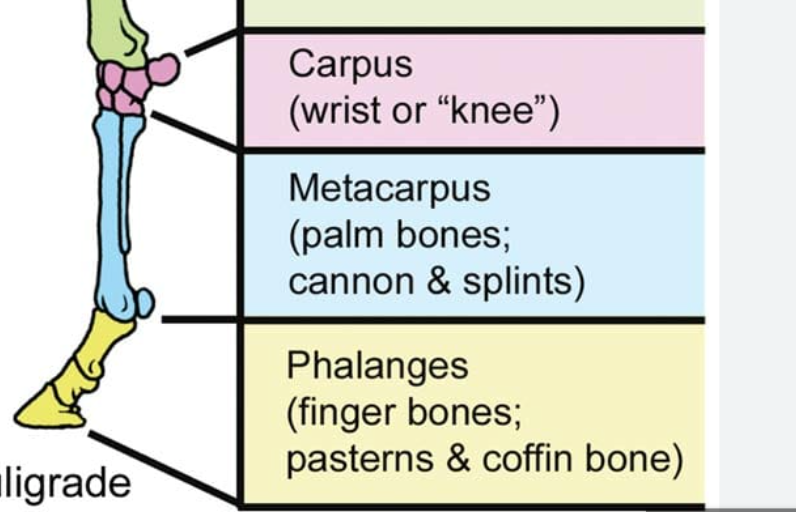
Phalanges
(singular “phalanx”) Each digit composed of 2 or 3 phalanges with up to 5 digits on each foot. Digits numbered 1 (most medial) to 5 (most lateral), In horses, phalanges are referred to as “pastern bone” (proximal), “short pastern bone” (middle), and “coffin bone” (distal)

Paradigits
Digits that don’t bear weight. Dewclaws

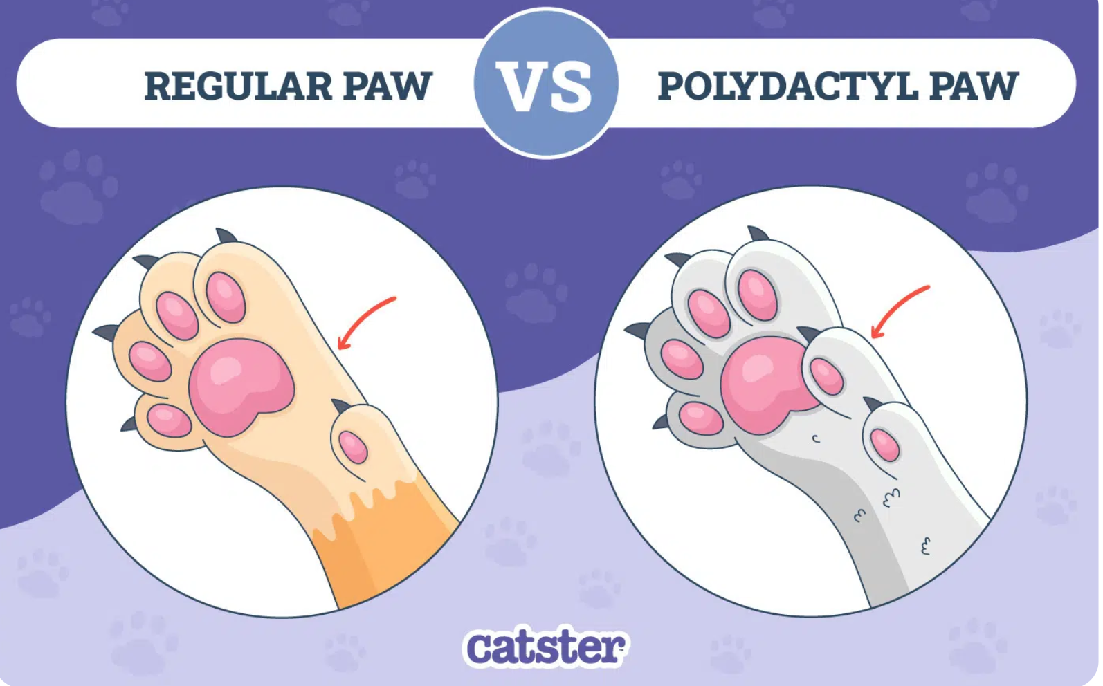
Polydactyly
The condition where an animal is born with extra toes on one or more paws. More common in cats (especially Maine Coons) but is sometimes seen in certain dog breeds like Great Pyrenees and Australian Shepherds. It is a dominant, autosomal trait. There are 3 types of polydactyly according to where the extra toes are found. POSTAXIAL (outside / lateral). PREAXIAL (inside / ventral) and MESOAXIAL ( middle )
Autosomal
In veterinary genetics, "autosomal" refers to traits or diseases determined by genes located on the non-sex chromosomes (autosomes), rather than the sex chromosomes (X and Y). Autosomal traits can be inherited in a dominant or recessive manner, affecting males and females equally.
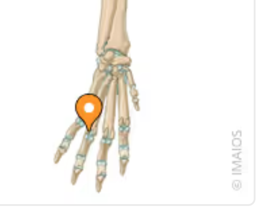
Sesamoid Bones
Small, bone-like structures embedded within tendons or muscles acting to reduce friction and increase leverage. Found in various locations in the limbs, often near joints. They help to improve the biomechanics of movement by altering the angle of tendons and reducing friction. In dogs - palmar sesamoid bones in the metacarpophalangeal joints and dorsal sesamoid bones in the extensor tendons. (Sesamoid Disease - a degenerative condition) is known in dogs, particularly larger breeds. In cats and larger dog breeds, a sesamoid bone can be found in the supinator muscle of the elbow joint.
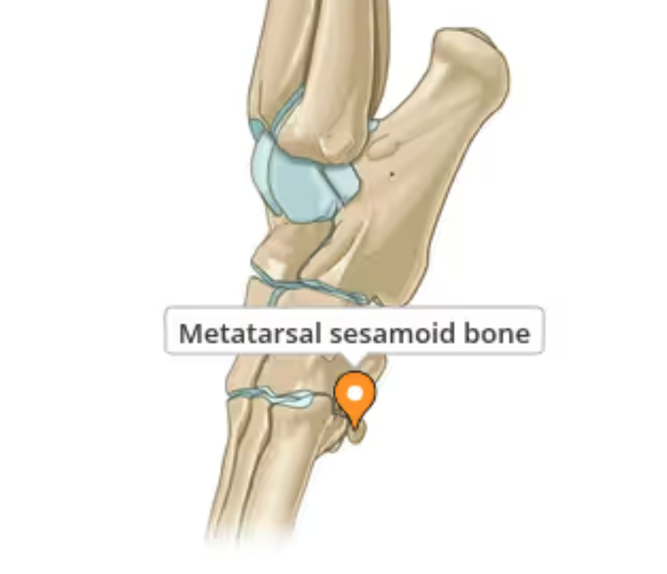
Proximal Sesamoid Bone
Located behind the joint and between the large metacarpal bone and the proximal phalanx joint in the large, digital, flexor tendons - or Fetlock Joint. Sometimes fracture in horses.
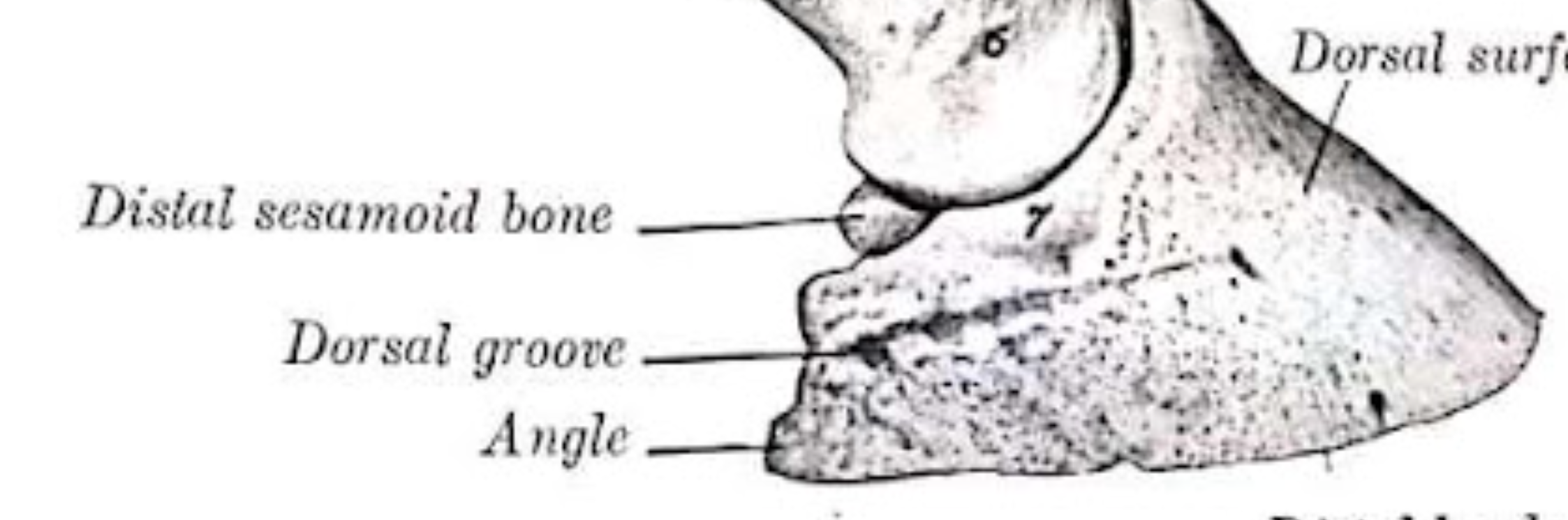
Distal Sesamoid Bone
“Navicular Bone”. Palmar surface behind the second phalanx ( P2) and in front of the third phalanx ( P3) - both forelimb and hindlimb. Rarely found in carnivores. Navicular Disease / Syndrome is a common condition in horses characterized by pain and lameness associated with the Navicular Bone and surrounding structures. It can involve inflammation, damage to the bone itself or problems with the deep digital flexor tendon (DDFT) or its bursa.
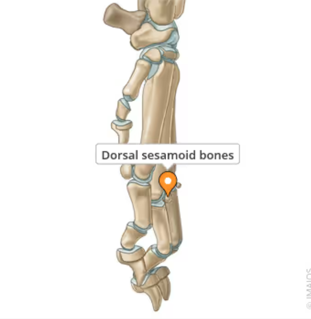
Dorsal Sesamoid Bones
small bones located on the dorsal surface of the paw, found within the extensor tendons of the metacarpophalangeal and metatarsophalangeal joints. In some animals (particularly dogs), the dorsal sesamoid bone in the carpus is a normal structure routinely found in radiographs. In other cases, they might be partialy or completely cartilaginous at birth.
Fundamental Bones of the Pelvic Limb
Ilium, Ischium, Pubis, Femur, Patella, Tibia, Fibula, Tarsal, Metatarsal, Phalanges
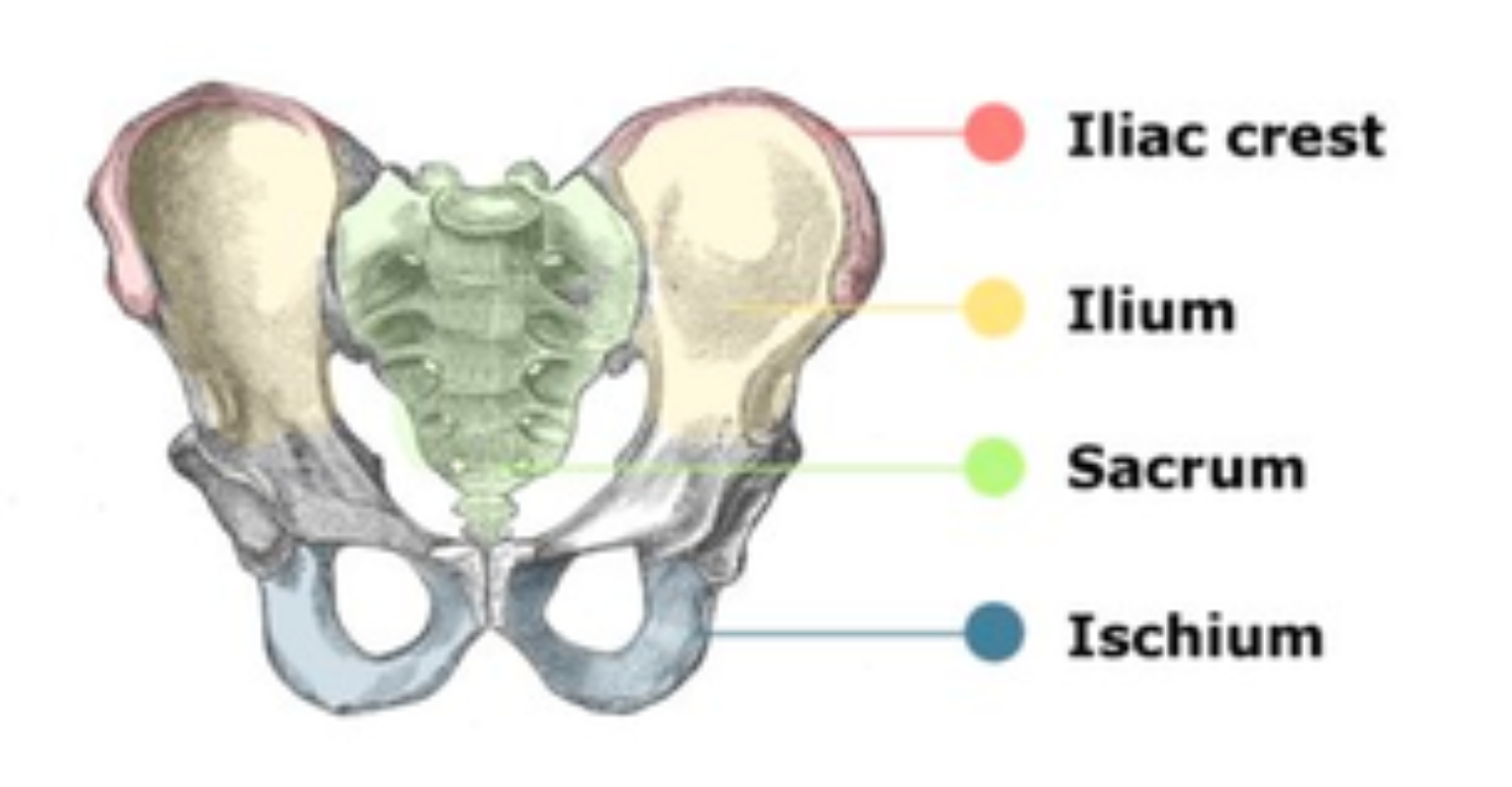
Pelvis
Girdle of bones that attach the spine to the pelvic limbs. Comprised of three pairs of bones - ilium, ischium, pubis (one of each pair on each side)

Ilium
Largest bone of the pelvis. Roughly flat, triangular bone that curves outward slightly at the cranial end.
Wing of the Ilium