Nucleus, cell cycle, apoptosis, autophagy, necrosis
1/16
There's no tags or description
Looks like no tags are added yet.
Name | Mastery | Learn | Test | Matching | Spaced |
|---|
No study sessions yet.
17 Terms
Nucles
contains genome
can display different shapes
is not present during the whole cell cycle
can be missing in some “cells” (blood red cells)
more than one nucleus can be present in a cell (e.g.syncytium *)
Nucleus structure
enveloped by a double-bilayer membrane - delimiting the perinuclear space called nuclear envelope.
consists of an inner and an outer membrane separated by a perinuclear cisternal space and perforated by nuclear pores
The outer membrane of the nuclear envelope is continuous with that of the rough-surfaced endoplasmic reticulum (rER) and is
often studded with ribosomes.
Chromatin
nuclear material organised as euchromatin or heterochromatin contains DNA associated with roughly and equal mass of various nuclear proteins - necessary for DNA to function
Nuclear lamina
formed by intermediate filaments and lies adjacent to the inner nuclear membrane
Function
supports the nucleoskeleton
Essential in processes like:
DNA replication
Transcription
Gene regulation
Major component - nuclear lamins - specialised intermediate filament and lamin-associated proteins
Nuclear pores
70 to 80 nm openings
Formed by merging the inner and outer membranes of the nuclear envelope
Forms nuclear pore complex - close-fitting or gated channel
Proteins directed to nucleus present specific signal sequences for nuclear import: Nuclear localisation signals (NLS)
Nucleolus
non membranous region of the nucleus
Granular appearance
surround transcriptionally active rRNA genes
free in nucleoplasm
Primary site - for ribosome production and assembly
Varies in size - but particularly well developed in cells in protein synthesis
Cell cycle
period of time between to cell division
Can last from hours to days
Yeast and bacteria: 2-4 hours
Intestinal, epithelial cells, erythroblast: 12 hours
Hepatocytes: 1 year
Neurones and muscle tissue never divide during adult life, howeve,r they contain stem cells that in case
STAGES
G0 - Quiescence, resting phase for undefined time. Cell division can be activated via internal stimuli or exogenous growth factors (damages for example).
G1 (8 hours) - Cellular growth, protein synthesis and duplication of organelles. The cell is preparing for division.
S (7-10 hours) - DNA duplication
G2 (2-5 hours) - Microtubules and mitotic spindle appearing, synthesis of cytoplasm components 5. M (1-2 hours) - Mitosis
Prophase
begins as the replicated chromosomes condense and become visible.
chromosomes continue to condense, each of the four chromosomes derived from each homologous pair can be seen to consist of two chromatids.
late prophase - the nuclear envelope begins to disintegrate into small transport vesicles and resembles the sER. The nucleolus, which may still be present in some cells, also completely disappears
Microtubules of the developing mitotic spindle attach to the chromosomes.
Telophase
marked reconstitution of a nuclear envelope around the chromosomes at each pole. The chromosomes uncoil and become indistinct except at regions that will remain condensed in the interphase nucleus.
The nucleoli reappear, and the cytoplasm divides (cytokinesis) to form two daughter cells. The separation at the cleavage furrow is achieved by a contractile ring consisting of a very thin array of actin filaments positioned around the perimeter of the cell.
Myosin II molecules are assembled into small filaments that interact with the actin filaments, causing the ring to contract. As the ring tightens, the cell is pinched into two daughter cells, genetically identical and containing the same kind and number of chromosomes. The daughter cells are (2d) in DNA content and (2n) in chromosome number.
3. Anaphase
begins at the initial separation of sister chromatids.
occurs when the cohesins that have been holding the chromatids together break down.
chromatin begin to separate and are pulled to opposite poles of the cell by the molecular motors (dyneins) sliding along the kinetochore microtubules toward the MTOC.
2. Metaphase
mitotic spindle, consisting of three types of microtubules, becomes organis around the microtubule-organising centres (MTOCs) located at opposite poles of the cell.
Microtubule are pulled toward the MTOC, where additional microtubules will attach. Microtubules and their associated motor proteins direct the movement of the chromosomes to a plane in the middle of the cell, the equatorial or metaphase plate.
Meisosi
Carried out by gonads - only cells that propagate genetic information to next generation
two nuclear divisions - followed by cell divisions that produce gametes
gamete - contains half the DNA and chromosomes found in Somatic cells
2 successive mitotic divisions without the S phase between the divisions
During the S phase that precedes meiosis, DNA is replicated forming sister chromatids (two parallel strands of DNA) joined together by
the centromere.
The DNA content becomes (4d), but the chromosome number remains the same (2n). The cells then undergo a reductional division (meiosis I) and an equatorial division (meiosis II).
Reduce chromosome number from diploid (2n) to haploid (n)
Ensure that each daughter cell has one
full set of chromosomes
Promote genetic diversity
Cell renewal
Static cell populations
consist of cells that no longer divide (postmitotic cells), such as cells of the central nervous system and skeletal or cardiac muscle cells.
Under certain circumstances, some of these cells (cardiac myocytes) may enter mitotic division.
Stable cell populations
consist of cells that divide episodically and slowly to maintain normal tissue or organ structure.
These cells may be stimulated by injury to become more mitotically active. Periosteal and perichondrial cells, smooth muscle cells, endothelial cells of blood vessels, and fibroblasts of the connective tissue may be included in this category.
Renewing cell populations
may be slowly or rapidly renewing but display regular mitotic activity. Division of such cells usually results in two daughter cells that differentiate both morphologically and functionally or two cells that remain as stem cells.
Daughter cells may divide one or more times before their mature state is reached. The differentiated cell may ultimately be lost from the body.
Types of tissue populations
Slowly renewing populations include smooth muscle cells of most hollow organs, fibroblasts of the uterine wall, and epithelial cells of the lens of the eye. Slowly renewing populations may actually slowly increase in size during life, as do the smooth muscle cells of the gastrointestinal tract and the epithelial cells of the lens.
• Rapidly renewing populations include blood cells, epithelial cells and dermal fibroblasts of the skin, and the epithelial cells and subepithelial fibroblasts of the mucosal lining of the alimentary tract.
These are differentiated cells, that differ from stem cells: after a stem cell becomes activated, it replicates into two daughter cells, one will go back to the rest (maintaining the pool stable) and the one will actually differentiate.
Also, the maintenance of the number of cells in a tissue is based on a balance of different processes: proliferation, differentiation, cell death. 18
Apoptosis
programed cell death
Mode of cell that occurs under normal conditions
Cells undergoing apoptosis show the
following characteristic morphologic and biochemical features:
DNA fragmentation - cut within nucleosomes
Cytoplasm modification - release of cytochrome C from mitochondria - activation of enzymes able to digest several cytoplasmic and nuclear substrates
Destruction of mitochondria physiology - depolarization of mitochondrial membrane potential - alteration of membrane permeability with release of cytochrome C in cytoplasm
Decreased cell dimension
Modification of plasma membrane - distribution of membrane phospholipids id modified - phosphatidylserine - translates from the cytoplasmic side to extracellular face of plasma membrane
Necrosis
cell death after injury
cell swells
cell lysis - break leading to tissue inflammation
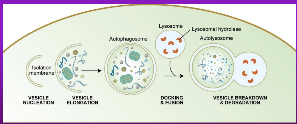
Autophagy
Role
get ride of aged and damaged organelles and molecules
autophagy generates fuel and re-utilises components for cellular renewal and repair
Autophagy - helps in the elimination of pathogens
Involved in protection against neoplastic transformation and other diseases
Process
Creation of phagophore - vesicle formed during initial phase of autophagy - engulf cytoplasmic components
Develop into autophagosome - an organelle that encloses parts of cytoplasm into a membrane
fuses with lysosome -releasess lysosomal hydrolase - content is degraded