Nervous System and Conditions
 Call Kai
 Learn
Learn Practice Test
 Spaced Repetition
 Match
Match Flashcards
 Knowt Play
1/26

There's no tags or description
Looks like no tags are added yet.
Name | Mastery | Learn | Test | Matching | Spaced | Call with Kai | Chat |
|---|
No analytics yet
Send a link to your students to track their progress
27 Terms
What are the three main functions of the Nervous system?
Sensory Function (perception)
Information Transfer and Processing
Motor function
Describe how the nervous system is seperated
Split into the Central (CNS) and Peripheral (PNS).
CNS includes brain and spinal cord. Receives information from nerves, analyses and processes this information and responds by sending impulses via nerves across the body to have an effect
PNS is network of peripheral nerves originating from brain or spinal cord.
The PNS is then separated into the Sensory (afferent) and Motor (efferent) division.
Sensory (afferent) division transmits impulses from the body to the CNS. It is composed of sensory receptors in the skin. Their nerve impulses are conveyed by somatic sensory fibres, as well as internally all around the body.
Motor (efferent) division transmits impulses from CNS to the body.
The Motor (efferent) division is again split into the Somatic and Autonomic divisions.
Autonomic (controls self regulation action of internal organs and glands)
Somatic (controls voluntary movement of skeletal muscles).
The Autonomic System is ALSO split into the Sympathetic (fight or flight) and Parasympathetic (rest and digest). Also the enteric division (neurons in GI tract, pancreas and gall bladder enable monitoring and modification of motility and secretions of the gut a controls diameter of blood vessels there).
What is a Neuron and what does it consist of?
The cells in the nervous system that generate electrochemical impulses enabling nervous system to function.
Consists of:
One Cell Body (where nucleus and other cellular organelles found)
Many Dendrites (receiving or input portions)
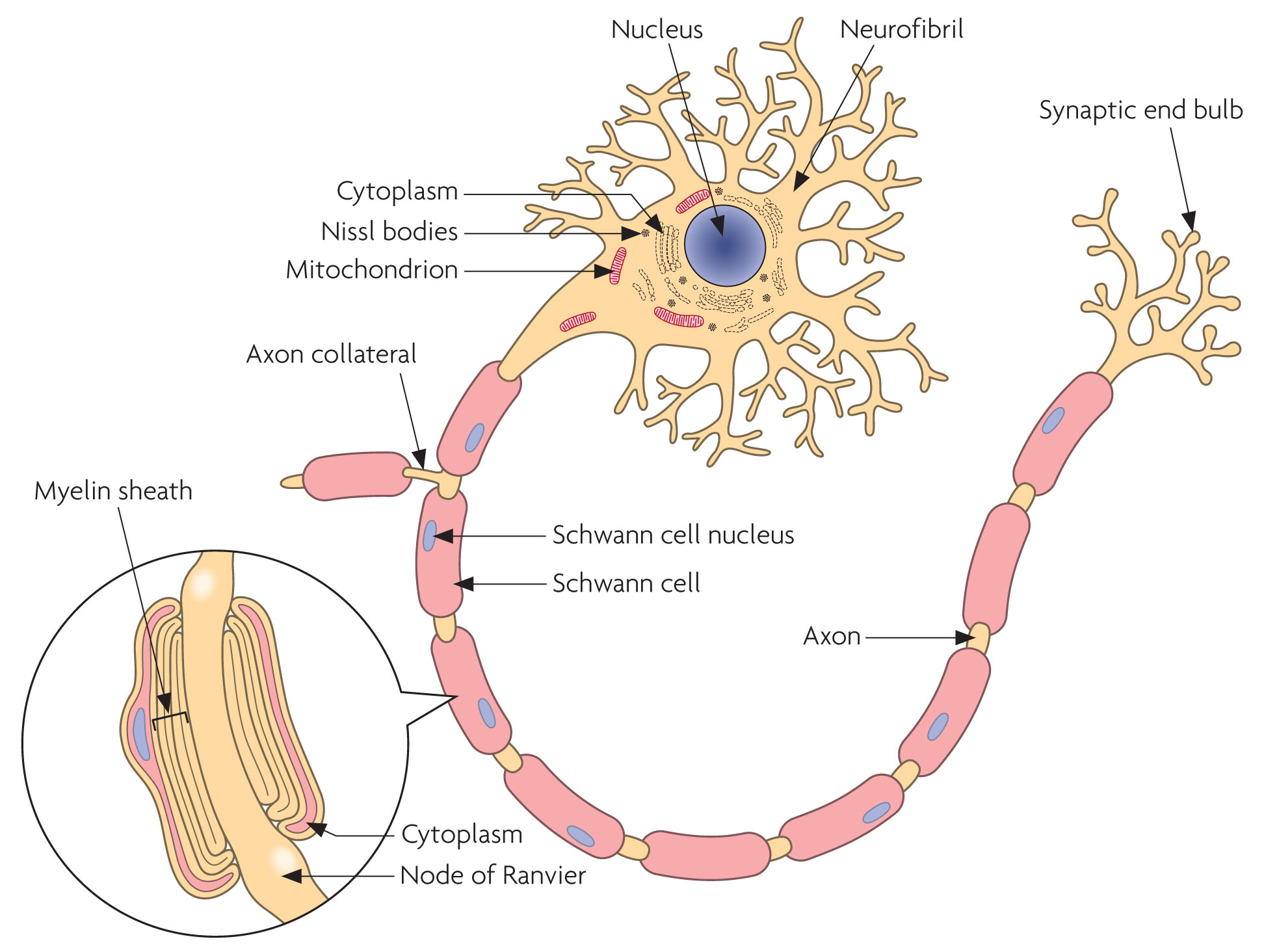
One Axon (thin, long portion transmits electrical signals from cell body to axon terminals. Neurons communicate with each other via chemicals called neurotransmitters, which are released from the synaptic end bulbs at the end of the axon.
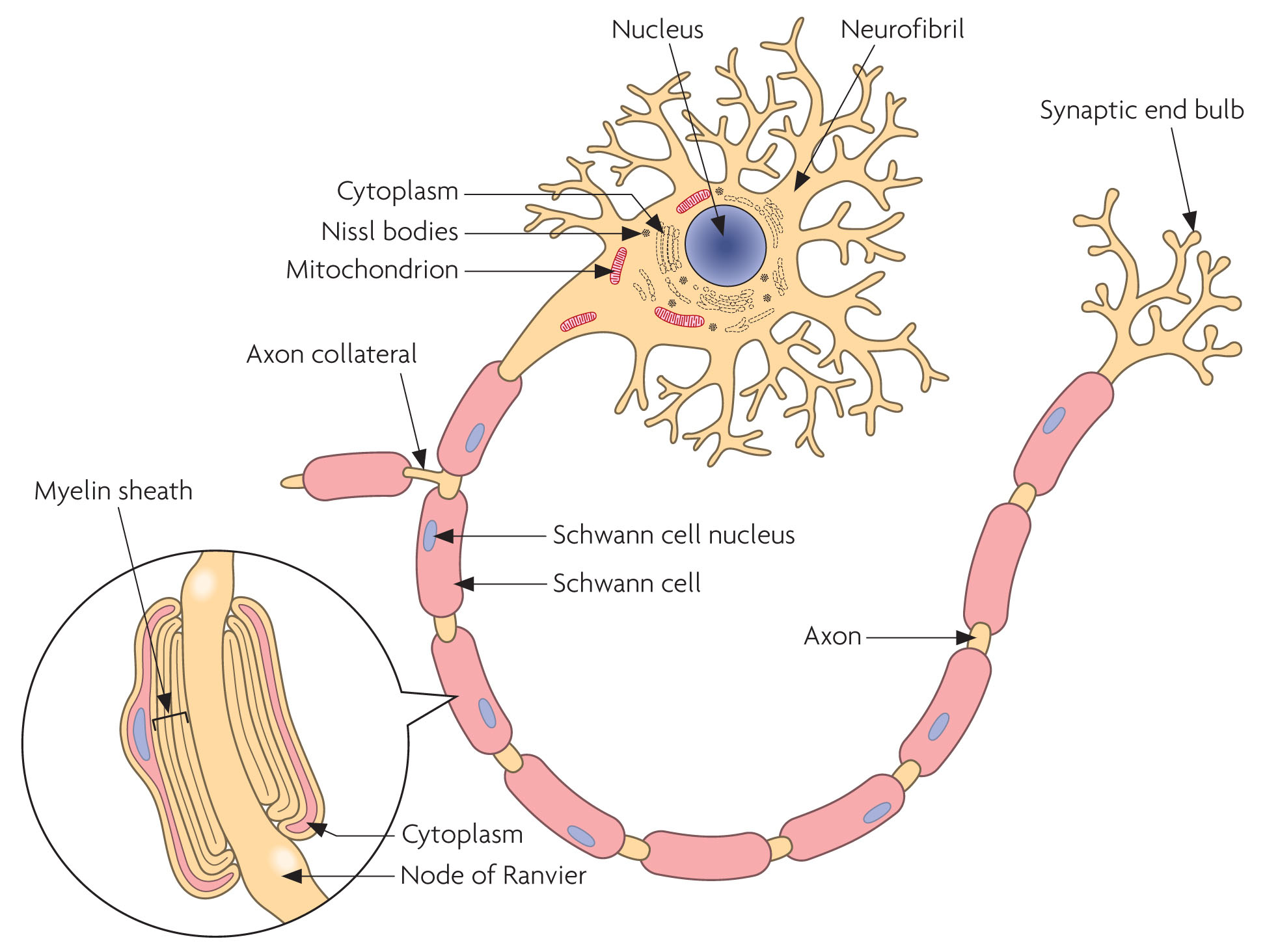
What are Myelin Sheaths and Nodes of Ranvier?
Some axons are covered in myelin sheaths which can conduct electrical impulses much faster than those that do not, and also helps to protect the nerve cells.
Myelin sheaths are formed by concentric Schwann cells. Nodes of Ranvier sit in between segments of Myelin Sheath to assist in the rapid transmission of nerve impulses.
What are Neuroglia?
The supporting, non impulse generating cels in the nervous system:
Provide structural support for neurons
Create blood brain barrier
Restrict movement of substances between blood and tissue fluids within the CNS
What is the cranial vault occupied by and what are the four major sections the brain is divided into?
Cranial vault occupied by
Brain 80%
Cerebral blood 12%
Cerebral Spinal Fluid 8%
Four major sections of the brain
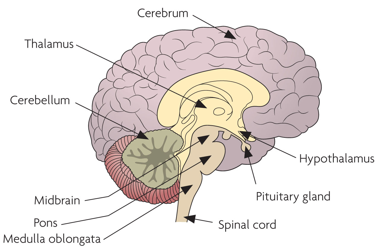
Brain stem (Midbrain, Pons, Medulla oblongata)
Cerebellum
Diencephalon (Consists of Thalamus and Hypothalamus)
Cerebrum
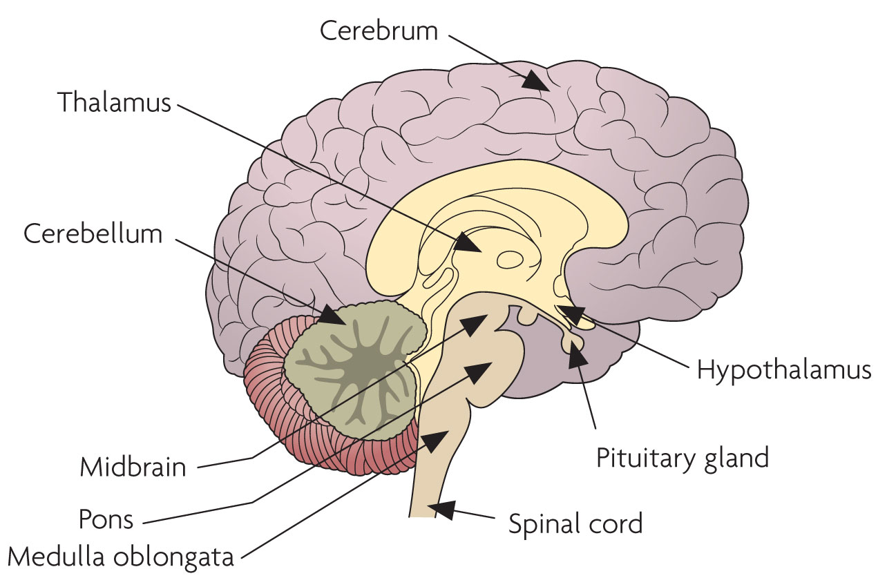
Describe the structure of the Brain Stem and what each component of the brain stem is responsible for
Consists of, moving from top to bottom:
Midbrain:
Relays motor output from cerebral cortex and sensory input from spinal cord to thalamus
Controls and coordinates movement
Contains origin of two cranial nerves
Pons:
Relays nervous impulses from one side of cerebellum to other and between medulla and midbrain
Contains origins of a number of cranial nerves
Contains centres to regulate breathing
Medulla Oblongata
Relays sensory and motor input between other parts of brain and spinal cord
Together with pons and midbrain, controls level of consciousness
Contains centres to manage HR, BP and Breathing
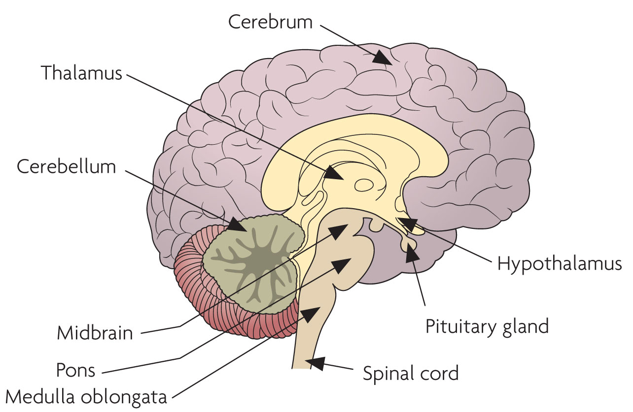
Describe the structure and function of the Cerebellum
Hemispheric, located posterior to brain stem and inferior to cerebrum
Responsible for coordinating complex and skilled movements
Regulates posture and balance
Describe the structure and function of the Diencephalon
Located deep within brain under cerebrum
Connects cerebrum with brain stem
Link between nervous system and endocrine system
Comprised of thalamus and hypothalamus
What is the function of the Thalamus
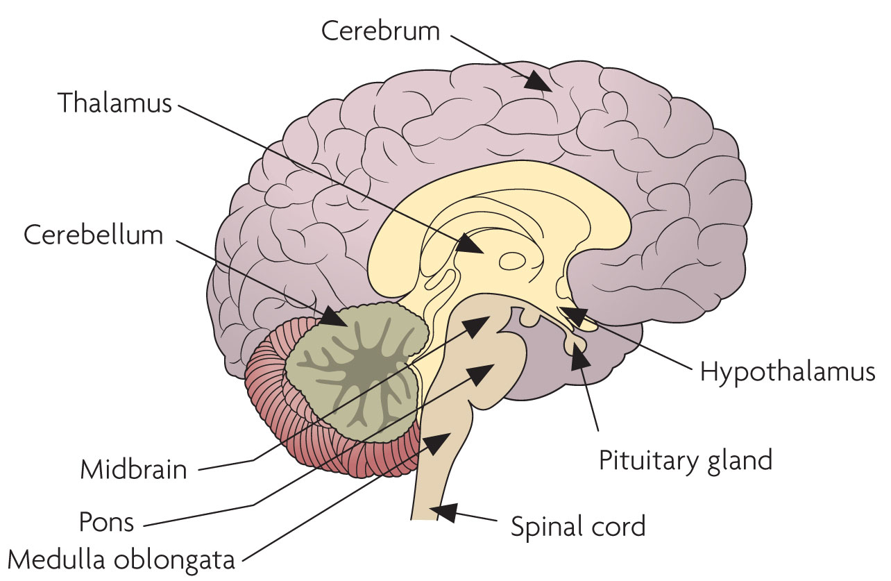
relays sensory input to the cerebral cortex
Provides perception of touch, pressure, pain and temperature
Describe the structure and function of the Cerebrum
Hemispheric, largest part of the brain
Outer rim of grey matter (contains the cerebral cortex which contains billions of neurons) and internal region of white matter
Split into four lobes
Frontal
Parietal
Temporal
Occipital
Allows you to read, write, speak
Various regions responsible for a range of other functions, including sensory perception, motor control, personality, memory and intelligence
Describe the structure and function of the Cerebrum
Hemispheric, largest part of the brain
Outer rim of grey matter (contains the cerebral cortex which contains billions of neurons) and internal region of white matter
Split into four lobes
Frontal
Parietal
Temporal
Occipital
Allows you to read, write, speak
Various regions responsible for a range of other functions, including sensory perception, motor control, personality, memory and intelligence
What is CSF?
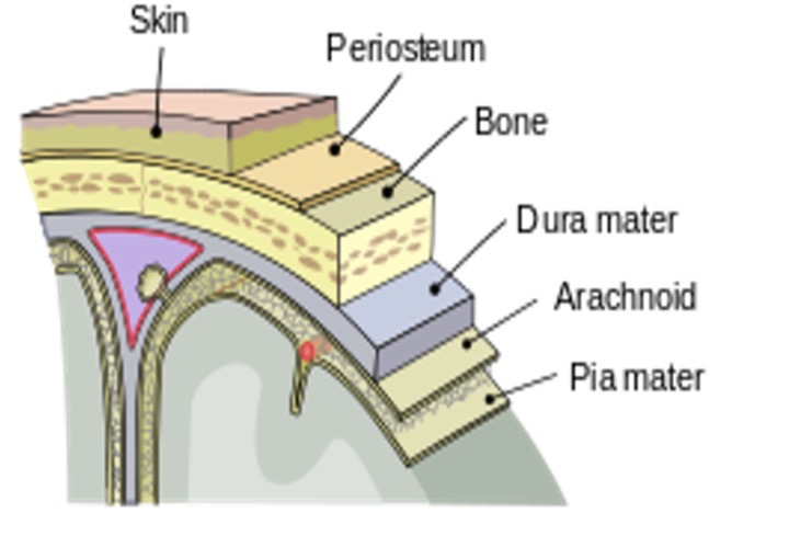
Cerebrospinal Fluid
Clear, colourless fluid
Protects brain and spinal cords from physical and chemical damage
Carries O2, glucose and other nutrients essential for normal brain function
Removes waste products
What is CSF?
Cerebrospinal Fluid
Clear, colourless fluid
Protects brain and spinal cords from physical and chemical damage
Carries O2, glucose and other nutrients essential for normal brain function
Removes waste products
Describe the spinal cord and spinal nerves
Spinal Cord:
Consists of a vertebral column consisting of 33 vertebrae, providing physical protection for spinal cord
The cord itself is cylindrical in shape, with a average diameter of 12mm
Extends from medulla oblongata to superior border of 2nd lumbar vertebrae, where it tapers to form a cone like shape called the conus medullaris. The nerves extending distal from the conus medullaris form the cauda equine (horses tail)
Contains both afferent and efferent fibres
Spinal nerves
31 pairs of spinal nerves branch out from the spinal cord
Describe the spinal cord and spinal nerves
Spinal Cord:
Consists of a vertebral column consisting of 33 vertebrae, providing physical protection for spinal cord
The cord itself is cylindrical in shape, with a average diameter of 12mm
Extends from medulla oblongata to superior border of 2nd lumbar vertebrae, where it tapers to form a cone like shape called the conus medullaris. The nerves extending distal from the conus medullaris form the cauda equine (horses tail)
Contains both afferent and efferent fibres
Spinal nerves
31 pairs of spinal nerves branch out from the spinal cord
What does the Sympathetic Nervous System do?
‘Fight or flight” prepares body to respond to stress and facilitate sudden strenuous exerciser and increased vigilance
Stimulates cardiac activity
Opens the airways
Dilates skeletal blood vessels
Reduces gastric activity
What does the Parasympathetic Nervous System do?
‘Rest and digest”
General ‘peacemaker’ effect
Stimulates digestion and obstruction of food
Slows down cardiac and respiratory activities
Describe Dementia
Disease that affects memory, thinking and daily life
1/14 over age of 65 have it
Risk groups are age, genetics, lifestyle
Controlled by medication but no known cure
Includes Alzheimer’s
Describe Parkinson’s Disease
Causes parts of brain to become damaged over many years
1/500 in UK
Men slightly more at risk of developing, though unclear what causes condition to occur
Treated with medication and physiotherapy
Describe Multiple Sclerosis (MS)
Affects brain and spinal cord, stopping nerve impulses from travelling correctly
Over 130000 in UK
Affects mainly woman and common disability in younger adults
Managed by medication but no cure
Describe Ataxia
Group of disorders affecting balance, coordination, speech
Mainly hereditary but can develop later in life
No cure, responds well to physiotherapy and medication
Describe paralysis
Defined as partial or complete loss of muscles function
Other terms include
-plegia (paralysis)
-paresis (muscle weakness or incomplete loss of muscle function)
Terms used to identify areas affected
Monoplegia - one limb
Hemiplegia - one side of body
Quadriplegia/tetrapelgia - all 4 limbs
Paraplegia - both lower limbs
What is a CVE and describe the different types and how they are differentiated
Cerebrovascular Event (CVE)
Stroke or TIA
Risk factors include Age, Sex, Ethnicity, Family Hx, Smoking, AF, Diabetes, Obesity, previous CVE or MI, Cholesterol, Diet, Hypertension
Stroke
Blood supply to brain interrupted
Signs and symptoms develop rapidly, last at least 24 hours
Bleed or blockage
TIA
Signs and symptoms of stroke but resolves within 24hrs
Anyone with continuing neurological signs during management - treat as stroke
Signs and symptoms
Numbness
Weakness or paralysis
Slurred speech
Blurred vision
Confusion/inability to communicate
Severe headaches
Management
FAST
If major ABC - nearest ED
If not but FAST+ HASU (If >10
Describe types of convulsions, fitting disorders and how they are managed
Convulsions are involuntary contractions and retractions of the muscles.
Generalised convulsion
Affect large part of cerebral cortex
Bilateral Tonic-Clonic seizures (BTCS)
Tonic phase - body wide rigidity, falls, crying out, tongue biting, jaw clenching
Clonic phase - regular jerking movements which eventually slow and stop - sometimes accompanied by incontinence
Prolonged period of confusion, disorientation, sleepiness or unconsciousness after convulsion called Post-Ictal phase
Absent convulsion
Staring/daydreaming/absent period
May be accompanied by repetitive motions
Last a few seconds with immediate recovery
Partial convulsion
Affects localised part of cerebral cortex, though may progress into generalised tonic clonic
Patient may be conscious or altered level of consciousness
May be changes in behaviour or mood
May be localised tonic clonic activity
Epilepsy
Uncontrolled firing of neurons in brain
Some well managed by medication, other struggle to manage
Can significantly limit cognitive development in children
Generally tonic clonic
Can progress to Status Epilepticus
Continuous convulsion lasting over 5mins
Series of convulsions (3 or more in an hour) close together with no recovery period in between
Non-epileptic seizures
FND (Functional Neurological Disorder) and PNES (Psychogenic non-epileptic seizures)
Usually last a lot longer
Usually triggered by psychological trauma
No tonic clonic period
Generally does not affect breathing or circulation
Other causes for non epileptic seizures
Hypoglycaemia
Kidney failure
Tumour
Infection
Alcohol
Eclampsia
Stroke
Management using the Primary Survey
R - if actively consulting, put on high-flow O2 and call for paramedic backup. If reduced LOC protect from injury and recovery position
A - Jaw may be clenched (trismus), may have bitten tongue, may be impossible to assess. Do NOT use OP if actively convulsing. Attempt NP if possible.
B - administer high flow, consider Capnogrophy if ventilating
C - oximetry may be difficult but can assess skin colour, tone, cap refill
D - reassess AVPU, any improvement? Convulsion caused by head injury? Check BM now! Could be Hypoglycaemia
E - look for medic alert bracelet, signs of underlying injection, obvious injury, protect pat from injury and protect dignity
Time critical if:
Major ABCD problem
Serious head injury
Status EP
Underlying infection
Remember when attending seizures
First, worst or longest should attend hospital
Describe meningococcal disease, symptoms of meningitis and septicaemia, and management
Bacterial infection of meninges caused by meningococcus bacteria. Presents as Meningitis, Septicaemia or combination of both. Can be fatal.
Meningitis symptoms:
Fever, vomiting, severe headache, rash (particularly non blanching rash), stiff neck, photophobia, sleepy/vacant, confusion/delirium, seizures
Septicaemia symptoms:
Fever/vomiting, limb/joint pain, cold and shivering, DIB, rash, sleepy/vacant, confusion/delirium
Management
Standard primary survey approach, look particularly for rash in E (however absence of rash does not exclude meningitis)
Time critical if
any major ABCD problem
Non-blanching rash
Any suspicion of meningitis/septicaemia
Describe coma and management
Coma is described as absence or loss of consciousness
GCS of 8 or below
May not always find cause, management is supportive
Causes
Stroke, convulsion, inadequate airway, inadequate breathing, hyper/hypoglycaemia, OD, meningitis, arrhythmias, carbon monoxide etc
Time critical if any major ABCD problem
Management
comprehensive obs, GCS, pupils
Thorough secondary survey
12 lead
Reassess
Scene for clues, med alerts, needle marks, consider trauma