Lecture #25: Male reproductive system
 Call Kai
 Learn
Learn Practice Test
 Spaced Repetition
 Match
Match Flashcards
 Knowt Play
1/156

There's no tags or description
Looks like no tags are added yet.
Name | Mastery | Learn | Test | Matching | Spaced | Call with Kai | Chat |
|---|
No analytics yet
Send a link to your students to track their progress
157 Terms
1. Sperm production
2. testosterone production.
What are the two main functions of the testes?
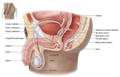
To propel sperm.
What is the role of the genital ducts in the male reproductive system?
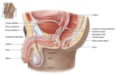
They produce substances needed for sperm activity and make up the semen.
What do the accessory glands contribute to the male reproductive system?
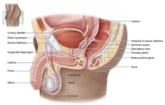
It serves as the copulatory organ.
What is the function of the penis in the male reproductive system?
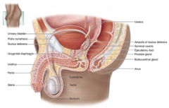
Surrounded by the tunica albuginea
What surrounds the testes?
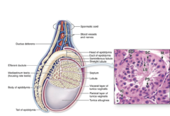
The thicker posterior portion of the tunica albuginea.
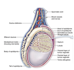
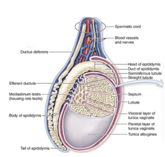
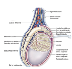
What is the mediastinum testis?
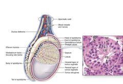
Around 250 pyramidal lobules containing sparse connective tissue, Leydig (interstitial) cells, and 1-4 highly convoluted seminiferous tubules.
How many testicular lobules are typically present, and what do they contain?
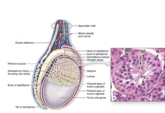
Dense, highly fibrous connective tissue.
What type of tissue forms the mediastinum testis?
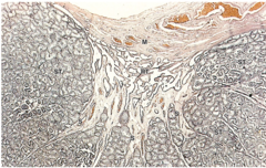
The septa that separate the testicular lobules.
What structure originates from the mediastinum testis?
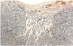
The rete testis.
What important network is contained within the mediastinum testis?
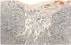
A serous sac surrounding the testes.
What is the tunica vaginalis?
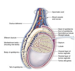
It is derived from the peritoneum during testicular migration from the abdominal cavity to the scrotum.
What is the embryologic origin of the tunica vaginalis?
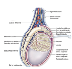
lines the scrotum.
What does the parietal layer of the tunica vaginalis line?
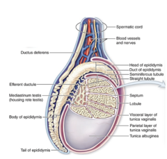
Covers the tunica albuginea
What does the visceral layer of the tunica vaginalis cover?
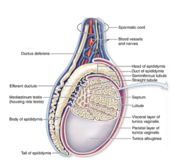
Sparse connective tissue.
What type of connective tissue makes up the interstitial tissue of the testes?
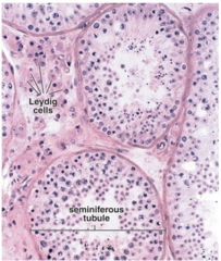
1. Fibroblasts
2. lymphatics
3. blood vessels
4. fenestrated capillaries
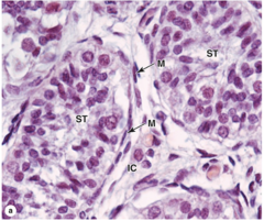
5. Interstitial (Leydig) cells
What cell types and structures are found in the interstitial tissue of the testes? (5)
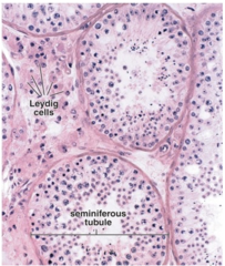
Testosterone production
What is the function of Interstitial (Leydig) cells?
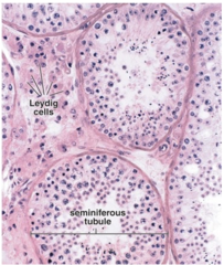
They are large, polygonal, steroid-secreting cells.
What is the shape and function of Leydig (interstitial) cells?
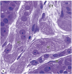
Cytoplasmic acidophilia
What staining characteristic is typical of Leydig cell cytoplasm?
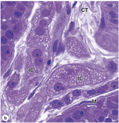
•Abundant SER and mitochondria
•Golgi apparatus
•Lipid droplets
•Microvilli
•Reinke crystals****
What organelles are abundant in Leydig cells?
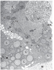
They are probably a protein product of the Leydig cell.
What is the likely composition of Reinke crystals?
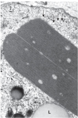
Abundant smooth endoplasmic reticulum (SER). (the arrows)
What cellular feature is often noted near Reinke crystals in histological images?
Sperm production.
What is the function of the seminiferous tubules?
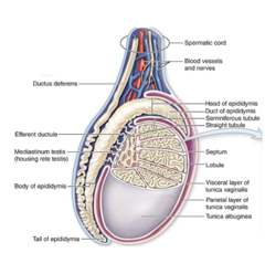
Through a straight tubule.
How is each seminiferous tubule connected to the rete testis?
Efferent ductules.
What connects the rete testis to the head of the epididymis?
SEVEN UP
1. Seminiferous tubules
2. Epididymis
3. Vas deferns
4. Ejaculatory duct
5. NOTHING
6. Urethra
What is the pathway of sperm?
Germinal (spermatogenic) epithelium, a specialized stratified epithelium.
What type of epithelium lines the seminiferous tubules?****
1. Sertoli (supporting) cells: DO NOT DIVIDE after puberty
2. Spermatogenic cells
What are the two main cell populations in the seminiferous epithelium?
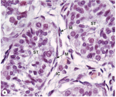
They replicate regularly and differentiate into mature sperm.
What is the role of spermatogenic cells?
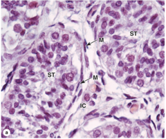
Fibrous connective tissue called the tunica (or lamina) propria.
What covers the basement membrane of the seminiferous tubules?
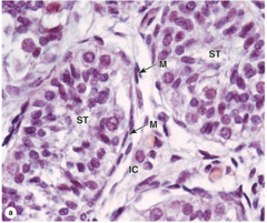
Smooth muscle-like myoid cells.
What type of cells form the innermost layer of the connective tissue surrounding the seminiferous tubules?
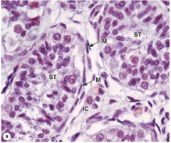
Columnar cells
What is the general shape of Sertoli cells?
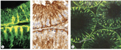
Extensive apical and lateral processes.
What type of cellular processes do Sertoli cells have?
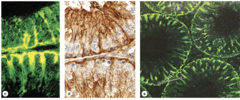
They help maintain the structural organization of the seminiferous tubules.
What structural role do Sertoli cells play in the seminiferous tubules?
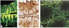
They provide physical and metabolic support.
What is the functional role of Sertoli cells for spermatogenic cells?
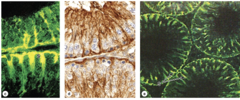
1. Replicate
2. Differentiate
What are the two main functions of spermatogenic cells?
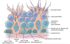
They are derived from primordial germ cells from the yolk sac.
What is the embryologic origin of spermatogenic cells?
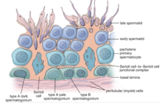
In four or more poorly defined layers.
How are spermatogenic cells organized within the seminiferous tubules?
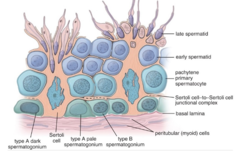
They gradually develop between Sertoli cells.
How do spermatogenic cells develop within the tubule?
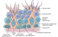
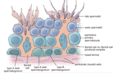
They rest on the basement membrane and are the most immature spermatogenic cells.
Where are spermatogonia located?
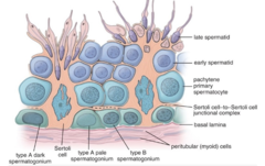
At the luminal border of the tubule, attached to the apical region of Sertoli cells; they are the most mature spermatogenic cells.
Where are spermatids located?
The process of sperm production.
What is spermatogenesis?
Begins during early puberty
When does spermatogenesis begin?
Pituitary gonadotropins (FSH and LH).
What hormones influence spermatogenesis?
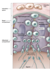
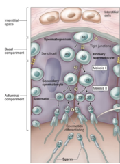
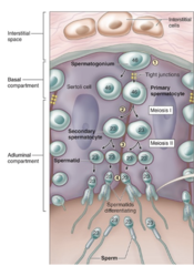
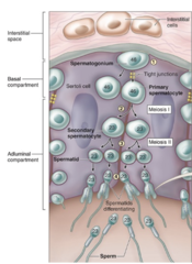
1. Spermatogonial phase (mitosis)
2. Spermatocyte phase (meiosis)
3. Spermatid phase (spermiogenesis)
What are the three phases of spermatogenesis?
Spermatogonial stem cells undergo mitosis to produce spermatogonial progeny.
What happens during the spermatogonial phase of spermatogenesis?
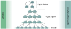
They act as reserve stem cells.
Features: Small, dark nucleus; divide at irregular intervals.
What is the role of Type A dark (Ad) spermatogonia?
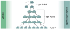
They act as progenitor (renewing) stem cells.
Features: Pale chromatin; divide regularly.
What is the role of Type A pale (Ap) spermatogonia?
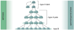
They are the final cells of the spermatogonial phase and give rise to primary spermatocytes.
What is the role of Type B spermatogonia?
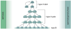
Daughter Ap spermatogonia are connected to each other.
What cells are connected to form the spermatogenic syncytium?
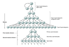
After the second meiotic division.
When do the cytoplasmic connections in the syncytium disappear?
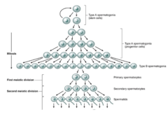
All progeny originating from a single pair of Ap spermatogonia.
Which cells remain connected throughout development in a syncytium?
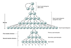
It ensures synchronous cell development of each clone derived from an Ap cell.
What is the purpose of the syncytium in spermatogenesis?
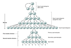
Primary spermatocytes, which are daughter cells of Type B spermatogonia.
What cells enter the spermatocyte phase (meiosis)?
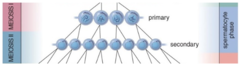
They undergo immediate DNA replication.
What happens to primary spermatocytes before entering meiosis?
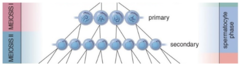
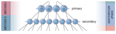
Secondary spermatocytes that are haploid with:
Chromosomes = n
DNA content = 2d
What is the result of Meiosis I in spermatogenesis?
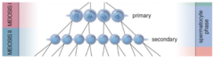
Spermatids that are haploid with:
Chromosomes = n
One single chromatid (DNA = 1d)
What is the result of Meiosis II in spermatogenesis?
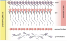
The phase of spermatogenesis in which spermatids undergo cellular remodeling and differentiate into mature sperm without cell division.
What is spermiogenesis?
Highly specialized junctions.
What keeps developing spermatids attached to Sertoli cells during spermiogenesis?
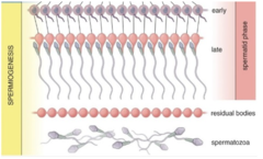
1. Golgi phase
2. Cap phase
3. Acrosome phase
4. Maturation phase
What are the four phases of spermiogenesis?
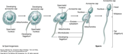
The Golgi apparatus, positioned adjacent to the nucleus.
What organelle is prominent during the Golgi phase of spermiogenesis?
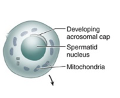
Proacrosomal granules produced by the Golgi.
What PAS-positive structures appear during the Golgi phase?
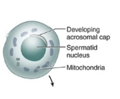
By the coalescence of proacrosomal granules.
How is the acrosomal vesicle formed in the Golgi phase?
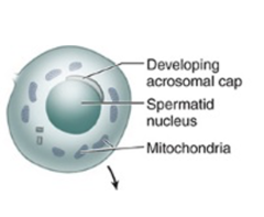
At the anterior pole of the cell.
Where is the acrosomal vesicle located on the developing spermatid during the Golgi phase?
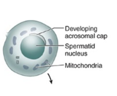
Numerous mitochondria and other organelles.
What organelles are abundant during the Golgi phase?
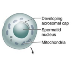
They migrate to the posterior pole of the cell.
What happens to the centrioles during the Golgi phase?
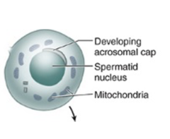
The axoneme of the flagellum.
What structure begins to form from the centrioles during the Golgi phase?
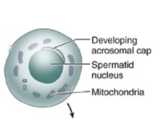
It spreads over the anterior half of the nucleus, forming the acrosomal cap.
What happens to the acrosome during the cap phase?
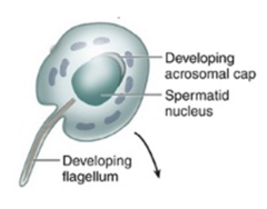
The nuclear envelope thickens locally.
What structural change occurs in the nuclear envelope during the cap phase?
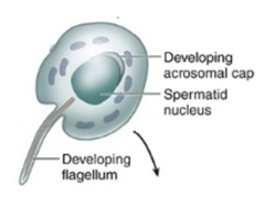
The nuclear content condenses.
What happens to the nuclear content during the cap phase?
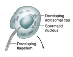
•Spermatid reorients
•Head deeply embedded into Sertoli cell with apex toward basement membrane
•Flagellum extends into the lumen
What happens to the spermatid during the acrosome phase of spermiogenesis?
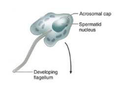
The nucleus elongates, and it along with the acrosome moves closer to the anterior cell membrane.
How does the nucleus change during the acrosome phase?
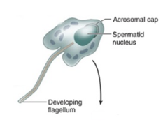
The cytoplasm is displaced posteriorly.
What happens to the cytoplasm during the acrosome phase?
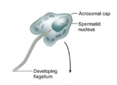
The manchette is a cylindrical sheath formed by reorganized microtubules, extending from the posterior rim of the acrosome to the posterior pole of the spermatid.
What is the manchette and when does it form in the acrosome phase?
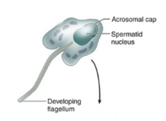
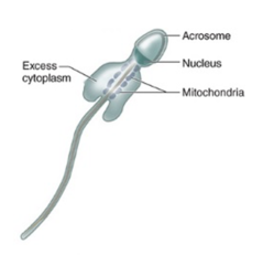
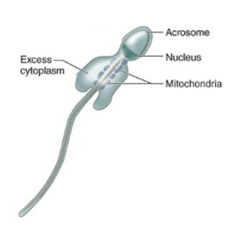
Excess cytoplasm around the flagellum is reduced, resulting in the formation of a spermatozoon.
What is the main cellular change during the maturation phase of spermiogenesis?
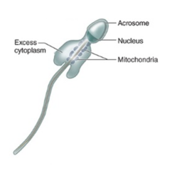
Sertoli cells phagocytose the excess cytoplasm (residual body)
What happens to the excess cytoplasm during the maturation phase?***
Spermatids are no longer attached to each other and are released from the Sertoli cells as mature spermatozoa.
When are spermatids fully released from Sertoli cells in the Maturation phase?
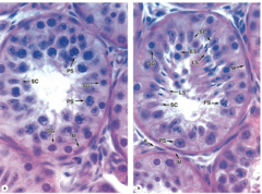
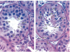
Sertoli cells are tall columnar epithelial cells located within the seminiferous tubules of the testis.
What type of cells are Sertoli cells and where are they located?
Support and nourish spermatogenic cells
What are the main functions of Sertoli cells?
Divide the seminiferous tubule into two compartments:
1. Basal
2. Adluminal
How do Sertoli cells divide the seminiferous tubule?
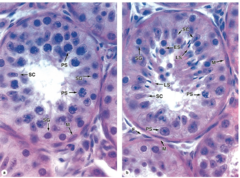
- Basal membrane adhered to the basement membrane of the seminiferous tubule
- Apical end extends to the lumen of seminiferous tubule
How is the basal and apical orientation of Sertoli cells structured?
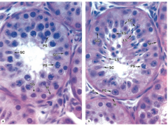
Abundant smooth ER (SER), some rough ER (RER), well-developed Golgi complexes, numerous mitochondria, and many lysosomes.
What organelles are abundant or well-developed in Sertoli cells?
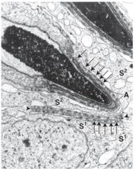
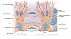
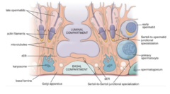
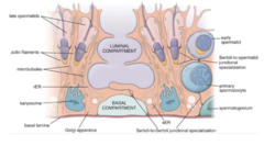
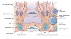
Sertoli cells form Sertoli-to-Sertoli and Sertoli-to-sperm specialized junctions.
What specialized junctions do Sertoli cells form?
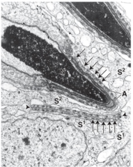
They form the blood-testis barrier
What is the function of the basolateral junctions between Sertoli cells?
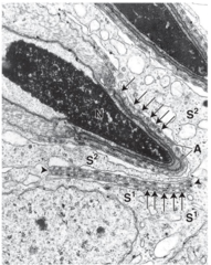
They support, protect, and provide nutrition to developing germ cells.
What is the primary role of Sertoli cells in relation to developing germ cells?
They have both exocrine and endocrine secretions
What types of secretions do Sertoli cells produce?
Androgen-binding protein (ABP).
What protein do Sertoli cells produce to bind and concentrate testosterone in the seminiferous tubule?
1. Production of inhibin
2. Production of Müllerian-inhibiting substance (MIS) in male fetus
Which hormones or substances are secreted by Sertoli cells?
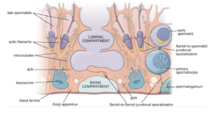
It is between the basement membrane and the Sertoli cell-to-Sertoli cell junctions and contains spermatogonia.***
What defines the basal compartment of the seminiferous tubule?
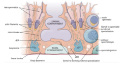
Primary spermatocytes move here, and all other developing germ cell stages are found in this compartment.
What defines the adluminal compartment of the seminiferous tubule?
1. Intratesticular ducts (inside the testis)
2. Extratesticular or excretory ducts (outside the testis).
What are the two main divisions of the male duct system?
They carry sperm from the seminiferous tubules to the epididymis.
What is the function of the intratesticular ducts?
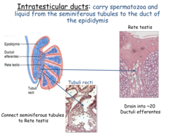
1. Straight tubules (tubuli recti)
2. Rete testis
3. Efferent ductules
Which ducts are considered intratesticular? (3)
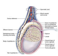
Transport sperm from scrotum to the penis during ejaculation
What is the function of the extratesticular (excretory) ducts?
1. Epididymis
2. Ductus (vas) deferens
3. Urethra
Which ducts are considered extratesticular (excretory) ducts? (3)
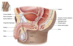
They connect the seminiferous tubules with the rete testis.
What is the function of the straight tubules (tubuli recti)?
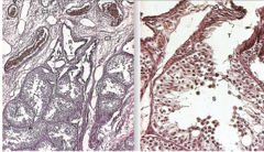
Sertoli cells only, with no germ cells.
What is the epithelial lining of the proximal segment of the straight tubules?
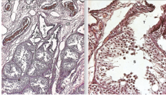
Simple squamous epithelium
What is the epithelial lining of the distal segment of the straight tubules (next to the rete testis).
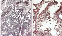
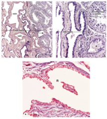
A network of interconnected channels within the testis that connects straight tubules to efferent ductules.
What is the rete testis?
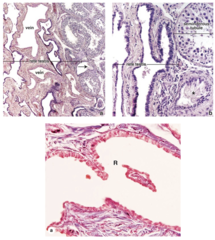
Lined with simple cuboidal epithelium
What is the epithelial lining of the rete testis?