13. Accommodation and Pupillary Function
1/72
There's no tags or description
Looks like no tags are added yet.
Name | Mastery | Learn | Test | Matching | Spaced |
|---|
No study sessions yet.
73 Terms
What is accommodation?
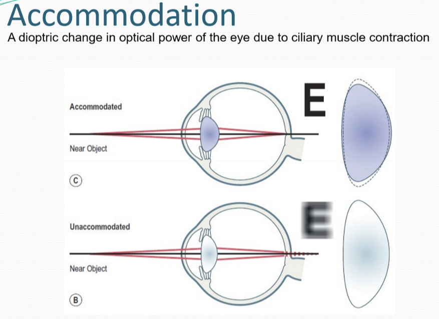
A dioptric change in optical power of the eye due to ciliary muscle contraction.
What is the basic mechanism of accommodation?
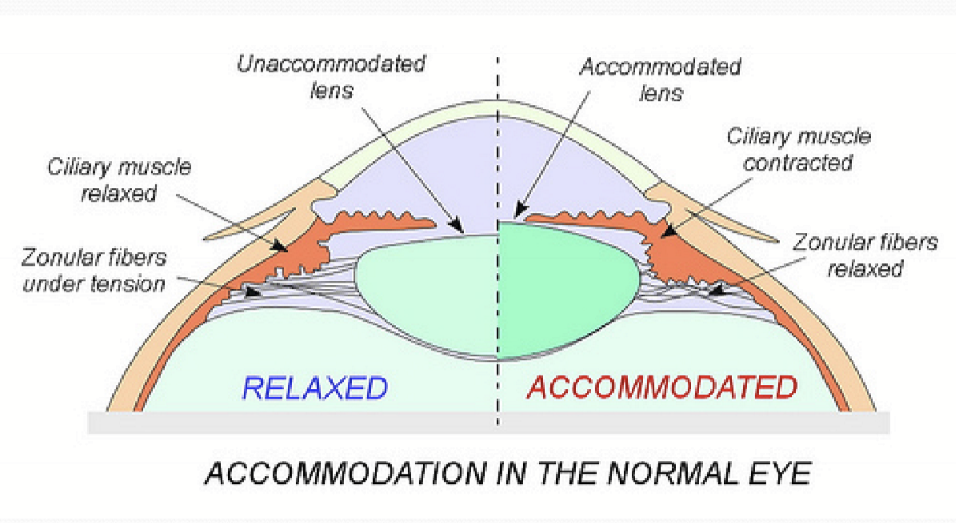
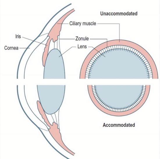
Ciliary msulces contraction moves the apex of the ciliary body towards the axis of the eye and releases resting zonular tension around the lens equator. When zolunlar tension is released, elastic lens capsule changes into a more spherical shape.
What happens when the lens changes into its accommodative shape?
Diameter decreases
Thickness increases
Anterior and posterior surface curvatures increase
What types of Ciliary muscles are there?
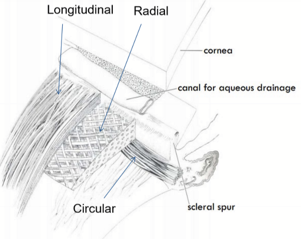
Longitudinal fibers
radial fibers
circular fibers
Where do ciliary muscles come from?
The ectoderm.
What does the longitudinal/radial muscles fibers do?
Anterior portion applies force to the scleral spur and open TM
Posterior portion applies force to pars plana moving it anteriorly
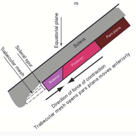
Where are radial fibers located?
Posteriorly to the elastic tendons of the choroid
What happens to the ciliary muscles when they contract?
Increase in thickness of the circular portion
decrease in thickness of the radial and longitudinal portions
Contraction pulls the anterior choriod forward, moves the apex of the ciliary processes towards the lens equator
What are the characteristics of Ciliary Muscle?
Has both sympathetic and parasympathetic innervation.
Parasympathetic M3 receptors mediates contraction.
Sympathetic Beta2-adrenergic receptors mediate relaxation.
How does Ciliary muscles respond to stimulus?
One stimulus causes nearly simultaneous contraction of all muscle groups
What is the embryological origin for the Iris dilator and sphincter muscles?
Ectoderm
What is the embryological origin of the ciliary body/muscle?
Ectoderm
What is the embryological origin of the smooth and skeletal muscle?
Mesodermal lineage
What parts of the ciliary muscle are “fast twitch” muscles?
Longitudinal fibers, with fewer mitochondria and larger neuron size than radial and circular portions of the ciliary body.
What are fast twitch fiber proteins needed for?
Fine movements
What is the function of the ciliary zonules?
Hold lens in place
Transmit tensile forces for accommodation shape change of the lens
Allows fluid to pass
Can ciliary zonules regenerate?
No
Where can the ciliary zonules insert?
On the lens: anterior, equatorial, and posterior. Ciliary Body: Pars plicata and Pars plana
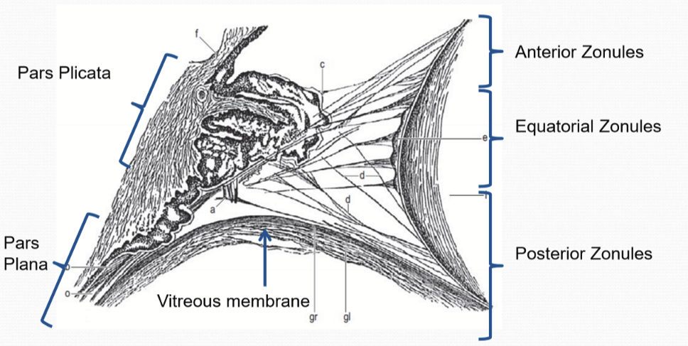
What part of the lens capsule do the ciliary zonules insert into?
Superficial layer of the lens capsule
When, during embryonic development, does the zonules form?
Late in embryonic development, non-pigmented ciliary epithelial cells synthesize the zonules.
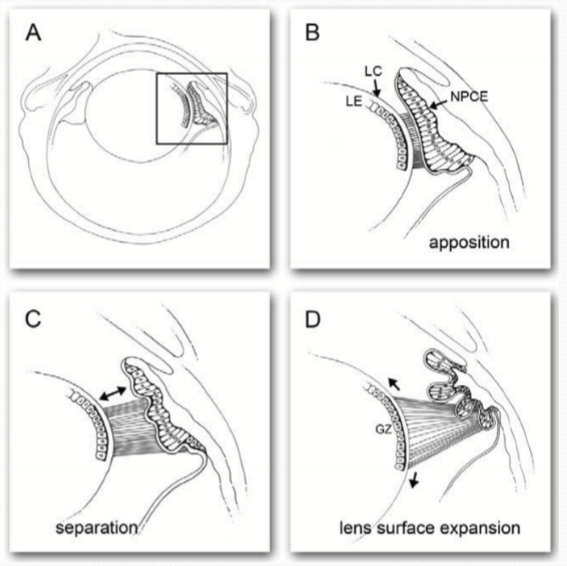
What are zonules fibers primarily made up of?
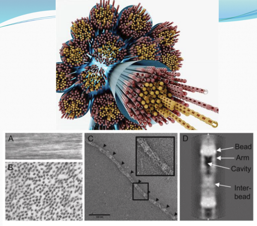
Glycoproteins: Fibrillin and Fibrillin like proteins (forms beads and strings)
MAGP-1 (microfibril-associated glycoprotein 1) and localized to beads only
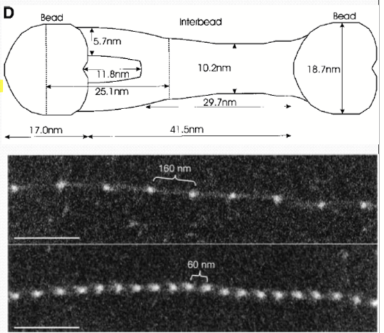
What are the properties of zonule fibers?
Very elastic
Non-collagenous
Non-elastin
What is fibrillin commonly used for?
Force-bearing structures such as blood vessels, lungs, and ligaments
What are fibrillins?
Large cycsteine-rich multidomain glycoprotein that polymerize in the extracellular space in head-to-tail manner to form microfibrils
How many people have marfan syndrome?
6 in 10
What does Marfan syndrome do to the eye?
Dislocate lenses in one or both eyes due to mutation in fibrillin gene
What is the lens capsule?
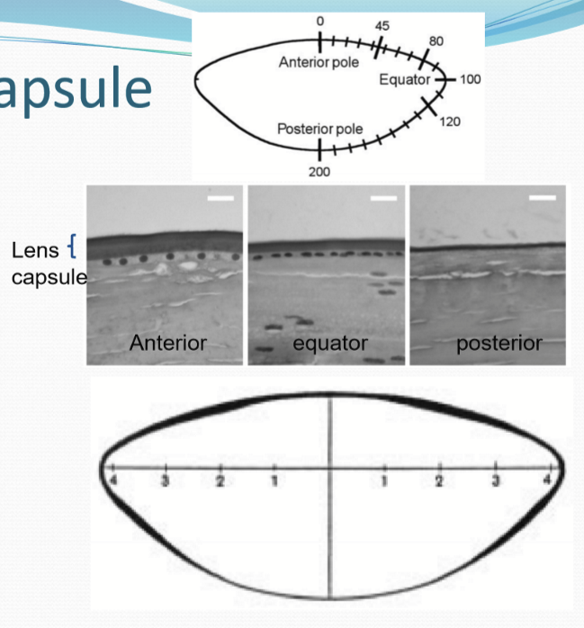
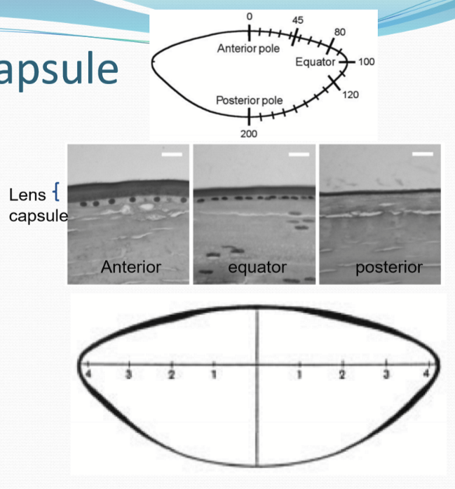
A thin transparent elastic membrane that is secreted by lens epithelial and fiber cells
Where is the lens thickest at?
Just anterior and posterior to the equatorial region. It is the thinnest posteriorly.
What is the lens capsule responsible for?
Reshaping lens upon zonule relaxation and is 2000x more elastic than the lens itself.
What is the lens capsule composed of?
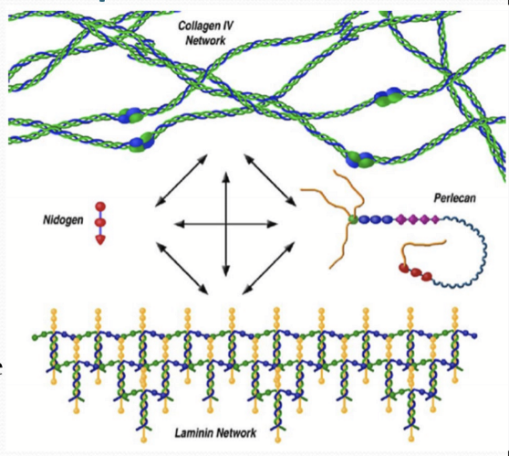
extracellular proteins with basement membranes
Collagen IV
Laminin
Heparin sulfate proteoglycans: perlecan, nidogen, collagen XVIII
lens integrins associate with laminin network of ECM
How long does it take for the ECM proteins of the lens capsule to turnover?
Months-years while other basement membranes of other epithelia take hours.
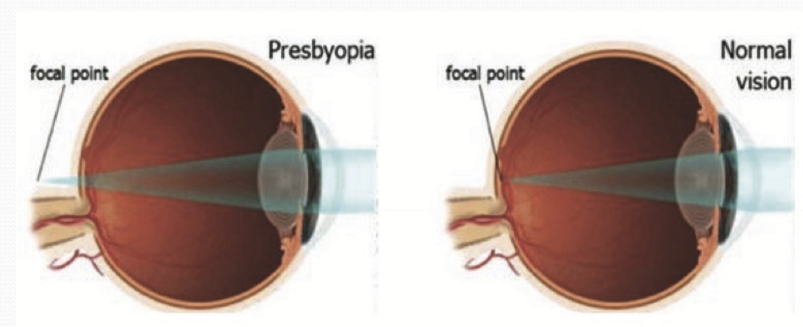
What is Presbyopia?
Gradual age related loss of accommodative amplitude. It is a linear decline of 2.5D/10 years.
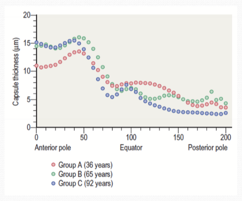
What happens to the lens capsule over time?
General increase in capsule thickness occurring anteriorly and posteriorly.
What causes the lens capsule to lose its elasticity?
Non-enzymatic glycation of collagen IV, increasing its stiffness. This effect is compounded by the slow turnover rates of lens capsule components.
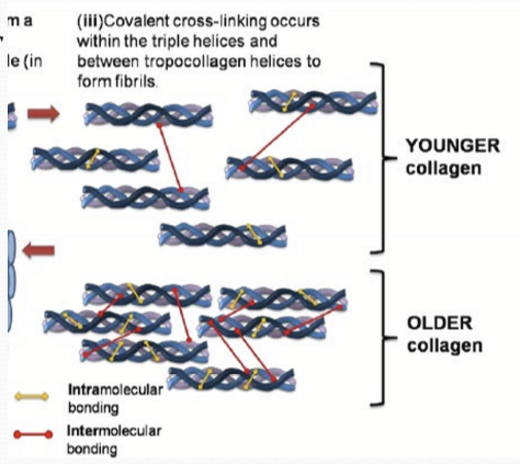
Why can induce intra and intermolecular cross-linking between lysine and amine groups (glycation)?
Increase in aqueous humor glucose.

Do individuals with type 1 diabetes have lower amplitude of accommodation in comparison with age-matched controls?
Yes, Collagen IV in lens capsule are susceptible to glycation.
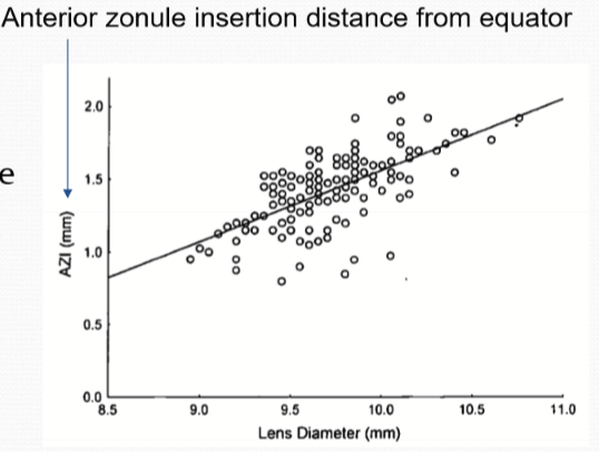
Do zonules change over time?
They do not have any elasticity change, but zonular/capsule insertion distance to the lens equator increases with age due to growth of lens
What happens to ciliary muscles with age?
loss of muscle fibers and increase in connective tissue
Contractile force stays constant, does not decrease
Movement of ciliary body still occurs after accommodative loss
What happens to the lens with age?
Linear increase in mass
Increase in axial thickness
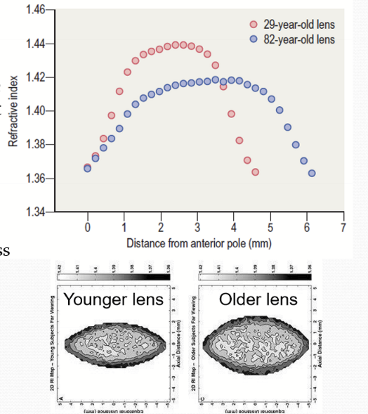
Increase in anterior and posterior curvature (more accommodation in terms of focus)
Equatorial lens diameter in disaccommodated state does not change
Changes in refraction: lens paradox
Stiffness
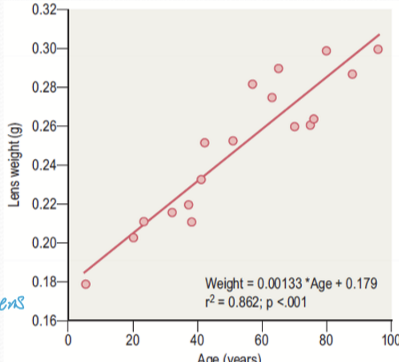
What is the lens paradox, and what causes it?
The lens paradox is the phenomenon where, despite increased lens thickness and curvature with age, there is little change in refractive power. It is caused by a decrease in the refractive index gradient, especially near the equatorial region, and an increase in lens thickness, which counteracts the expected increase in power.
How can the older lens inability to change form be demonstrated?
The removal of the lens capsule can demonstrate differences in the lens’s ability to change shape in the absence of the constraining capsule.
What are the accommodative triad?
3 physiological responses occur during accommodation
ciliary muscle contraction
pupil constriction
convergence
What stimulates the accommodative reflex?
Blur cues: blur presented to one or both eyes induce both eyes to accommodate; blur also induces bilateral pupil constriction
Convergence: Isolating convergence with base-out lenses; induces both eyes to accommodate and bilateral pupil constriction
How are the components of the accommodative reflex linked?
They are neuronally coupled via supranuclear connections, which coordinate their activation.
Can the components of the accommodative reflex occur independently?
Yes, while they are usually linked, each is controlled by distinct neural pathways and can occur separately in certain situations.
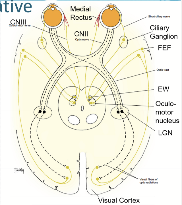
What are the afferent pathway of the accommodative reflex?
Optic nerve axons project to and synapse in the LGN (lateral geniculate nucleus)
Neurons project from LGN to visual cortex
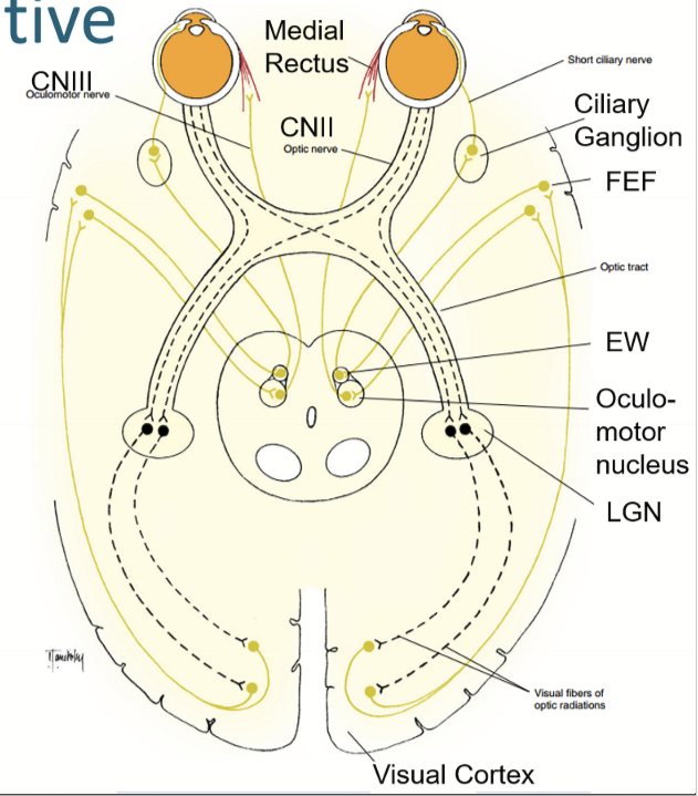
What are the interneurons of the accommodative reflex?
From visual cortex to frontal eye fields (FEF)
From FEF, to Edinger-Westphal nucleus (EW) and oculomotor nucleus
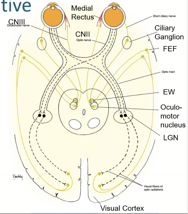
What are the efferent pathway of the accommodative reflex?
From EW to ciliary ganglion along CNIII
From ciliary ganglion to ciliary muscle
Parasympathetic: Stimulates M3 receptors in fast twitch muscle for fine control
What is convergence?
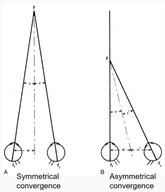
Simultaneous and synchronous adduction in both eyes. Can be voluntary or involuntary (typically involuntary for normal use of eyes).
What stimulates convergence?
Contraction of medial rectus muscle. Can be symmetrical or asymmetrical.
What are the neural pathway for convergence?
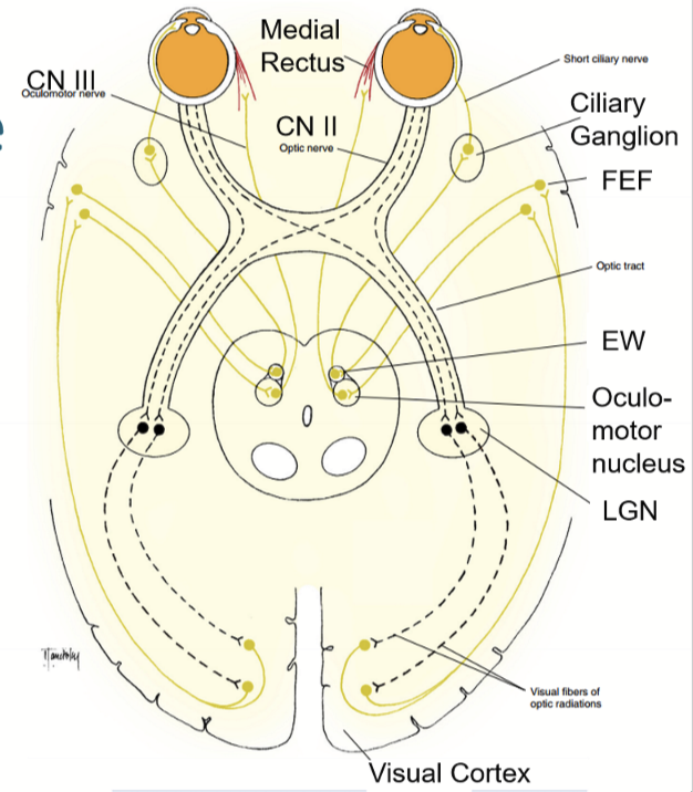
Oculomotor nerve receives signals from oculomotor nucleus to stimulate medial rectus muscles
Supranuclear signals from the FEF and visual cortex couple ciliary muscles and medial rectus co-contraction
What are the functions of the pupil?
Control of retinal illumination: aids in optimizing retinal illumination to maximize visual perception
Reduction of optical aberrations, reducing the degree of chromatic and spherical aberration
Improve depth of focus/field: smaller pupil increases depth of field
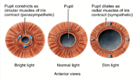
How is the dilator muscle oriented?
Radially, and associated with pigmented epithelium.
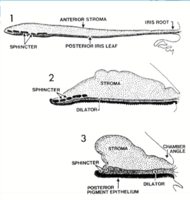
What happens when the dilator muscle contracts?
It pulls the pupillary margin towards ciliary body peripherally (mydriasis)
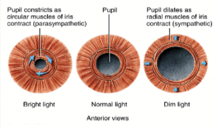
Where is the sphincter muscle?
Encircles the pupillary margin and is separated from pigmented epithelium
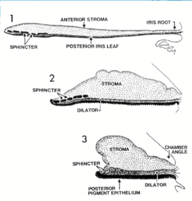
What happens when the sphincter muscle contracts?
Reduction in pupil size/miosis
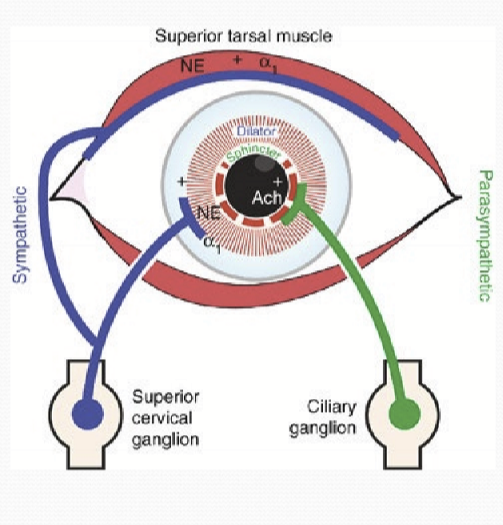
What innervates the iris muscles?
Sphincter muscle is innervated by parasympathetic; ACh and muscarinic receptors; innervation occurs in a segmental distribution (~20 segments)
Dialtor muscle is innervated by sympathetic; norepinephrine and alpha1 receptors
What stimuli does the pupillary response respond to?
light/brightness
near-reflex/accommodation
Is the efferent pathway control for light/brightness stimuli the same as the near-reflex stimuli?
Yes, parasym control of iris sphincter and sym control of dilator. However, the supranuclear control over each response is different.
Can miosis occure without change in retinal luminance?
Yes
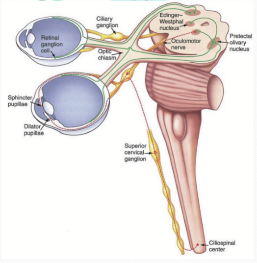
What is the afferent pathway for the pupil light reflex?
Retinal ganglion cell axons project to pretectal olivary nucleus.
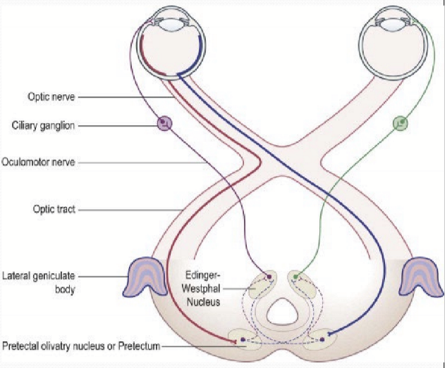
What cells are part of the afferent pathway?
Rod and cone photoreceptors
Intrinsic photoreceptive retinal ganglion cells (ipRGCs)
Contain Melanopsin
Can trigger action potential
What is the interneuron pathway for the pupil light reflex?
From the pretectal olivary nucleus to EW nucleus
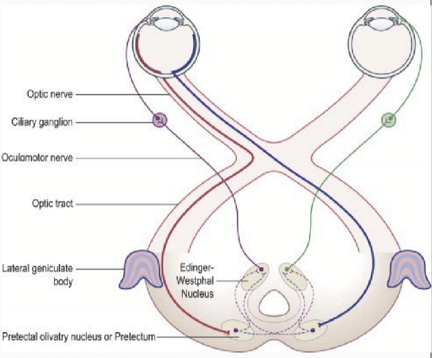
What is the efferent pathway for the pupil light reflex?
From EW to ciliary ganglion. Then from ciliary ganglion to iris sphincter for parasympathetic stimulation.
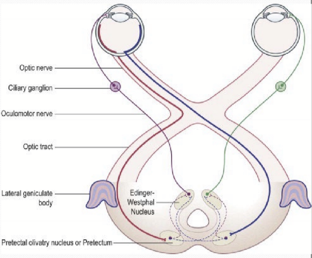
How do the pupils react to light?
Symmetrically between both eyes due to interneurons sending signals to left and right EW
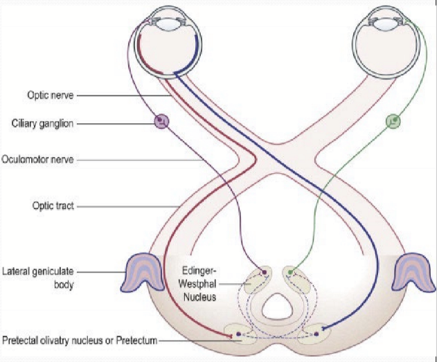
How is the iris size regulated?
Regulation of iris sphincter contraction by two competing signals. Illumination induced increases parasym nerve stimulation and continuous supranuclear inhibition of parasym nerves.
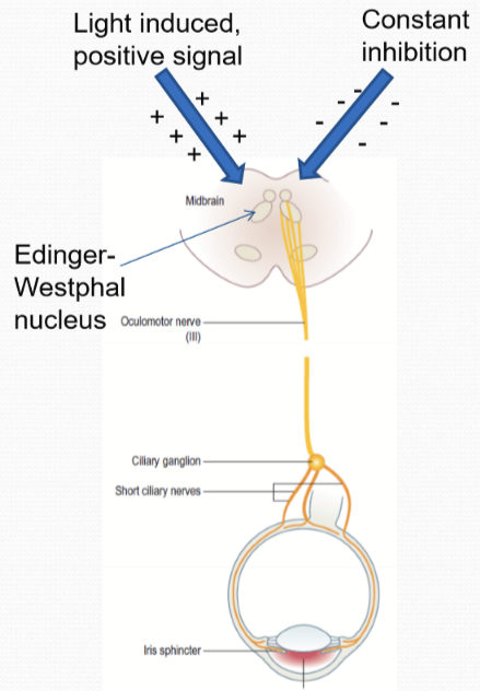
What happens in the pupil light reflex when there is significantly reduced light stimulus?
Inhibitory pathways prevents sphincter contraction: CNS/brainstem inhibitory input is constant, parasym nerve to sphincter muscle is not stimulated, inhibitory signals comes from a sympathetic never. Result is mydriasis.
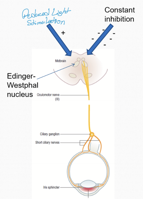
What happens in the pupil light reflex when there is some light stimulus?
Some competition to inhibitory signal, so some stimulation along parasympathetic nerve to iris sphincter. Result: some miosis occurs.
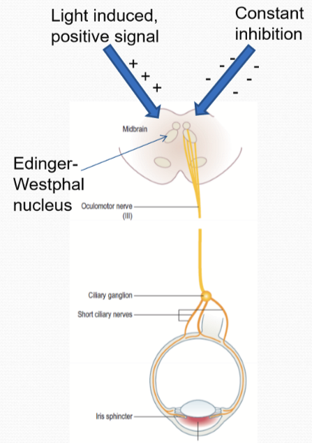
What happens in the pupil light reflex when there is more light stimulus?
More competition to inhibitory signal, so more stimulation along parasym nerve to iris sphincter. Result: miosis occurs
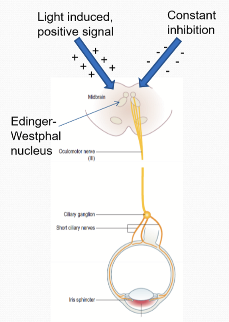
When can a lack of inhibition occur?
Sleep
anethesia
sympathetic inhibition is suppressed
Baseline level of positive signal from light-induced response can cause miosis
What controls the iris dilator?
Sympathetic nerves from the superior cervical ganglion and the axons pass through the ciliary ganglion to innervate the dilator muscle?
There is a secondary mechanism for pupil dilation. What is its purpose?
Enhances speed of maximal pupil diameter.
What also regulates the iris muscles?
Circulating catecholamines released from adrenal glands in the blood may act directly on iris dilator causing mydriasis.
Sensory innervation to iris from CN V. mechanical and chemical irritants of the eye can cause strong miotic response. It fails to reverse without autonomic drugs.