Cell Biology Final Material
1/99
There's no tags or description
Looks like no tags are added yet.
Name | Mastery | Learn | Test | Matching | Spaced |
|---|
No study sessions yet.
100 Terms
What is the cell cycle
It is the ordered events of a cell to duplicate its material and divide into two
Cell growth/Duplication
Chromosome segregation
Cell division
Does every cell have the same duration of the cell cycle?
No, not every cell has the same duration of cell cycle
for examples gut cells divide very quickly and nerve cells very slowly
they have different generation times
What are the 4 phases of the cell cycle
G1 - Gap 1 - cell gets ready to do the rest of the work - it grows and metabolises, preparing for S
S phase - DNA is replicated - commital phase - if cell enters S it is commited to dividing (becuase if it produces twice the DNA and doesnt divide it is lethal)
G2 - Gap 2 - gets the cell ready for M
M - division phase - subcategories
Nuclear division
Cytoplasmic division
G1, S, G2 make up interphase
If cell cycle was on a clock
23 hours in interphase
1 hour in division phase
Is the cell cycle regulated
Yes, there is a control system that monitors the process of the cell cycle
it is one of the most tightly regulated processes
How does a cell pass the checkpoint
at each phase every cell is assessed and it needs to pass a checkpoint to go to the next phase
Between G1 and S
It is assessed whether the environment is favorable and if it has everything to successfully move on
Between G2 and M
Assess whether all DNA got replicated
DNA polymerase check if things are done right
In M
makes sure things go well like if all chromosomes attacked to proper spindle
What are checkpoints
chemical in nature
What are the control molecules
Activity is regulated by the complex of two molecules joining
Cyclin
Cyclin dependent Kinases (CDK) - it adds a phosphate to something to phosphorylate target protein
Names of the CDK’s
The ones that control Cell into M would be named (M- CDK activity or M- cyclin concentration) with S it would be S-…
etc…
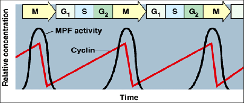
M- CDK activity
When M-CDK activity high the cell is undergoing mitosis and this is when cyclin is high
you cant get activity of CDK without enough of the paired cyclin
Concentration first goes down (cyclin) then the activity (CDK - phosphorylation - changes shape and function) and then it moves out of the phase
What does it mean that the activity of individual CDKs varies during the cell cycle?
Each CDK is active only at specific stages of the cell cycle because it must bind to a specific cyclin. Cyclin levels rise and fall, so CDK–cyclin complexes form and activate at different times to control each phase (e.g. S-CDK and M-CDK active at different times).
How does CDK work
Transfers a phosphate from ATP to the target protein
What do cyclin molecules do
they bind to the CDK’s as a step towards activation
Life cycle of CDK
Active CDK is when cyclin is binded
At the right time when it wants to be inactivated Anaphase-Promoting Complex/Cyclosome (APC/C) becomes active
APC/C adds a chain of ubiquitin molecules to the cyclin (ubiquitylation)
ubiquitylated cyclin is then recognized and degraded by the proteasome
Inactivated CD
What else is CDK’s function controlled by
Addition and removal of phosphate is also necessary for activity
They need to take out the inhibitory phosphate to function
What else can CDK be controlled by
CDK can also be blocked by inhibitor proteins
for example P27 is one
what does a phosphatase do
it removes a phosphate
Levels of CDK and cyclin through cycle
LOOK at notes for specific ones
Movement from G1 to S
CDC6 (not a CDK but a licensing factor) sits at a replication origin called the ORC
Helicase binds near cdc6
once binds cdc6 is phosphorylated for helicases to move
the phosphorylation is done by S-cdl
cant get movement out of helicase until it is phosphorylated by S-CDK
P53
It is a tumor suppressor gene
A regulatory protein whose activity is increased due to DNA damage (especially UV light)
50% of non inherited cancer is due to mutations in P53 - dont get P21
Normal activity: turn on when DNAA damage, turn on P21 which inactivates CDK (this is correct since damage leads to no S- dont want to bring damaged DNA in S)
DNA damage
Increase P53
Increase P21
P21 binds CDK complex and arrests in G1
GO
Cells can withdraw from the cell cycle to GO
this can happen in cells with realling long cell cycle tumes
if a cell withdraws they are called terminally differentiated
What is organ and body size regulated by
It is regulated by the interplay of 3 processes
Cell growth
Cell proliferation (division)
Cell death
Apoptosis
Programmed cell death for the purpose of
Developing structures
forming digits - fingers/toes
ear lobe - free (they had an apoptotic event occur)
Very important for body plants
In embryos very common
Regulation of cell numbers
no increase or decrease in the size of an internal organ
it is cleaned cell death - beneficial not problematic
difference between apoptosis and necrotic events
Necrotic event - expulsion of cellular material - an explosion
Apoptosis - everything implodes
building skyscraper imploding
very regulated
How does apoptosis work
It is a highly regulated program it uses the caspase family of proteases cleaves the laminar proteins, causes breakdown of nuclear membrane
cytoskeleton collapse
nuclear envelope dissassembles
DNA fragments
Cell surgace altered (cell wont look normal so macrophages engulg)
Macrophages engulf
What are the external signals
Survival factors
Mitogens
Growth factors
Negative control factors
1-3 are stimulating
4 is inhibitory
Survival factors
promote cell survival
there to make sure cell stays viable
for example nerve cell. some stay (good connection to target cell) and get survival factor and others apoptosis (bad connection)
so the survival factor is a chemical signal molecule that blocks apoptosis so the cell survives
Mitogens
stimulate cell proliferation (division)
When activated it activates a CDK phosphorylate Rb (usually blocks DNA replication. its an inhibitor so when inactivated it turns on the DNA replication) which inactivates it allowing cell to start transcription
Growth factors
stimulate cell growth
inhibit protein degradation
OR stimulate protein production
Both stimulate growth
Negative Control factors
inhibit the survival factors
growth, cell division, cell death
structural and organizational changes a cell undergoes to prepare for division.
1. chromosome condensation (first sign moving into M phase)
2. nuclear envelope breakdown
3. ER and golgi reorginize cuz close to nuclear envelope
4. cell loosens attachment (surrounding neigboring cells)
5. cytoskeleton reorginzation
Division necessities
DNA replication
Cytoskeleton structures appearing
Centrosomes Duplicated
Dynamic intability
Microtubule instability
DNA replicated
each chromosome is replicated and copied parts remain together until segr
cell division wont happen of replication doesnt
Cohesin - make sure sisters stay close
Condensins- part of looping domains in condensation
Whats a chromosome pair
one from mom one from dad can be replicated or not
Are their homologous chromosomes in G1
Yes
Cytoskeleton structures appear
Mitotic spindle
contractile ring- myoson and actin filaments in animals
Centrosomes duplicated
Centrosome: region of the cell where the centrioles are. there may or may not be centrioles there
Plant cells have centrosomes not centrioles
Centrioles run perpendicular to each other = 2 centrioles
locate organizing site
in nucleation site
Interphase: 1 centrosome, 2 centrioles
After duplication (mitosis):2 centrosomes, 4 centrioles
Centrosome life cycle
Nucleation site for microtubules of spindle
Pair of centrioles in animal cells
Duplication - movement centrosome cycle
g1 1 pair S-G2 2 pairs (next to each other) M- centrosomes move to either side and place spindle (good placement poles)
NOT IN PLANT CELLS
Microtubules
long, hollow and stiff tubes of protein
straw
Each microtibule has a polarity (plus end beta and minus end alpha)
Molecules of tubulin = dimer of an alpha and a beta tubulin
Tubulin dimers stack to form protofilaments (alpha and beta)
Wall has 13 protofilaments alpha and beta on each ends determining polarity
Assembly of a microtubule at centrosome begins with an initial ring of 13 tubulin molecules (Y tubulin rings) addition occurs faster to the plus end then the minus end
LOOK AT NOTES DRAWING
Dynamic instability
the growth and dissasembly of individual microtubules at any single time allows for movements of chromosomes
Microtubule instability
GTP cap loss
at the ends of it has a lot stabilized to growth
if there is no cap causes the breakdown of microtubules and dimers are released
this is how u can get growing microtubules to shrink
if growth faster than hydrolysis → tubule grows
Variety of microtubule associated proteins regulated microtubules
growth/shrink/stable/unstable
What can mitosis be defined as
A continuous process that can be defined as moving through 6 phases
Reorganization of microtubule arrays
Large number of microtubules
shorter microtubules
Depolymerization rate 20X faster than normal interphase stage
Change occurs due to activites of MAPs(microtubule associated proteins)
Kinesisna and Dyneins
Kinesins are motor proteins that move toward the plus end of microtubules, helping separate spindle poles and move chromosomes toward the cell center.
Dyneins move toward the minus end of microtubules, pulling chromosomes toward spindle poles and positioning the spindle within the cell.
Prophase
centrosome separate and move to poles
microtubules extend from 1 centrosome to other and this interaction stabilizes the spindle
interpolar microtubules - between 2 poles - stabilize spindle cuz connected to each other
chromosomes condense
Prometaphase
breakdown of nuclear membrane
spindle microtubule bind to the chromosome at kinetochores
Each chromatid bound by microtubule from opposite poles
kinetechore proteins bound to centromere which microtubule binds to
microtubule not directly bound to chromosome cuz then not able to shrink if it were directly attached
types of microtubules
Astral microtiules - attach to same pole side
Kinetichore - move chromosomes
interpolar- non kinetechore
Metaphase
chromosomes align at equatorial plate of spindle = metaphase plate
chromosome are under tension
Anaphase
release of cohesins
sister chromatids begin to move to poles
protein breaks down cohesins (a protease called separase) → happens really quickly
A. kinetechore microtubules shorten - remove tubulin subunits at the kinetechore
B. overlapping interpolar microtubules move past each other, pushing poles further apart so they slide past each other
Telophase
prophase backwards
Nuclear membrane reforms including pores
Decondense so transcription begins again
How does nuclear membrane breakdown/reformation happen
It breaks in a bunch of packets
phosphorylation
Comes back together by dephosphorylation
Cytokineses process and timing
begins in late anaphase and doesn’t end until late telophase
Utilizes a contractile ring (animals) of actin and myosin that sever the 2 cells from each other
like a drawstring on a pair of pants - pinch action
First evidence of cytokinesis is puckering and cleavage furrow
In plants a fragmoplast forms in the middle of the cell which forms the start of the cell wall which grows out dividing the cells into two
How does division of organelles occur
either double the number and then divide
Binary fission - chloroplasts and mitochondria
Fragment and reassemble in new cells
Golgi and ER
Somatic Cell
Fully differentiated body cell
all have pairs of chromosomes
Germ cell
Gametes, sex cells. Produced by meiosis
Homologous pairs
maternal and paternal
Same gene at same locus (position)
Different allele can occur
Diploid
pairs of homologous chromosomes
somatic cells
Haploid
One chromosome of each type
Gametes
Even if replicated still haploid cuz no pairs
Meiosis
Production of haploid cells (gametes) from a diploid cell
In preparation chromosomes duplicate
Meisosis 1
Homologous pairs find each other (synapsis) and form a bivalent/tetrad in prophase 1
cohesins keep sisters together
synaptonemal complex keeps bivalent together
Recombination/crossing over occurs and formation of chiasmata
double stranded breaks occur then new bindin within the homologous chromosome
+ variability
Sister crhomatids locked by cohesins and homologous tied by chiasma
Split homologous pairs
Rnadom assortment - way homologous chromosomes line up on metaphase plate to generate variability
2n-n
Meiosis 2
Duplicated chromosomes separate (sisters)
What does each cell remain with after meisosis
it ends up with 1 chromosome from each homologous pair
Difference between meiosis and mitosis
mitosis produces identical diploid daughter cells
Meiosis produces non identical haploid cells
Cancer genes
oncogens
tumor supressor genes
Oncogenes
send constant divide signals
tumor supressor genes
prevent cancer by stopping cell division,
Whats a neoplasm
A neoplasm is an abnormal mass of cells that grows uncontrollably — basically another word for a tumor, which can be benign (non-cancerous) or malignant (cancerous).
Benign
Essentially normal cells which stays at the site of origin
A little mass
dont have a real removal plan unless large enough to affect other organs
a wart is techincall a benign tumor
Malignant
Mass of cancerous cells that displaces normal tissue during its growth
leak out into blood vessels so spread throughout body
more dangerous
Metastatic
came from another place
How does the inside of a tumor function
inside the tumor is a microenvironment - may not participate in the normal function of that region of the body
can have its down blood vessels cuz needs blood supply
Number of mutations
most cancers require more than one mutation to occur
inactivation of P53
get rapid accumulation of other mutations
P53 is a tumor suppressor gene
Why do most cancers appear later in life
cuz it takes time for a cell to develop numerous mutations
Can cancer be inherited
Yes and no
If inherit one mutation and the cancer requires 4 you have a predisposition to that cancer - doesnt necessarily mean u will get it
Some cancers can also develop during life from factors and arent passsed down ex. lung cancer from smoking
Nomal tissue organization
cells organize into tissues and tissues into organs
epithelia cells, smooth muscle cells, connective tissues in distinct layers
Many tissues organize into epithelial (sheets) of cells
To coordinate function, cells connect via junctions (Tight junction, Adherens junction, Desmosome, Gap junction, Hemidesmosome)
They are polarized- each side diff function
Tissues/organs are mixtures of cell types
Renewal rats of normal cells
renewal rates of different cell types differ
most terminally differentiated cells cant divide, they go to. G0
These cells are replaced by precursor cells
Precursor cells come from step cells (undifferentiated cells)
Precursor cells
Totipotent - can become any part of the body
Pluripotent - already walked down a lineage - produce 1 type of organ
Characteristics of cancerous tissues
Cant really see boundaries, unorganized, weird shape/nuclei size
uncontrolled cell division
loss of cell specialization
cancerous cells change shape
loss of contact inhibition
leads to plasma membrane changes (ruffling)
loss of contact with neighbors (malignant)
Genetic instability (# of chromosomes in cells differ)
from dividing abnormally
invade normal tissue
can move into the blood stream and colonize a new site (metastasis)
contact inhibition
in normal cells
when one cell grows next to another cell it will stop dividing- why it forms a single layer in the petri dish
Patterns of metastasis
Breast cancer → brain liver, bones, lungs
prostate → bones
Colon → liver
What abilities do cancercells evolve
break through tissues
Angiogenesis- creation of blood vessles
tumor full of cells needs to maintain themselves by developing their own blood vessles in tumor to support cell division
General traits of cancer cells
reduced dependance on signals from other cells
survive stress and internal changes that would cause normal cells to undergo apoptosis
can proliferate indefinitely
Genetically unstable
abnormally invasive
abnormally avid for nutrients cuz always dividing- very metabolically active
can colonize inappropriate locations
can modify the cells behaviour in surrounding connective tissue
How many tissues in body
270 - for each is a type of cancer
Types of cancers
carcinomas
sarcomas
leukemia
lymphomas
Carcinomas
originate in coverings of the body or glandular tissues (skin, lining of intestines, breast, liver)
Sarcomas
Arise in connective tissue (bone/muscle)
Leukemia
Arise from the bone marrow or blood forming tissues
Lymphomas
arise in the immune system
Carcinogens
agents that contribute to the development of cancer
development of learning about carcinogens over time
1761 - john hill noticed increased nasal cancers in men with excessive tobacco snuff
1775 - percival pott- reported skin cancers in scrotum of adolescent ment who in youth worked as chimney sweeks
1915- chemically linked by Dr. yamagiwa - used coal tars to inducecancers in ears of rabbits
3 types of skin cancers
basal cell carcinoma, squamous cell carcinoma, malignant melanoma
in order from less bad to worst
UV radiation
causes breaks between A-T base pairs and forms TT dimers
Chemical carcinogens
Smoke, red dye 2, asbestos
can u recover from smoking and not get cancer
yes the cells can recover and your risk of cancer can go down
Finding out that viruses are carcinogens
chicken with sarcoma in breast tissue → removed from it an grinded it with sand → filtered it and colected filtrate → inject filtrate in young tissue → observe sarcoma in injected chicken
Smth smaller than bacteria cause cancer - this is a virus
how oncogenic viruses contribute to cancer development
Insertion of an oncogene → produces viral oncogene protein or produces a protein that can influence adjacent genes
Induce genetic instability → disrupt DNA repair mechanisms
Disrupt cell cycle
Infection causes chronic inflammation → changes the microenvironment
Causes immunosuppression