(AnaPhy) Topic 1-3
1/177
Earn XP
Description and Tags
Human Organism, Cells, Tissues
Name | Mastery | Learn | Test | Matching | Spaced |
|---|
No study sessions yet.
178 Terms
Organelles
specialized structures in cells that perform
specific functions
Example: nucleus, mitochondria, ribosomes
Cytoplasm
jelly-like substance that holds organelles
Cell membrane / Plasma membrane
a structure that encloses the cytoplasm
It forms a boundary between material in inside the cell and the outside.
• It acts as a selective barrier.
Functions of the Cell
Smallest units of life
cell metabolism and energy use
Synthesis of molecules
Communication
Reproduction and inheritance
fluid-mosaic model
the model used to describe the cell membrane structure.
The membrane contains phospholipids, cholesterol, proteins, and carbohydrates.
Phospholipids form a bilayer.
Phospholipids contain 2 regions: polar and nonpolar.
Polar region
Exposed to water around the membrane.
Hydrophilic (water-loving)
Non-polar region
Facing the interior of the membrane.
Hydrophobic (water-hating)
Found inside the cell
enzymes, glycogen, and potassium
Found outside of the cell
sodium, calcium, and chloride
Passive membrane transport
does not require the cell to expend energy.
diffusion, osmosis, and facilitated diffusion.
Active membrane transport
Does require the cell to expend energy, usually in the form of ATP.
Diffusion
Involves movement of substances in a solution down a concentration gradient.
Solutes, such as ions or molecules, tend to move from an area of higher concentration of a solute to an area of lower concentration of that same solute in solution.
Concentration Gradient
the difference in the concentration of a solute in a solvent between two points divided by the distance between the two points.
Osmosis
is the diffusion of water (a solvent) across a selectively permeable membrane from a region of higher water concentration to one of lower water concentration.
Hypotonic
has a lower concentration of solutes and a higher concentration of water relative to the cytoplasm of the cell.
If the cell swells enough, it can rupture / burst
Isotonic
has the same solute concentrations inside and outside the cell.
The cell will neither shrink nor swell. (stays the same)
Hypertonic
Has a lower solute concentration and higher water concentration than the surrounding solution.
Resulting in cell shrinkage, or crenation
Facilitated diffusion
is a carrier-mediated transport process that moves substances across the cell membrane from an area of higher concentration to an area of lower concentration of that substance.
High to low, but needs help
Leak channels
constantly allow ions to pass through.
Gated channels
limit the movement of ions across the membrane by opening and closing.
Carrier molecules
are proteins within the cell membrane involved in facilitated diffusion.
They exhibit specificity; only specific molecules are transported by the carriers.
Active Transport
is a carrier-mediated process, requiring ATP, that moves substances across the cell membrane from regions of lower concentration to those of higher concentration against a concentration gradient.
Low to high, needs help, uses energy
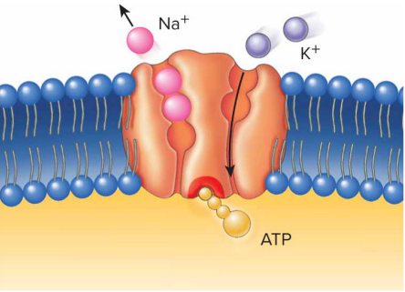
Sodium-Potassium Pump
A major example of active transport
moves (Sodium) Na+ out of cells and (Potassium) K+ into cells.
The result is a higher concentration of Na+ outside cells and a higher concentration of K+ inside cells.
Secondary Active Transport
uses the energy provided by a concentration gradient established by the active transport of one substance, such as Na+ to transport other substances.
Sumasabay
cotransport
the diffusing substance moves in the same direction as the initial active transported substance.
countertransport
the diffusing substance moves in a direction opposite to that of the initial active transported substance.
Endocytosis
is a process that that brings materials into cell using vesicles.
Receptor-mediated endocytosis
occurs when a specific substance binds to the receptor molecule and is transported into the cell.
Phagocytosis
often used for endocytosis when solid particles are ingested.
Pinocytosis
has much smaller vesicles formed, and they contain liquid rather than solid particles.
Exocytosis
involves the use of membrane-bound sacs called secretory vesicles that accumulate materials for release from the cell.
Attaches to plasma membrane to remove waste
Cell Nucleus
Usually located near the center of the cell
Bounded by a nuclear envelope with outer and inner membranes
Contains nuclear pores through which materials pass
Contains 23 pairs of chromosomes consisting of DNA and proteins
Ribosomes
components are produced in the nucleolus.
are the organelles where proteins are produced.
may be attached to other organelles, such as the endoplasmic reticulum.
Endoplasmic Reticulum
is a series of membranes forming sacs and tubules that extends from the outer nuclear membrane into the cytoplasm.
Rough ER
involved in protein synthesis and is rough due to attached ribosomes.
Smooth ER
no attached ribosomes and is a site for lipid synthesis, cellular detoxification, and it stores calcium ions in skeletal muscle cells.
Golgi Apparatus
Consists of closely packed stacks of curved, membrane bound sacs.
Collects, modifies, packages, and distributes proteins and lipids manufactured by the ER.
Lysosomes
Are membrane-bound vesicles formed from the Golgi apparatus.
Contain a variety of enzymes that function as intracellular digestive systems (They clean out waste products)
Key role: Recycling, defense
Peroxisomes
are small, membrane-bound vesicles containing enzymes that break down fatty acids, amino acids, and hydrogen peroxide (H2O2).
Key role: Detoxification, lipid metabolism
Mitochondria
Are small organelles responsible for producing considerable amounts of ATP by aerobic (with O2) metabolism
Powerhouse of the cell
Cytoskeleton
Gives internal framework to the cell
consists of protein structures that support the cell, hold organelles in place, and enable the cell to change shape.
These protein structures are microtubules, microfilaments, and intermediate filaments
Microtubules
Hollow structures formed from protein subunits.
Perform a variety of roles, including helping to support the cytoplasm of cells, assisting in cell division, and forming essential components of certain organelles, such as cilia and flagella.
Microfilaments
Are small fibrils formed from protein subunits that structurally support the cytoplasm, determining cell shape.
are involved with cell movement, and enable the cells to shorten, or contract.
Intermediate Filaments
are fibrils formed from protein subunits that are smaller in diameter than microtubules but larger in diameter than microfilaments.
They provide mechanical support to the cell.
Centrosome
A specialized area of cytoplasm close to the nucleus where microtubule formation occurs.
contains two centrioles, which are normally oriented perpendicular to each other
Cilia
Project from the surface of certain cells.
Are responsible for the movement of materials over the top of cells, such as mucus.
Are cylindrical structures that extend from the cell and are composed of microtubules.
Flagella
Have a structure similar to that of cilia but are much longer, and they usually occur only one per cell.
Microvilli
Are specialized extensions of the cell membrane that are supported by microfilaments.
They do not actively move
Mitosis
the process by which a cell replicates its chromosomes and then segregates them, producing two identical nuclei in preparation for cell division.
2 nuclei that result in identical to the original cell (Somatic cells)
Meiosis
a type of cell division in sexually reproducing organisms that reduces the number of chromosomes in gametes.
Results in 4 nuclei, each having half the number of chromosomes of the original cell (Sex cells)
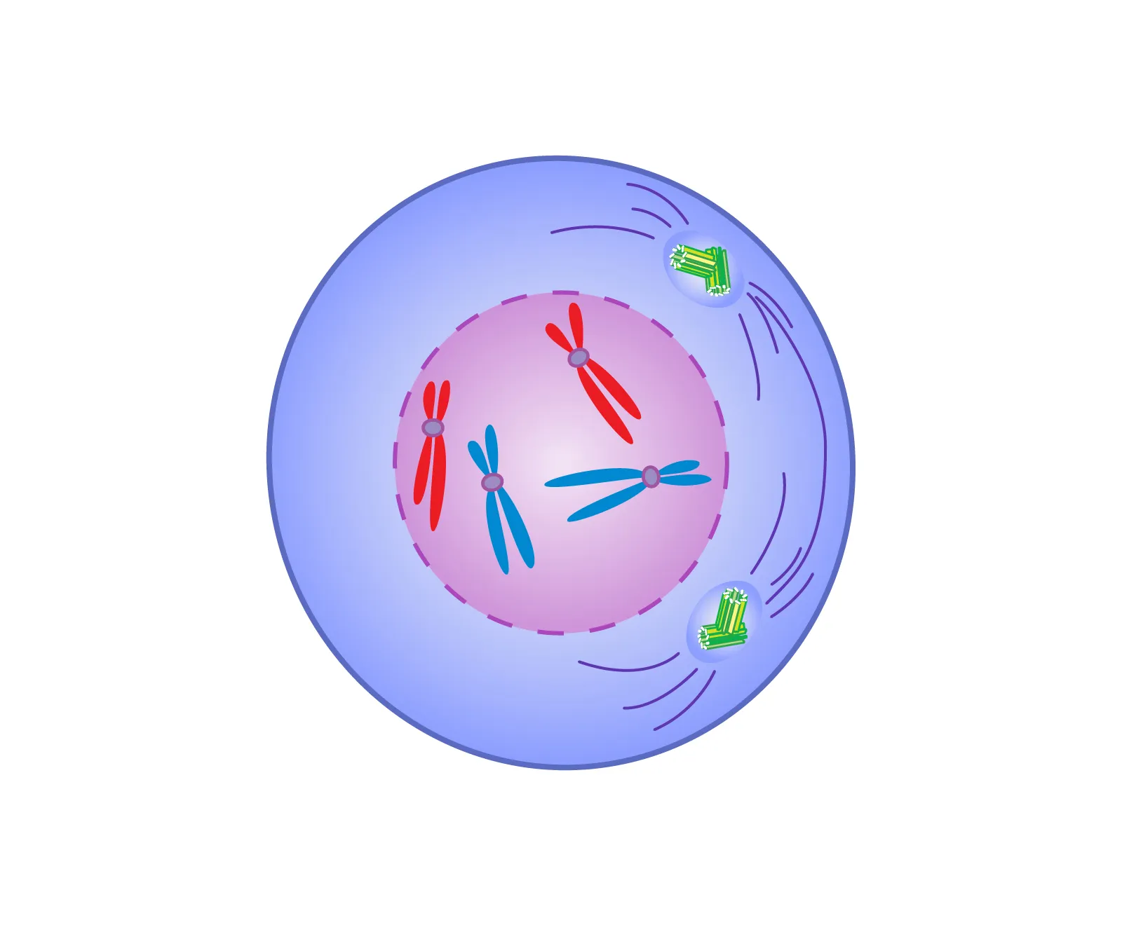
Prophase
Long and thread-like chromatids during interphase start to coil, the nuclear membrane dissolves and spindle fibers are formed and centrosomes migrate at the opposite poles of the cell.
the first phase of mitosis, the process that separates the duplicated genetic material carried in the nucleus of a parent cell into two identical daughter cells.
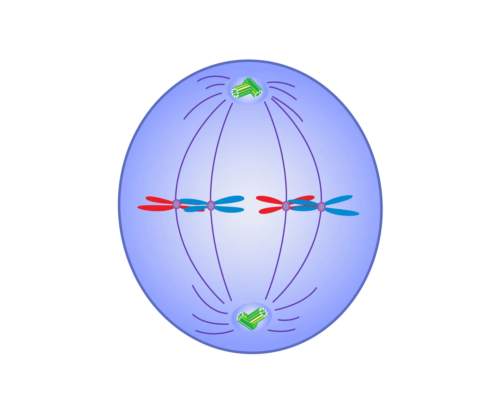
Metaphase
Chromosomes move and align themselves at the center of the cell called metaphase plate. Spindle fibers connect each chromosome on its centromere to the centrioles located at opposite poles of the cell.
individual chromosomes are spread out in the cell nucleus.
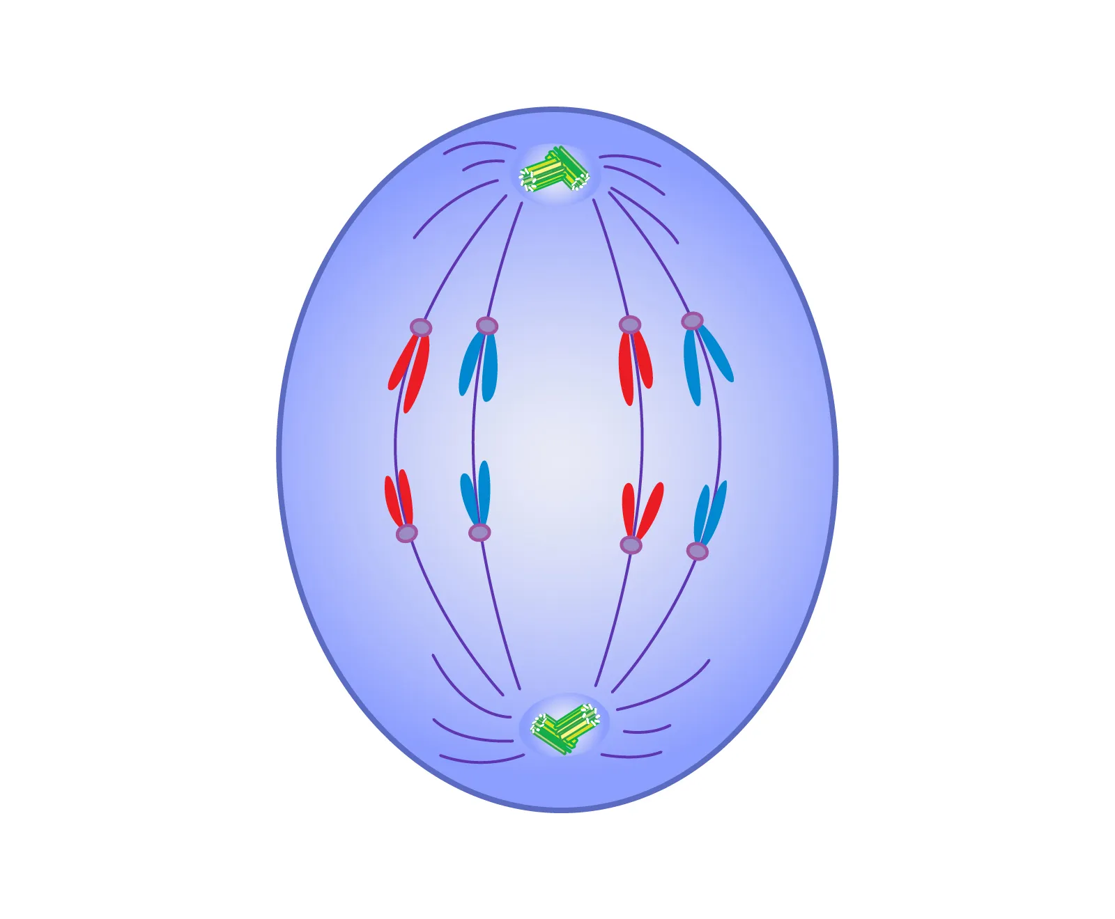
Anaphase
The sister chromatids of each chromosome divide or split and move toward opposite poles due to shortening of the spindle fibers.
the process that separates the duplicated genetic material carried in the nucleus of a parent cell into two identical daughter cells.
Telophase
The chromosomes are located on opposite poles of the cell and the spindle fibers disappear. At the poles, the chromosomes uncoil and the nucleolus and nuclear membrane begin to reform.
The process that separates the duplicated genetic material carried in the nucleus of a parent cell into two identical daughter cells.
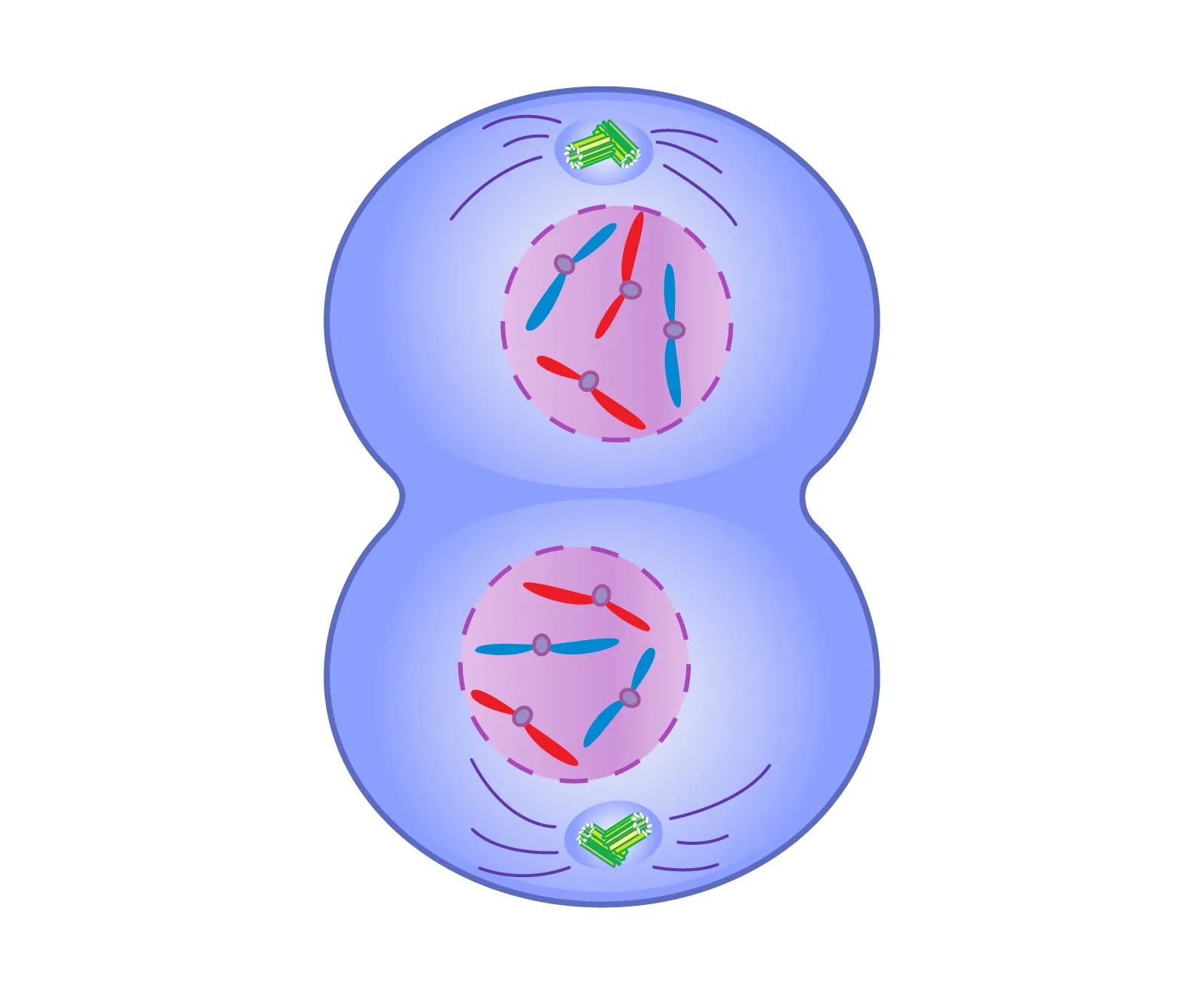
Cytokinesis
the physical process of cell division, which divides the cytoplasm of a parental cell into two daughter cells.
Histology
the study of tissues.
Epithelial Tissues
covers and protects surfaces, both outside and inside the body
exocrine and endocrine glands
Epithelial Tissue Characteristics
Mostly composed of cells
Covers body surfaces
Has an exposed surface
Attaches at the basal surface
Specialized cell connections and matrix attachments
Avascular
Capable of regeneration
Simple epithelium
consists of a single layer of cells, with each cell extending from the basement membrane to the free surface.
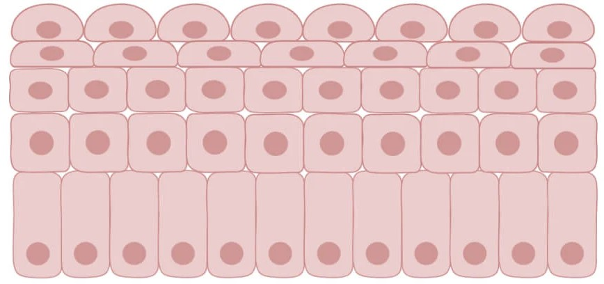
Stratified epithelium
consists of more than one layer of cells, but only the basal layer attaches the deepest layer to the basement membrane.
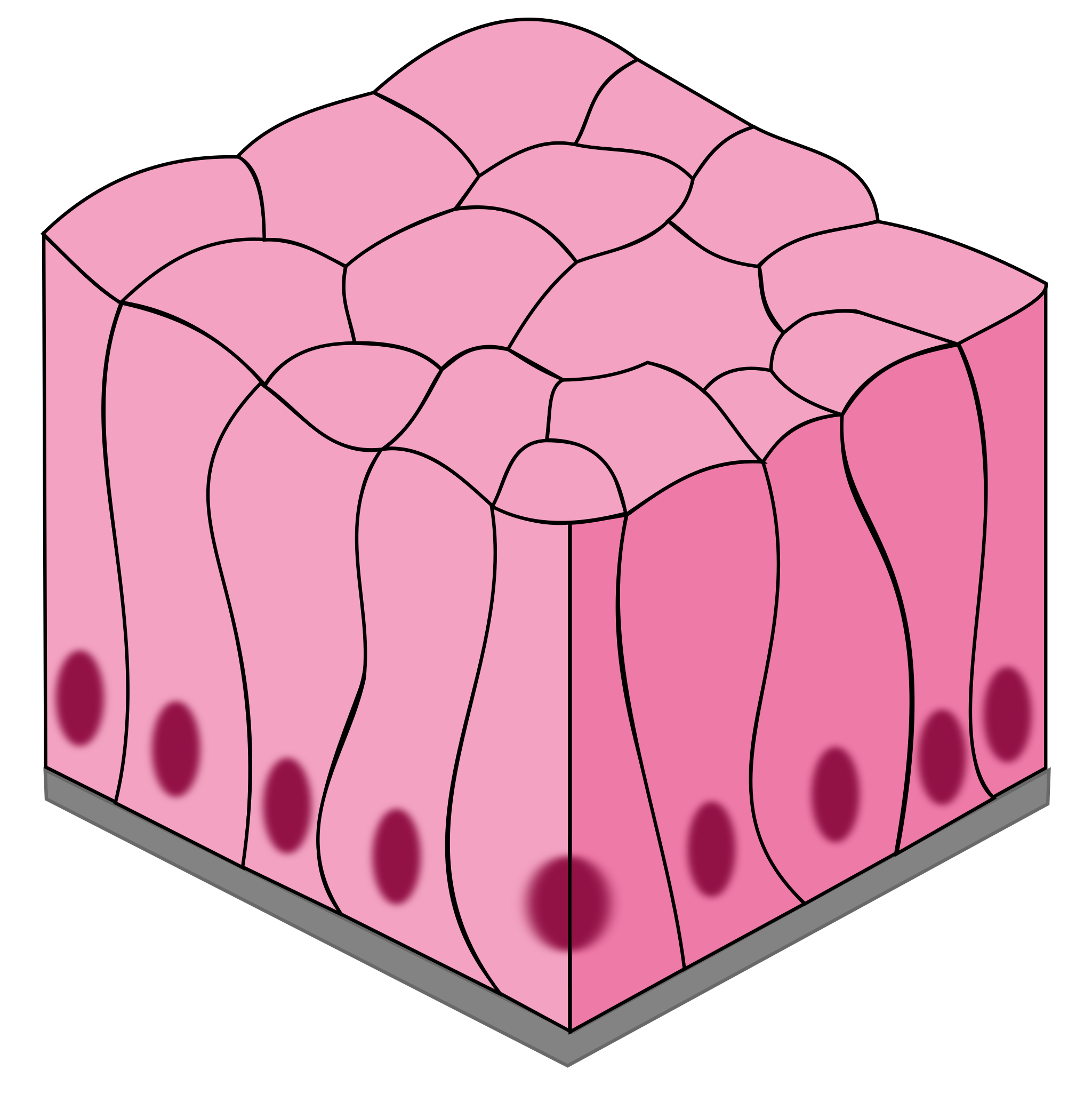
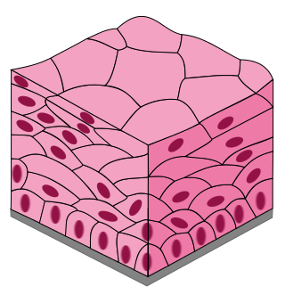
Pseudostratified columnar epithelium
s a special type of simple epithelium, that appears to be falsely stratified.
It consists of one layer of cells, with all the cells attached to the basement membrane.
Squamous cells
flat or scalelike
Cuboidal cells
cube-shaped—about as wide as they are tall.
Columnar cells
tend to be taller than they are wide.
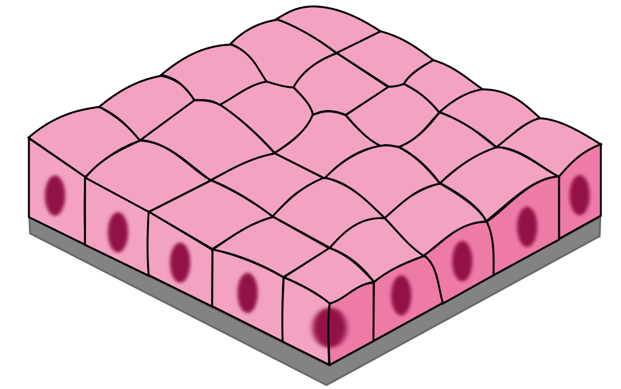
Simple Cuboidal
1 layer of square-shaped cells
Secretion
glands, ovaries, kidneys
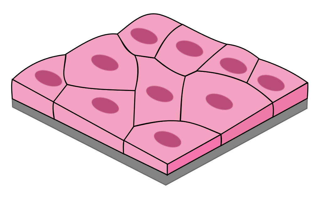
Simple Squamous
1 layer of flat, tile-like cells
good for diffusion & filtration
blood vessels, lungs, heart, kidneys
Pseudostratified Columnar
1 layer of tall, narrow cells appears stratified but isn’t
secretes mucus and propel debris out of respiratory tract (cilia)
nasal cavity and trachea
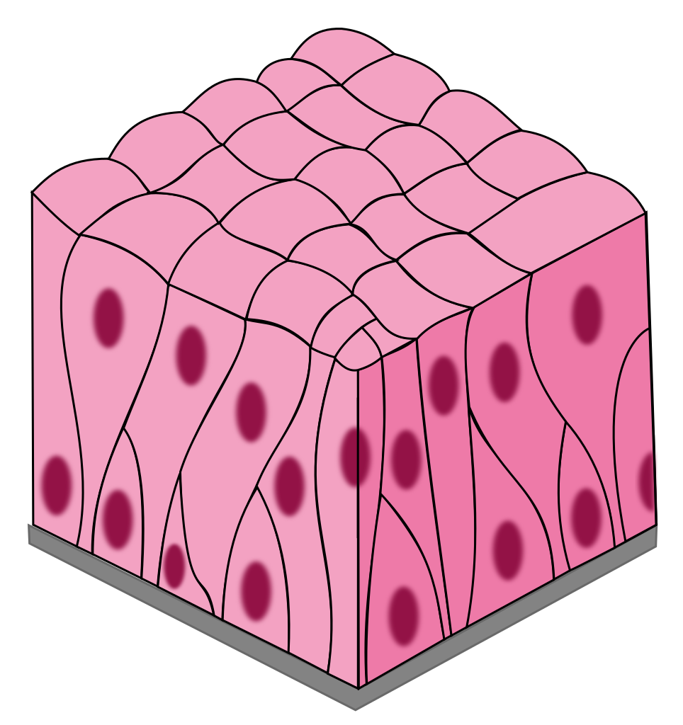
Simple Columnar
layer of tall, narrow cells
secrete mucus and absorption
stomach, intestines, resp. tract
Stratified Squamous
many layers of flat, tile-like cells
protect and acts as a barrier
skin, mouth, throat, esophagus
Transitional
special type of stratified epithelium; changes shape -stretched squamous.
hold fluids
urinary bladder
Keratinized Stratified Squamous Epithelium
The outer layer of the skin
The keratin reduces the loss of water from the body.
Nonkeratinized Stratified Squamous Epithelium
Of the mouth
Provides protection against abrasion and acts as a mechanical barrier.
Gastrulation
a critical stage in early animal development when a blastula reorganizes into a gastrula, a multi-layered embryo.
Ectoderm
The outermost layer.
Gives rise to:
Outer epithelium
Neural tube (spinal cord)
Neural crest (nervous system)
Mesoderm
The middle layer.
Gives rise to:
Notochord (bone of spine)
Digestive & urogenital system
Endoderm
The innermost layer.
Gives rise to:
Digestive system: Lining of the stomach, intestines, liver, pancreas.
Respiratory system: Lungs.
Endocrine glands: Thyroid, pancreas.
Tight junctions
cell connection structures that form barriers and anchor cells to each other.
Adhesion belts
Are found just below the tight junctions, and help tight junctions anchor epithelial cells to each other.
They prevent the passage of materials between epithelial cells because they completely surround each cell.
Endocrine glands
are ductless glands; they secrete their products (termed hormones) into the bloodstream.
Exocrine glands
glands with ducts; type of gland that release substances through ducts onto the body's surfaces.
Dense Regular Connective Tissue
Characterized by tightly packed, parallel collagen fibers, providing high tensile strength in a single direction. (limited space / orderly)
Found in:
Tendons: Connect muscles to bones.
Ligaments: Connect bones to bones at joints.
Aponeuroses: Sheet-like tendons that connect muscles to other muscles or bones.
Dense Irregular Connective Tissue
Collagen fibers are interwoven in a random arrangement, providing strength and resistance to stress in multiple directions. (Scattered & random)
Found in:
Dermis of the skin: Provides strength and resilience.
Joint capsules: Surround and support joints.
Organ capsules: Enclose organs such as the liver and kidneys.
Elastic connective tissues
are a type of dense connective tissue that are characterized by a high proportion of elastic fibers.
The presence of abundant elastin fibers within the extracellular matrix. Elastin is a protein that allows the tissue to stretch and recoil, returning to its original shape.
Areolar Connective Tissue
This is the most common type.
It has a loose arrangement of collagen, elastic, and reticular fibers. (connective tissue glue)
Contains various cell types like fibroblasts, macrophages, and mast cells.
Found beneath epithelia, surrounding organs, and in the dermis of the skin.
Adipose Connective Tissue
Primarily composed of adipocytes (fat cells).
Stores energy in the form of triglycerides.
Provides insulation and cushioning.
Found beneath the skin, around organs, and within bone marrow
Reticular Connective Tissue
Contains a network of reticular fibers that form a supportive framework for cells.
Found in lymphoid organs like lymph nodes, spleen, and bone marrow.
Provides a supportive framework for immune cells.
Forms the internal framework of stroma
Cartilage
Firm but flexible.
Provides support, cushioning, and smooth surfaces for joints.
Avascular (lacks blood vessels), so nutrients diffuse through the matrix.
Bone
Hard and mineralized (calcium and phosphorus).
Provides support, protection, and movement.
Stores minerals (calcium) and produces blood cells (in bone marrow).
Blood
Fluid connective tissue.
Transports oxygen, nutrients, hormones, and waste products.
Hyaline Cartilage
Most common type.
Found in:
Ends of bones in joints (articular cartilage)
Nose
Trachea
Ribs
Provides smooth surfaces for joint movement, flexibility, and support.
Elastic Cartilage
More flexible than hyaline cartilage due to the presence of elastic fibers.
Found in:
External ear
Epiglottis (flap of tissue that covers the windpipe)
Parts of the larynx (voice box)
Provides support and maintains the shape of structures that need to bend and flex.
Fibrocartilage
Contains a high density of collagen fibers.
Found in:
Intervertebral discs (between vertebrae)
Menisci (cartilage pads in the knee)
Pubic symphysis (joint where the two pubic bones meet)
Provides support and shock absorption in areas that experience high stress.
Osteoprogenitor
Stem cells: These are the precursor cells for all other bone cells, and can differentiate into osteoblasts.
Osteoblasts
Bone-building cells: They are responsible for synthesizing and secreting the organic matrix of bone (collagen) and initiating the process of mineralization (hardening of the bone)
Osteocytes
Mature bone cells: These are the most abundant type of bone cell.
Function: They maintain the bone matrix and regulate bone turnover.
They also play a role in sensing mechanical stress on the bone.
Osteoclasts
Bone-resorbing cells: They are large, multinucleated cells that break down bone tissue.
Function: This process is crucial for bone remodeling, repair, and calcium homeostasis/
Red Blood Cells (Erythrocytes)
Primarily responsible for transporting oxygen from the lungs to all the cells in the body and carrying carbon dioxide back to the lungs to be exhaled.
White Blood Cells (Leukocytes)
Key components of the immune system, fighting infections.
Platelets (Thrombocytes)
Function: Essential for blood clotting.
Role: When a blood vessel is injured, platelets clump together to form a plug, helping to stop bleeding.
Skeletal Muscle
Appearance: Striated (has visible stripes)
Control: Voluntary
# of Nuclei: Multinucleated
Location of Nuclei: Peripheral