anatomy bojrab ch 4
 Call Kai
 Learn
Learn Practice Test
 Spaced Repetition
 Match
Match Flashcards
 Knowt Play
1/197

There's no tags or description
Looks like no tags are added yet.
Name | Mastery | Learn | Test | Matching | Spaced | Call with Kai | Chat |
|---|
No analytics yet
Send a link to your students to track their progress
198 Terms
true or false: Individual body cells are specialized
Each type performs specific functions that maintain homeostasis
true
tissues
-Groups of cells similar in structure that perform common or related function
histology
-Study of tissues
4 basic tissue types
epithelial, connective, muscle, and nervous tissue
nervous tissue overview
internal communication
- brain
- spinal cord
- nerves
muscles tissues overview
contracts to cause movement
- skeletal muscles
- cardiac muscles
- smooth muscles
epithelial tissue overview
forms boundaries between different environments, protects, secretes, absorbs, filters
- lining of digestive tract organs
- glands
- skin surface (epidermis)
connective tissue overview
supports, protects, binds other tissues together
- bones
-tendons
-fat and other soft padding tissue
fixed
tissue is preserved with solvent to be viewed
sectioned
cut into slices thin enough to transmit light to electrons to be viewed
stained
to enhance contrast, although artifacts detract from what the sample looks like in living tissues
artifacts
distortions
light microscopy staining
colored dyes
Electron Microscopy staining
uses heavy metal salts
TEM
transmission electron microscopy, shows a section
SEM
scanning electron microscopy, shows a surface
epithelial tissue definition
is a sheet of cells that covers a body surface or lines a body cavities
2 main forms of epithelial tissue
covering and lining epithelia, glandular epithelia
Covering and lining epithelium
-forms the outer layer of skin, lines open cavities, and covers walls and organs of the ventral body cavity
▪Skin
▪Lining of urogenital, digestive, and respiratory systems
Glandular epithelium
-forms glands of the body
▪Salivary glands
functions of epithelial tissue
protection, absorption, filtration, excretion, secretion, and sensory reception
5 distinguishing characteristics of epithelial tissue
1.Polarity
2.Specialized contacts
3.Supported by connective tissue
4.Avascular but innervated
5.Regeneration
apical surface of epithelial tissue
-upper free side, is exposed to surface or cavity
▪Most apical surfaces are smooth, but some have specialized fingerlike projections called microvilli
basal surface of epithelial tissue
lower attached side, faces inwards toward body and attaches to basal lamina
basal lamina
▪an adhesive sheet that holds basal surface of epithelial cells to underlying cells
apical and basal layers of epithelial tissue (picture)

specialized contacts of epithelial tissue
-Epithelial tissues need to fit closely together
▪Many form continuous sheets
-Specialized contact points bind adjacent epithelial cells together
lateral contacts of epithelial tissue include...
tight junctions and desmosomes
true or fale: All epithelial sheets are supported by connective tissue
true
reticular lamina
deep to basal lamina, consists of network of collagen fibers
basement membrane
made up of basal and reticular lamina, reinforces epithelial sheet, resists stretching and tearing, defines epithelial boundary
cancer cells and the epithelium
•Cancerous epithelial cells are not contained by the basement membrane boundary like other cells
•They penetrate the boundary and invade underlying tissues, resulting in spread of cancer
epithelial cells are avascular but innervated
-No blood vessels are found in epithelial tissue
▪Must be nourished by diffusion from underlying connective tissues
-Epithelia are supplied by nerve fibers, however
regeneration of epithelial tissue
-Epithelial cells have high regenerative capacities
-Stimulated by loss of apical-basal polarity and broken lateral contacts
-Some cells are exposed to friction, some to hostile substances, resulting in damage
▪Must be replaced
▪Requires adequate nutrients and cell division
Classification of Epithelial Tissue
•All epithelial tissues have two names
-First name indicates number of cell layers
-Second name indicates shape of cells
Simple epithelia (name)
▪consists of a single cell layer
-Located where thin barrier is desirable
Stratified Epithelia (name)
▪consists of two or more cell layers
-Located in high-abrasion areas (example: lining of mouth)
squamous
flattened and scale-like

cuboidal
box-like, cube
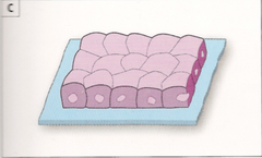
columnar
tall, column-like

true or false- cell shape is named according to basal layer
false- In stratified epithelia, shape can vary in each layer, so cell shape is named according to the cell shape in apical layer
simple epithelia
involved in absorption, secretion, or filtration processes
simple squamous epithelium
Cells are flattened laterally, and cytoplasm is sparse
Function where rapid diffusion is priority
Example: kidney, lungs
Endothelium
simple squamous epithelia- lining of lymphatic vessels, blood vessels, and heart
Mesothelium
simple squamous epithelia- serous membranes in the ventral body cavity
simple cuboidal epithelium
-Single layer of cells
-Involved in secretion and absorption
-Forms walls of smallest ducts of glands and many kidney tubules
simple columnar epithelium
Single layer of tall, closely packed cells
Involved in absorption and secretion of mucus, enzymes, and other substances
Found in digestive tract, gallbladder, ducts of some glands, bronchi, and uterine tubes
simple columnar epithelium and mucus
Some cells have microvilli, and some have cilia
Some layers contain mucus-secreting goblet cells
Ciliated cells move mucus
Pseudostratified columnar epithelium
Cells vary in height and appear to be multi-layered and stratified, but tissue is in fact single-layered simple epithelium
Many cells are ciliated
Involved in secretion, particularly of mucus, and also in movement of mucus via ciliary sweeping action
Located mostly in upper respiratory tract, ducts of large glands, and tubules in testes
"Pseudo" means...?
false
stratified
-Involve two or more layers of cells
-New cells regenerate from below
▪Basal cells divide and migrate toward surface
-More durable than simple epithelia because protection is the major role
Stratified squamous epithelium
-Most widespread of stratified epithelia
-Free surface is squamous, with deeper cuboidal or columnar layers
-Located in areas of high wear and tear (example: skin)
-Keratinized cells found in skin; nonkeratinized cells are found in moist linings
Stratified cuboidal epithelium
-Quite rare
-Found in some sweat and mammary glands
-Typically, only two cell layers thick
Stratified columnar epithelium
-Also, very limited distribution in body
-Small amounts found in pharynx, in male urethra, and lining some glandular ducts
▪Usually occurs at transition areas between two other types of epithelia
▪Only apical layer is columnar
transitional epithelium
-Forms lining of hollow urinary organs
▪Found in bladder, ureters, and urethra
-Basal layer cells are cuboidal or columnar
-Ability of cells to change shape when stretched allows for increased flow of urine and, in the case of bladder, more storage space
gland
One or more cells that make and secrete an aqueous fluid called a secretion
how are glands classified?
site of product release, relative number of cells forming the gland
endocrine gland meaning
▪ductless glands that produce hormones
exocrine gland meaning
▪secrete products onto body surfaces or into body cavities
-Example: sweat glands
example of unicellular gland
goblet cells
example of multicellular gland
salivary
endocrine glands
-Ductless glands
▪Secretions are not released into a duct; are released into surrounding interstitial fluid, which is picked up by circulatory system
-Secrete (by exocytosis) hormones
-Target organs respond in some characteristic way
hormones
messenger chemicals that travel through lymph or blood to their specific target organs
exocrine glands
-Secretions are released onto body surfaces, such as skin, or into body cavities
-More numerous than endocrine glands
-Secrete products into ducts
-Can be:
▪Unicellular
▪Multicellular
examples of exocrine glands
mucous, sweat, oil, and salivary glands
important unicellular gland
mucous cells and goblet cells, found in epithelial linings of intestinal and respiratory tracts and produce mucin
mucin
a sugar-protein that can dissolve in water to form mucus, a slimy protective, lubricating coating
multicellular exocrine glands
-Multicellular exocrine glands are composed of a duct and a secretory unit
-Usually surrounded by supportive connective tissue that supplies blood and nerve fibers to gland
▪Connective tissue can form capsule around gland, extend into gland, and divide it into lobes
how are multicellular exocrine glands classified?
structure and mode of secretion
Simple exocrine glands versus. compound glands structure
Structure- Simple exocrine glands have unbranched ducts versus. compound glands have branched ducts
3 forms of secretory units
tubular, alveolar, tubuloalveolar
tubular
-if the secretory cells form tubes
alveolar
-if the secretory cells form sacs
Tubuloalveolar
have both types of secretory units
merocrine
▪secrete products by exocytosis as secretions are produced
-Most common and does not alter secretory cells
examples of merocrine
most sweat glands, pancreas, salivary glands
holocrine
-accumulate products within until rupture
-Replaced by division of underlying cells
examples of holocrine
-sebaceous (oil) glands
apocrine
▪accumulate products within, but only apex ruptures
-Cell repairs damage and repeats
-Controversy if present in humans
examples of apocrine
-Mammary glands are closest example, but most scientists classify mammary glands as merocrine glands
what is the most abundant and widely distributed of the primary tissues?
connective tissue
major functions of connective tissue
binding and support, protecting, insulating, storing reserve fuel, transporting substances within the body (blood)
4 main classes of connective tissue
connective tissue proper
cartilage
bone
blood
connective tissue proper subclases
loose connective tissue (areolar, adipose, reticular)
dense connective tissue (regular, irregular, elastic)
connective tissue proper components
cells: fibroblasts, fibrocytes, defense cells, adipocytes
matrix: gel- like ground substance, all three fiber types (collagen, reticular, elastic)
connective tissue proper general features
Six different types; vary in density and types of fibers
Functions as a binding tissue
Resists mechanical stress, particularly tension
Provides reservoir for water and salts
Energy (fat) storage
cartilage subclasses
Hyaline cartilage, elastic cartilage, and fibrocartilage.
cartilage components
cells: chondroblasts (found in growing cartilage), chondrocytes
matrix: gel-like ground substance, fibers (collagen and elastic fibers in some)
cartilage general features
Resists compression because of the large amounts of water held in the matrix
Functions to cushion and support body structures
Avascular (lacks blood vessels)
bone tissue subclasses
compact bone and spongy bone
bone tissue components
cell: osteoblasts, osteocytes
matrix: Gel-like ground substance calcified with inorganic salts, fibers (collagen)
bone tissue general features
Hard tissue that resists both compression and tension
Functions in support
blood components
cells: red blood cells/erythrocytes, white blood cells/leukocytes, platelets
matrix: plasma
blood general features
-a fluid tissue
- functions to carry O2, CO2, nutrients, wastes, and other substances (such as hormones)
Two characteristics make connective tissues different from other primary tissues:
extracellular matrix and common origin
extracellular matrix of connective tissue
-Extracellular matrix of nonliving portion of connective tissues in which the living cells are suspended in
▪Supports cells so they can bear weight, withstand tension, endure abuse
▪All other primary tissues are composed mainly of cells, not extracellular matrix
common origin of connective tissue
arise from mesenchyme (embryonic tissue)
3 main components of all connective tissues
ground substance, fibers, cells
true or false: Cells and fibers together make up the extracellular matrix
false- Ground substance and fibers together make up the extracellular matrix
true or false: Composition and arrangement of these three elements vary considerably in different types of connective tissues
true