Bones of the pelvic limb
1/54
There's no tags or description
Looks like no tags are added yet.
Name | Mastery | Learn | Test | Matching | Spaced | Call with Kai |
|---|
No analytics yet
Send a link to your students to track their progress
55 Terms
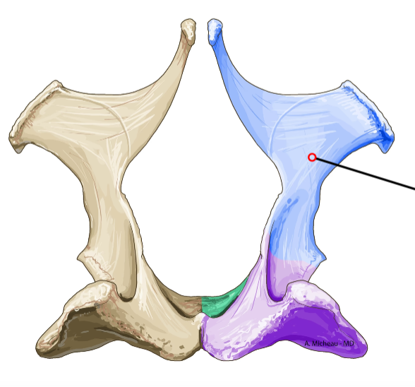
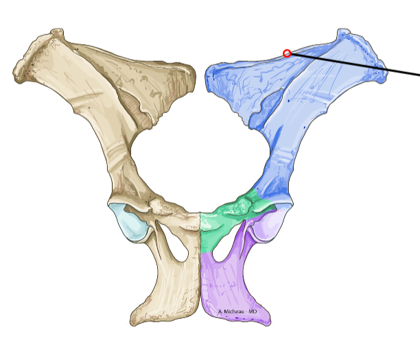
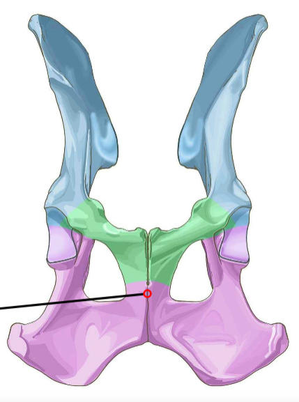
Os ilium
Wing-shaped cranial part of pelvis; forms sacroiliac joint dorsally.
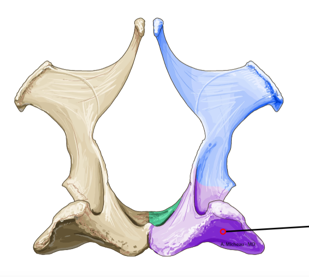
Os ischium
Caudal ventral part of pelvis; forms ischiatic tuberosity for hamstring attachment.
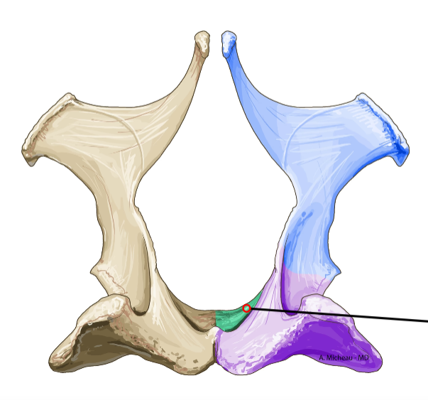
Os pubis
Cranial ventral part of pelvis; forms pubic symphysis medially.
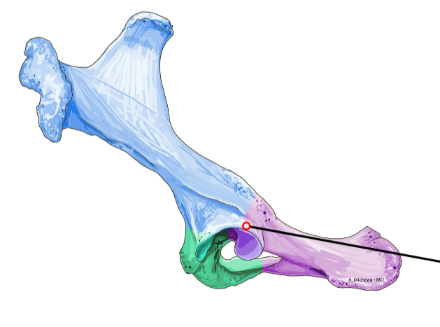
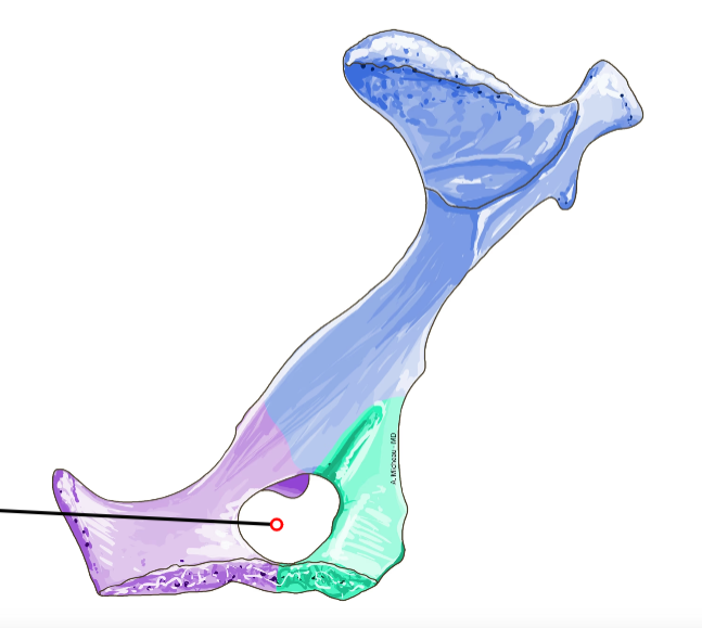
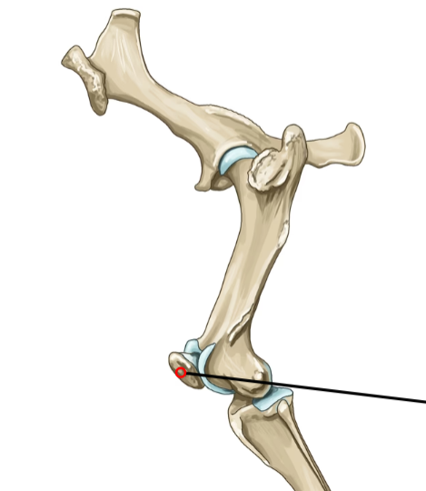
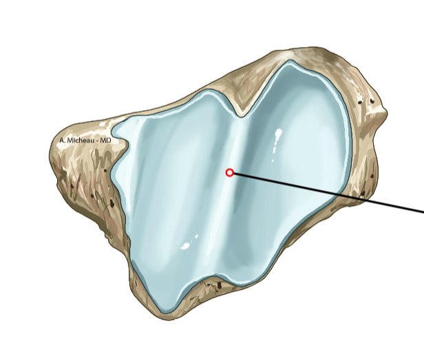
Acetabulum
Deep socket formed by ilium, ischium, pubis; articulates with femoral head.
Foramen obturatorium
Large oval opening between pubis and ischium; passage for obturator nerve.
Crista iliaca
Dorsal border of ilium; attachment for abdominal muscles.
Tuberositas iliaca
Rough prominence on ventral ilium; origin of rectus femoris.
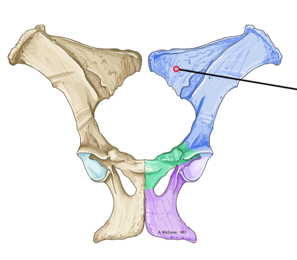
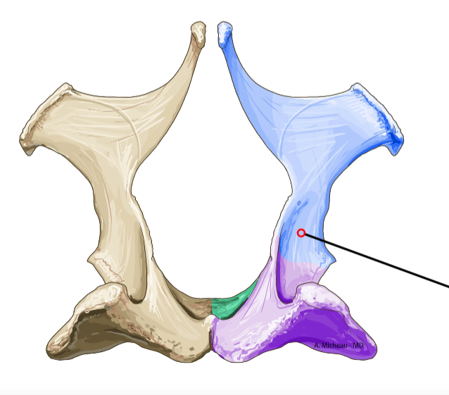
Corpus ossis ilii
Body of ilium; connects to sacrum.
Arcus ischiadicus
Arch formed by ischium; borders pelvic outlet.
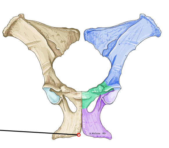
Symphysis pelvina
Midline fusion of pubes and ischia; forms pelvic floor.
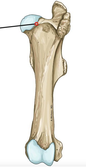
Caput femoris
Rounded proximal head; fits into acetabulum.
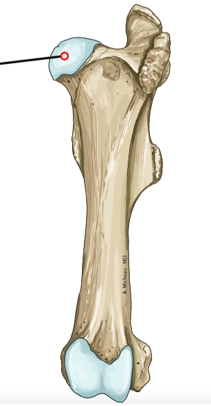
Collum femoris
Neck connecting head to shaft.
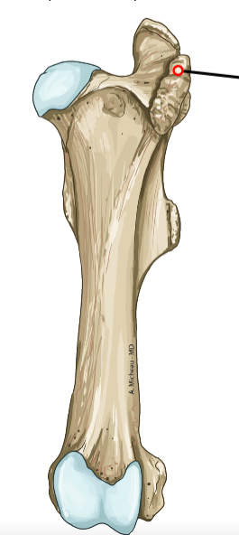
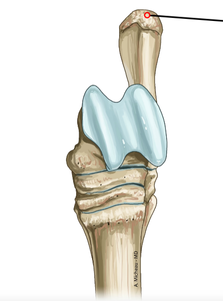
Trochanter major
Large lateral prominence proximal; insertion of gluteal muscles.
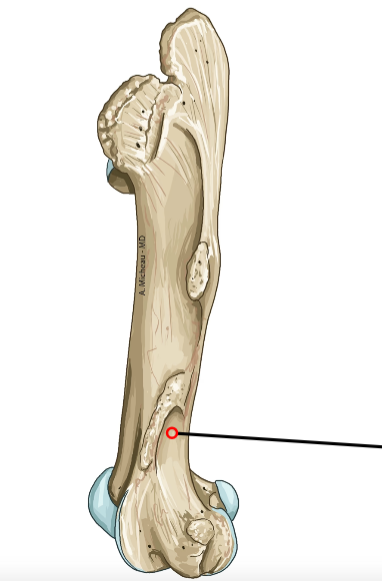
Trochanter minor
Smaller medial prominence proximal; insertion of iliopsoas.
Fossa trochanterica
Depression between trochanters; insertion of obturator externus.
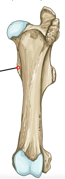
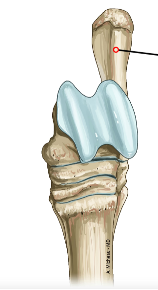
Facies aspera
Rough caudal ridge on shaft; attachment for adductors.
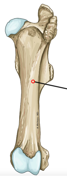
Corpus femoris
The body (Shaft) of femur is the diaphysis of the femur.
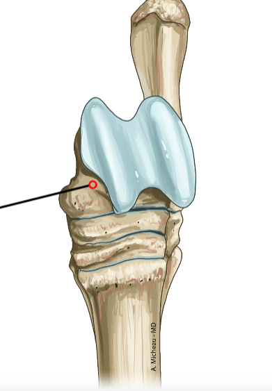
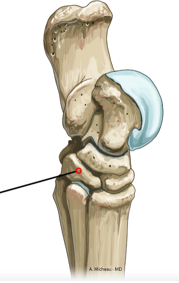
Condylus lateralis
Lateral distal condyle; articulates with tibia.
Condylus medialis
Medial distal condyle; articulates with tibia.
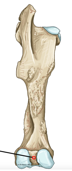
Fossa intercondylaris
Deep groove between condyles; cruciate ligament attachment.
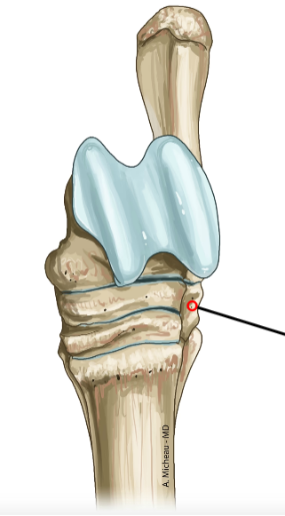
Trochlea femoris
Cranial pulley for patella; guides extensor mechanism.
Fossa supracondylaris
Shallow depression above lateral condyle; origin of gastrocnemius.
Patella
Sesamoid bone in quadriceps tendon; articulates with femoral trochlea.
Basis patellae
Proximal broad end; insertion of quadriceps.
Apex patellae
Distal pointed end; attachment of patellar ligament.
Facies articularis
Caudal smooth surface; glides on femoral trochlea.
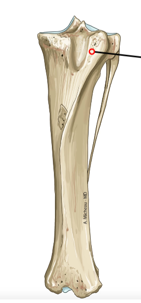
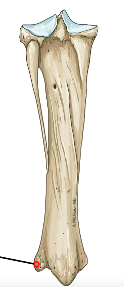
Tuberositas tibiae
Cranial prominence proximal; insertion of patellar ligament.
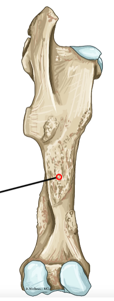
Corpus tibiae
Triangular shaft; caudal flat surface.

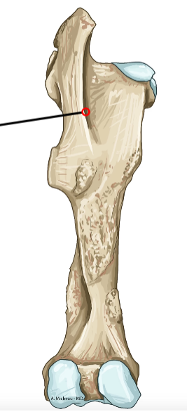
Crista tibiae
Sharp cranial border; palpable subcutaneously.
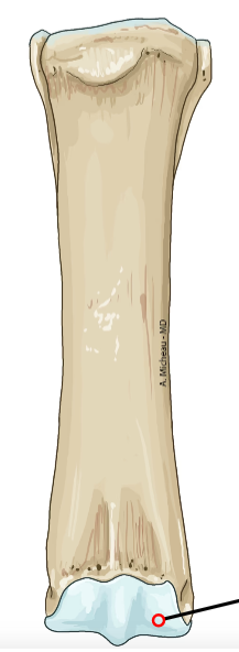
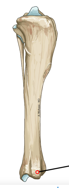
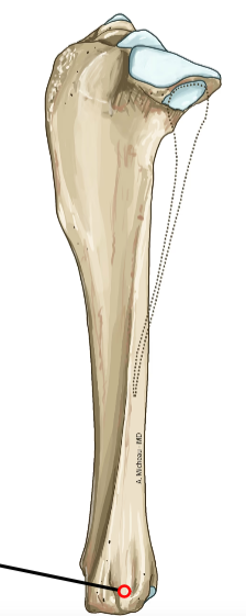
Malleolus medialis
Medial distal prominence; ligament attachment.
Cochlea tibiae
Distal grooved surface; articulates with talus.
Sulcus malleolaris lateralis
Lateral groove distal; peroneal tendon passage.
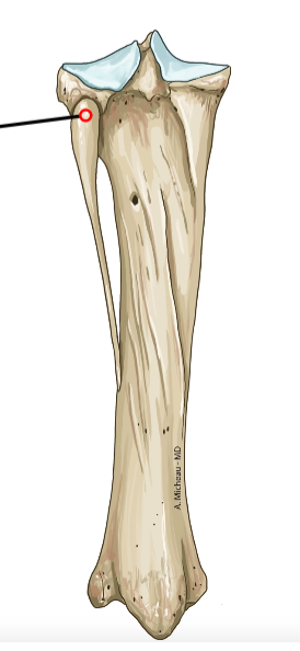
Caput fibulae
Proximal head; articulates with tibia laterally.
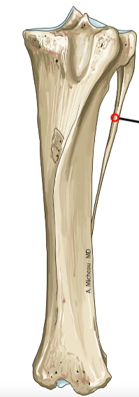
Corpus fibulae
Slender shaft; minimal weight-bearing.
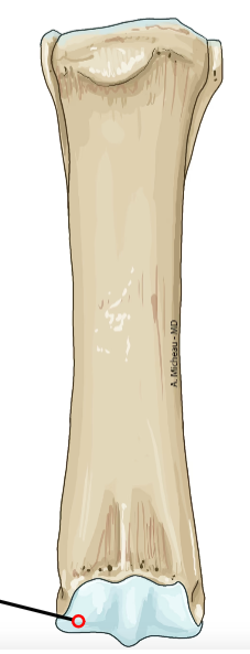
Malleolus lateralis
Distal lateral prominence; ligament attachment.
Facies articularis distalis
Distal facet; articulates with talus laterally.
Talus
Proximal tarsal bone; articulates with tibia and fibula proximally.
Trochlea tali
Proximal pulley; fits into tibial cochlea.
Collum tali
Neck connecting trochlea to body.
Caput tali
Distal head; articulates with central tarsal.
Calcaneus
Largest tarsal bone; forms hock point caudally.
Tuber calcanei
Proximal prominence; insertion of Achilles tendon.
Processus coracoideus
Cranial projection on calcaneus; ligament attachment.
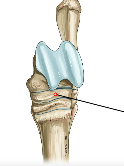
Os tarsale centrale
Central tarsal bone; connects proximal and distal rows.
Os tarsale I
First distal tarsal; supports Mt1 if present.
Os tarsale II
Second distal tarsal; supports Mt2.
Os tarsale III
Third distal tarsal; supports Mt3.
Os tarsale IV
Fourth distal tarsal; supports Mt4 and Mt5.
Os metatarsale II
Medial weight-bearing metatarsal; supports digit 2.
Os metatarsale III
Central longest metatarsal; main weight axis.
Os metatarsale IV
Lateral metatarsal; supports digit 4.
Os metatarsale V
Outermost metatarsal; supports digit 5.
Basis metatarsalis
Proximal base; articulates with distal tarsals.
Corpus metatarsale
Slender shaft; smooth plantar surface.
Caput metatarsale
Distal head; articulates with proximal phalanx.