Exam 4
1/77
There's no tags or description
Looks like no tags are added yet.
Name | Mastery | Learn | Test | Matching | Spaced | Call with Kai |
|---|
No analytics yet
Send a link to your students to track their progress
78 Terms
M ganglion cells
From peripheral rods - detect motion in periphery
High convergence 10-50 rods → 1 bipolar
Multiple bipolar → 1 M ganglion cell
P ganglion cells
From center/surround fields
Form + color detection
No convergence, high spacial resolution
Intrinsically Photosensitive Retinal Ganglion Cells
Detect light, but no conscious perception
No rods/cones - no rhodopsin - instead melanopsin
Why blind people still have circadian rhythm + pupillary reflex
From eye to optic tract
Ganglion axons form optic nerve (CN II)
Leave fovea at blind spot
Both sides reach optic chiasm
Temporal axons travel ipsilaterally into optic tract
Nasal axons travel contralaterally into optic tract
Recall nasal axons responsible for temporal optic field and temporal for nasal
Right optic tract = right temporal + left nasal axons = entire left visual field
Optic tract synapses onto
Hypothalamus - circadian rhythm
Superior colliculus - turning head to stimuli
Midbrain → pretectum - pupillary reflex
Lateral geniculate nucleus of thalamus → primary visual cortex
If light on left
Light falls onto left nasal + right temporal
Forveating
Looking through both foveas
If light in center, falls on both foveas at same location
Lateral geniculate nucleus of thalamus structure
6 sections
1&2 - M class 1 = contralateral, 2 = ipsilateral
3-6 - P class - 3&5 = ipsilateral, 4&6 = contralateral
Segregation important for depth perception
LGN has receptive fields like ganglion and a threshold for sending info to V1
LGN to V1
1&2 of LGN → 4Calpha of V1 (where/motion) n
3-6 of LGN → 4Cbeta of V1 (form + color)
*Still segregated by layers
Automatic gain control
LGN measures relative light intensity so light changes don’t feel too bright
Optic radiations
LGN sends optic radiations to V1 - relate to fovea
Top of fovea sees ground and bottom of fovea sees sky
So superior radiations see ground and run through upper bank
Inferior radiations see sky and run through lower bank
to V1
Layer 4C synapses onto
spiny or smooth stellate cells → Pyramidal cells
4C → 4B → layers 1&2 → V2, V3, etc
1&2 → 5&6 → SC, LGN, pons, etc
Pathway from 1&2 LGN
Dorsal pathway, magnocellualr inputs
The “where/mvmt” info
→ 4Calpha of V1 → V2 → middle temporal association area (MT) → Parietal lobe
Pathway from 3-6 LGN
Ventral pathway, parvocellular inputs
What/object pathway
→ 4Cbeta of V1 → V2 → V4 → Temporal lobe → Fusiform gyrus for faces
V1 - edges
V2 - general shape
Fusiform gyrus
Identify/associate an object
Complex visual connections, 1 cell receives unput from thousands of ganglion cells
Snakes simulate specific set of cells only for snakes before even know what a snake is
Specific cells for different parts/orientations of faces—respond to key face contrasts: ex: :)
V1 receptive fields
Not responsive to spots of light
Recognizes edges/light at angles - fundamental to how brain identifies objects
No response to horizontal bar - more vertical = more response
Ovular receptive field with bar in middle made of multiple overlapping cells/fields converging onto 1 pyramidal cell
Have simple cells for every orientation
Most firing at ideal angle but some firing at non ideal
Stellate → simple → pyramidal
Direction of mvmt
Stellate and simple cells responsible for sensing direction of light.
Inhibitory neurons connect stellate to simple cells so simple cells only fire if light moves in specific direction, otherwise inhibited
Ocular dominance in 4C
Ocular dominance columns in layer 4C. L or R eye column—changes every .5 mm
Segregation from LGN continues into 4C
Zero disparity cells
In V1
Fire only when analogous ganglion cells fire simultaneously on both retinas. Allows depth perception of objects in a straight line
Closer = light on temporal sides of retina
Further = light on nasal sides of retina
Different cells fire based on where on retina/depth
Binocular disparity cells
In V1
Fire when light strikes each retina at a different place
Cell for every possible conformation, also allow for depth perception`
V1 Structure
All angle pyramidal cells for both eyes and all color cells and all disparity cells in 1×1×2-4 mm cube of cortex
Takes output from LGN and breaks inro contrasting bars of light, then sends to higher order brain regions
Fovea gets largest SA of V1
Parasympathetic nervous system basics
Preganglionic neurons release Ach a ciliary ganglion onto post ganglionic neurons
Sympathetic nervous system basics
Preganglionic neurons release Ach, most post ganglionic use NE (sweat uses Ach)
Note: Preganglionic synapses directly onto adrenal gland
Prevertebral sympathetic ganglion
Celiac ganglion - stomach, liver pancreas
Superior mesenteric ganglion - colon
Inferior mesenteric ganglion - bladder, genitails
Lower extremities spinal nerves
Where does the sympathetic nervous system arise from?
Sympathetic preganglionic cell bodies arise from T1-L6 in the intermediolateral cell column in the gray matter between the dorsal and ventral horn.
Paraganglia leave through the ?lateral horn → through the white ramus → sympathetic trunk
@ Trunk:
Can go up to form cervical/chain ganglia
Leave through gray ramus to innervate blood vessels + skin
Synapse at trunk to lower motor neuron
Leave trunk and go to prevertebral ganglia
Prevertebral ganglia
close to organs they supply
Sympathetic chain ganglia
Chain of ganglia close to the cervical spinal cord
Where does the parasympathetic nervous system arise from?
From the cranial spine/brain stem
Edinger-westphal nucleus (in midbrain) → CN III (to ciliary ganglion for pupillary reflex)
Salivatory nuclei (upper medulla) → CN VII (facial nerve) + CN IX (saliva + tears)
Dorsal motor nucleus of vagus + Nucleus ambiguus (cardiac, larynx, pharynx) → CN X (HR, lung constriction)
And from the sacral spine (S1-S5) - intermediate gray zone b/t horns like the sympathetic nervous system - leaves spine to enter pelvic splanchnic nerve to synapse at ciliary ganglion
Regulation of HR
Carotid body (detects blood chemical comp) afferents in CN IX → nucleus of solitary tract
Baroreceptors (sense stretch by heart) afferents also → nucleus of solitary tract
Nucleus of solitary tract → nucleus ambiguus → cardiac plexus → SA node of heart
Also nucleus of solitary tract → preganglionic neurons in sympathetic nervous system → sympathetic chain ganglia → post ganglionic neuron → SA node
To inc/decrease HR/BP
To dec HR,
Carotid body or baroreceptors send APs to nulcues of solitary tract → APs to nucleus ambiguus → release Ach onto cardiac plexus → release Ach onto SA node → dec APs
NEED SYMPATHETIC INHIBITION AT THE SAME TIME
Nucleus of solitary tract → inhibits preganglionic neuron firing → inhibits post ganglionic neuron firing → decreases norepinephrine released onto heart
Regulation of core temp
Thermoreceptors all over body converge at comparator of hypothalamus
Preoptic area of thalamus sends APs to comparator based on set point
Comparator compares APs/sec from preoptic area and thermoreceptors to determine to inc or dec body temp and sends signals to rest of hypothalamus
During fever, pyrogens released by pathogens increase set point in preoptic area
Inc/dec core temp
Lateral + medial preoptic nuclei (anterior hypothalamus) - responsible for heat dissapation
Posterior area of hypothalamus - responsible for heat conservation
When we want to move
Motor cortex plans → sends info to basal ganglia and cerebellum
Basal ganglia and cerebellum send info back to excite the motor cortex enough to send info to motor neurons
Note that motor cortex dampens reflexes
Local circuit neurons
Receive sensory info and integrate w/ lower motor neurons near DRG
Motor neuron pools
Each major muscle = large poll of lower motor neurons coming from spinal cord
Organized spaciotopically - proximal regions more medial in spinal cord and distal regions more lateral in spinal cord
Flexor muscles more dorsal
Extensor muscles more ventral
Innervation
Single motor neuron innervates multiple muscle fibers, but single muscle fiber innervated by only 1 neuron. Lower innervation ratio for more precise mvmt
Eyes, ears: 1:10
Hands: 1:100
Posture: 1:180
Jumping: 1:1-2k
EPPs
Lower (alpha) motor neurons release Ach onto muscle fiber’s nicotinic ach receptor. Large SA in muscle fiber and very large EPSP from Ach
Muscle fiber types
Slow twitch - small forces over long time
Fast fatigue-resistant - large force for a long period, but not as long as slow twitch
Fast fatigable - large, explosive force but tire quickly
Graded recruitment
First recruit slow twitch, then fast fatigue-resistant, then fast fatigable
Slow twitch fibers have the smallest motor neurons (fast fatigable = largest neuron). Same ion channel density, but small neuron has smaller SA so higher R. V=IR, so small I produces large V in smaller neuron, firing AP, but doesn’t produce big enough V to fire AP in large neuron.
Muscle fiber summation
1 AP = 1 twitch. Multiple APs close together = twitches sum together to generate greater force
Unfused tetanus - so many APs, plateau of force, but can see individual twitches
Fused tetanus - so many APs, cannot see individual twitches
Proprioceptors
Sensory afferents give feedback about muscle + joint position
Extra + extrafusal muscle fibers
Extrafusal - produce tension, alpha motor neurons
Intrafusal - run parallel to extrafusal inside, wrapped in Ia spindle afferents
When extrafusal muscle stretches, stretches intrafusal and Ia spindles, generating AP
Ia afferents
Very fast APs, a lot of myelin
Output to lower motor neurons, local circuit neurons, dorsal nucleus of Clarke, somatosensory cortex (not conscious)
How keep arm at same height when pouring liquid?
Contract bicep + conscious initial setting of position
When add weight, extrafusal muscle fibers stretch so intrafusal muscle fibers stretch so Ia afferents send APs to DRG
Ias synapse onto flexor alpha motor neurons and send APs to activate more flexion. Ias also synapse onto inhibitory interneurons that synapse onto extensor alpha motor neurons and inhibit extension.
Gamma motor neurons
Control intrafusal muscle fibers/set tension
Usually contract intrafusal muscle fibers the same as extrafusal
If didn’t contract intrafusal, intrafusal couldn’t be stretched enough to send APs
Tension set up by gamma motor neurons determines threshold for adjustment
Glogi tendon organ
Series element between muscle and tenson
Has Ib afferent neurons + collagen fibrils in capsule
Measures shortening of muscle
Afferent output to same places and Ia neurons.
Does not respond to stretch
Acts in same way as Ia to prevent too much flexion
Nocioceptive reflexes
If step on glass, lift foot up + shift weight reflexively
APs from nociceptors → DRGs → local circuit neurons → excite flexor + inhibit extensor on one side and excite extensor + inhibit flexor on other side to lift up foot and shift weight
Tonic control while walking
Swing/flexion phase and stance/extension phase
When cat spinal cord cut, continues to walk and change pace
Can walk w/out input from brain
Central pattern generators in local spinal circuit that provide involuntary phasic control while walking
Medial motor neurons
Neurons to axial muscles that control posture
Colliculospinal/Tectospinal tract
Involuntary control of neck (turn head to sight/sound)
Superior (vision) + inferior (sound) colliculus (midbrain) → DEUSSATE in midbrain → down cervical spinal cord → branch bilaterally when synapse onto medial motor neuron in cervical spine
Reticulospinal tract
Colliculus → deussate in midbrain → synapse at pontine reticular formation → synapse bilaterally at target alertness/broader posture
Vestibulospinal tract
Begin in lateral and medial vestibular nuclei (medulla and pons) → synapse bilaterally at target → maintaining balance
Anterior/ventral corticospinal tract
Voluntary control of posture
Upper motor neurons in medial motor cortex → internal capsule → cerebral peduncle (midbrain) → pontine fiber bundles (middle pons) → synapse bilaterally (branch bilaterally at pyramidal deussation in spine) at lower motor neuron
Corticorecticulospinal tract
Involuntary, feed-forward posture - shift weight for movement before it happens
Primary motor cortex + medial + lateral premotor cortex → synapse bilaterally at reticular formation → synapse at lower motor neurons bilaterally
Lateral corticospinal tract
Voluntary movement of limbs
Movement planned in medial (internal cues) or lateral (external cues) premotor cortex → primary motor cortex → upper motor neurons → internal capsule → cerebral peduncle (midbrain) → pontine fiber bundles (middle pons) → pyramidal DECUSSATION at caudal medulla → synapse at ventral horn onto lower motor neurons
Motor cotex mapping
Somatotopic mapping like somatosensory cortex
Corticobulb tract
Voluntary face + neck movement
lateral portion of precentral gyrus → primary motor cortex → internal capsule → cerebral peduncle (midbrain) → middle pons →
synapse at contralateral trigeminal motor nucleus of CN V (jaw) or CN VII (face) (middle pons)
OR
pontine fiber bundles → middle medulla → synapse contralaterally at hypoglossal nucleus CN XII (tounge) or CN XI (trapezius + sternocleidomastoid muscles)
Facial expressions tract
Lower face:
Facial representation in primary motor cortex → pons → contralateral facial nucleus → facial nerve (VII)
Upper face:
Both sides of cortex have two paths
1 path: face representation in cingulate motor area → synapse ipsilaterally at facial nucleus
2nd path: face representation in cingulate motor area → synapse contralaterally at facial nerve
So damage on one side won’t affect upper face unless after facial nerve
Targeting limbs in voluntary movement
Small groups of upper motor neurons = many muscle fibers
Encode movement by using multiple lower motor neuron pools, not only one
Ex: money reaching 180 degrees uses 90 degrees, 135, 180, and 275 muscles
Sum multiple vectors to get desired movement
Motor areas
Basic movements use motor + somatosensory cortex
More complex movements add in supplementary motor area
Can also rehearse movement with supplementary motor area only
Basal ganglia
Suppress inappropriate motor programs
= Striatum = putamen (body) + caudate nucleus (head, neck, eyes)
Decides what to suppress from info from frontal association and motor association cortex
Ventral anterior + lateral complex of thalamus (VA/VL)
Relay info to motor cortex
Caudate nucleus
Inputs from multimodal association cortex, frontal lobe, and eye movement areas
Putamen
Inputs from somatosensory, visual, premotor, and motor cortex
Somatotopic mapping
Direct caudate pathway
Caudate → Substantia nigra pars reticulata (SNPR) → superior colliculus (SC)
Direct putamen pathway
Putamen → Globus pallidus internal segment (GPIS) → VA/VL thalamus → motor cortex
Indirect putamen pathway
Putamen → Globus pallidus external segment (GPES) → subthalamic nucleus (SN) → GPIS → VA/VL → motor cortex
Basal ganglia when we don’t want to move
No excitation of striatum → no GABA release onto SNPR or GPIS → SNPR and GPIS active → release GABA onto SC or VA/VL → SC or VA/VL inhibited
Basal ganglia when we do want to move
Excitation of the striatum → GABA release onto SNPR or GPIS → inhibition of SNPR or GPIS → no GABA release onto VA/VL or SC → VA/VL or SC active
Substantia nigra pars compacta (SNPC)
High density of dopaminergic neurons + communicate directly with striatum because cortical inputs alone not large excitement in striatum, need more excitement from SNPC
Acts on D1 and D2 pathway
D1 Pathway
SNPC releases dopamine into excitatory synapses in direct pathway. Increases activity in striatum → more GABA release onto SNPR or GPIS → less GABA release onto VA/VL or SC → more activity
D2 pathway
SNPC releases dopamine onto inhibitory synapses in indirect pathway. Decreases activity in striatum → decreased GABA on GPES → GPES active → GPES releases GABA onto SN → SN not active → does not excite GPIS → no inhibition of unwanted movement
Parkinson’s
Little wanted or unwanted movement
Degeneration of dopadenergic receptors in SNPC
Huntington’s disease
Degradation of striatum neurons onto GPES → can’t inhibit GPES → GPES active → GABA onto SN → SN inactive → GPIS inactive → unwanted movement + poor emotion regulation
How refine + coordinate movement?
Cerebellum integrates sensory info and coordinates precise timing of ongoing mvmt
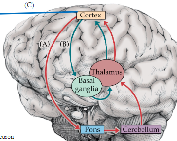
Cerebellum Input Pathways
Info from primary, pre, and supplementary motor, and somatosensory cortex (to pontine nuclei → deussates and enters through middle peduncle) + info from clarke’s nucleus and cuneate nucleus (through inferior peduncle) to coordinate timing
Info from sensory motor association cortex (to pontine nuclei → deussates and enters through middle peduncle) to refine movement
Info from vestibular nuclei (through inferior peduncle) for balance
Info from inferior olive (through inferior peduncle) for motor learning
Cerebellum output pathways
Out through superior peduncle
To red nucleus to inferior olive or
to VL complex of thalamus to primary + premotor cortex
Motor pathway with cerebellum decussations
Motor cortex → internal capsule → pontine nuclei → DECUSSATE through middle cerebellar peduncle → cerebellum → deep cerebellar nuclei → superior cerebellar peduncle → DECUSSATE → VL complex of thalamus → primary + premotor cortex) → DECUSSATE again before lower motor neurons
Left cerebellum for left movement