A&P1 Lab Test #4
1/218
There's no tags or description
Looks like no tags are added yet.
Name | Mastery | Learn | Test | Matching | Spaced |
|---|
No study sessions yet.
219 Terms

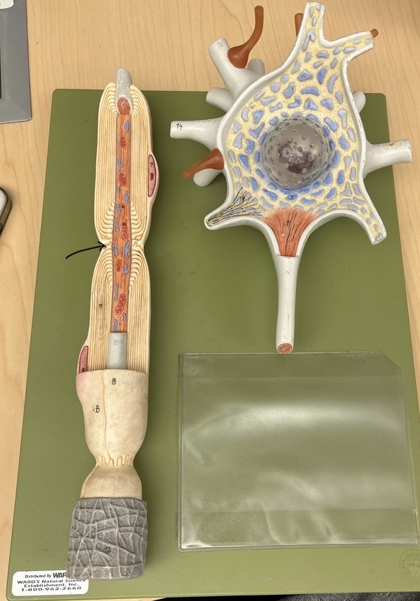
What does this model represent?
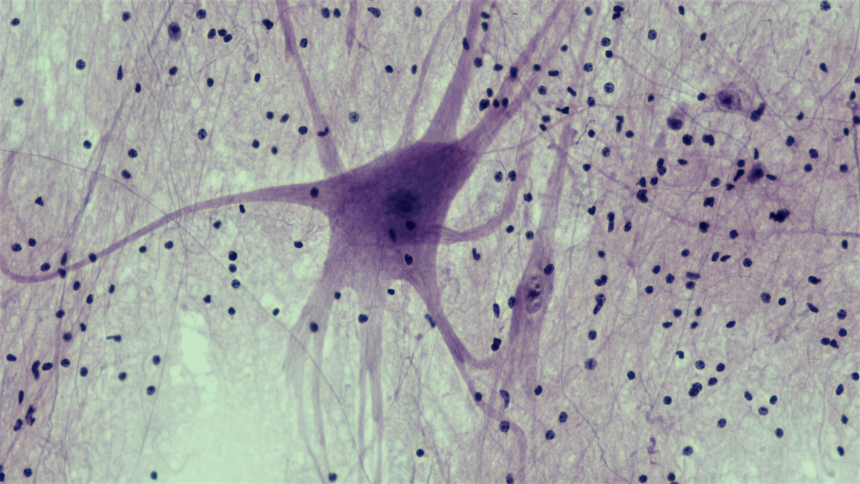
somatic motor neuron; multipolar


What is the blue part? Function?
Nissl bodies; to produce proteins


synaptic end bulbs

What is this area?
soma (cell body)

nucleus


dendrites

axon hillock

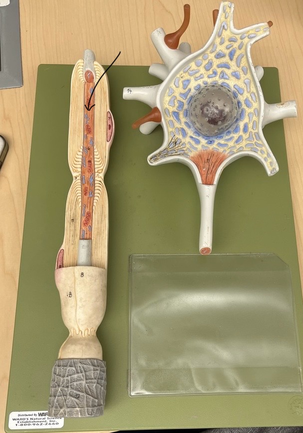
axon


What is this? Function?
myelin sheath; speeds up action potential


nucleus of schwann cell


neurolemma

node of ranvier


endoneurium


soma (cell body)


neuroglial cells

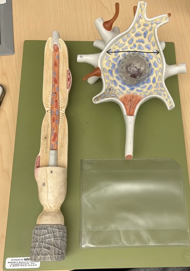
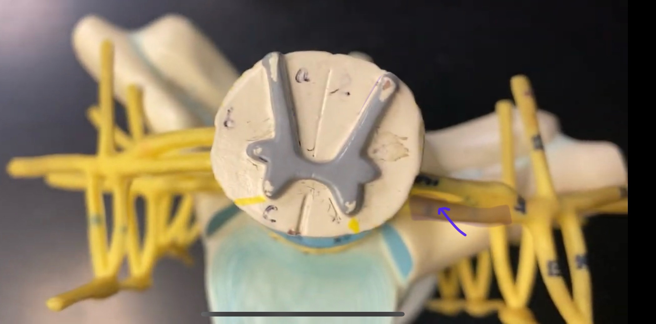
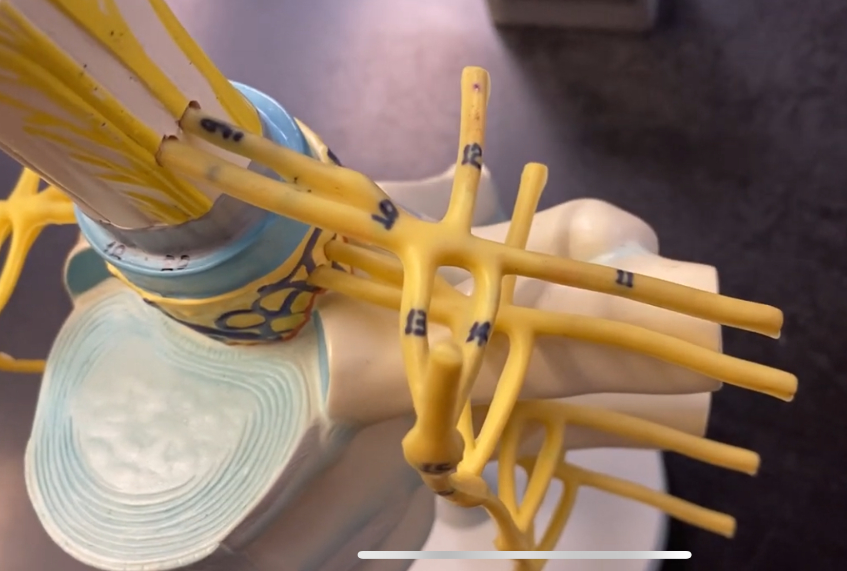
What type of spinal nerve is this? How many pairs?
cervical (8 pairs)

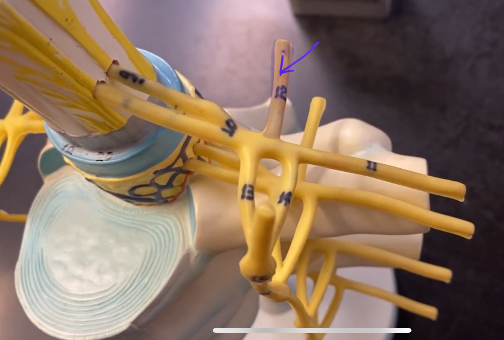
What type of spinal nerve is this? How many pairs?
thoracic (12 pairs)

What type of spinal nerve is this? How many pairs?
lumbar (5 pairs)


What type of spinal nerve is this? How many pairs?
sacral (5 pairs)

What type of spinal nerve is this? How many pairs? (not pictured)
coccygeal (1 pair)
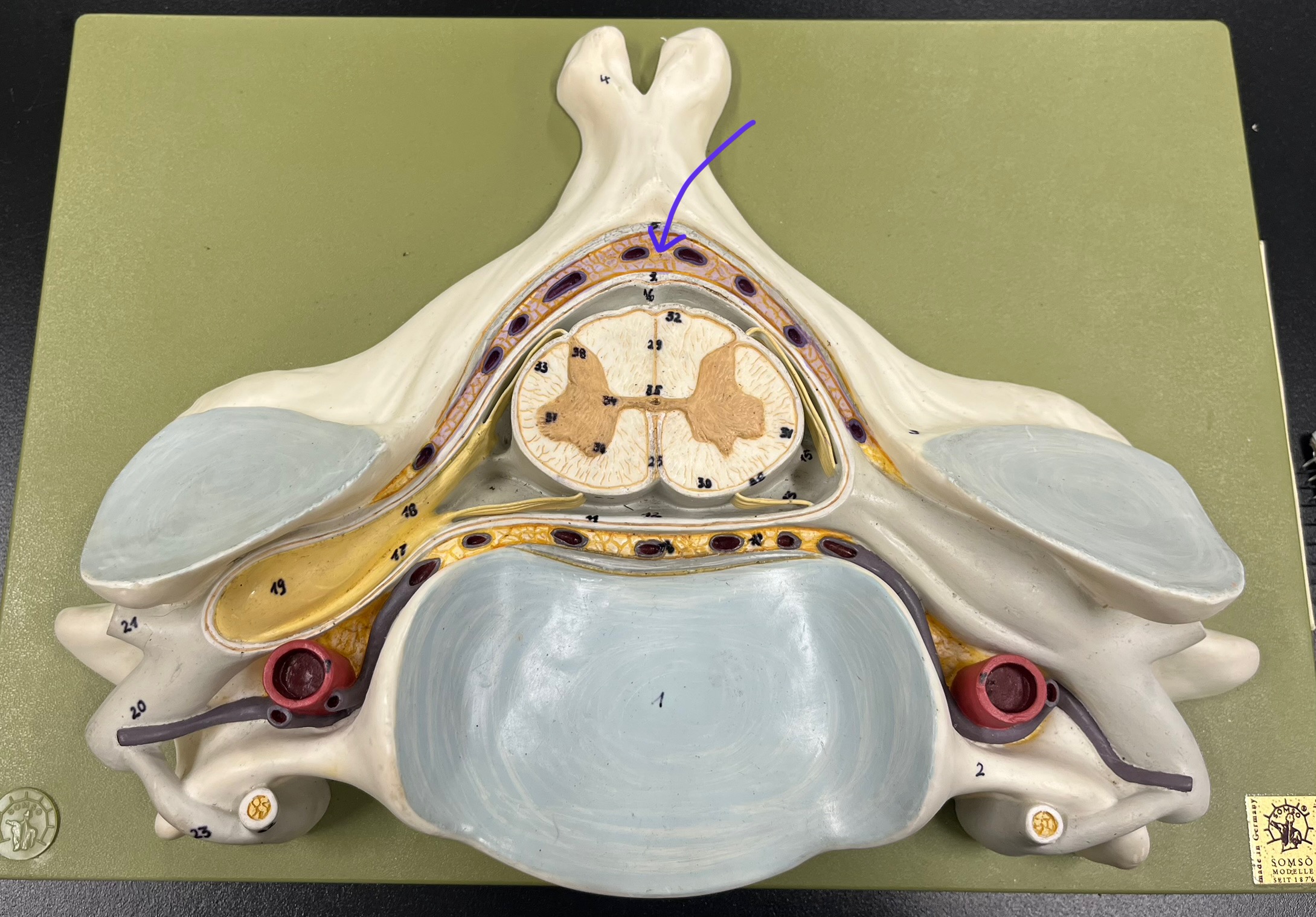
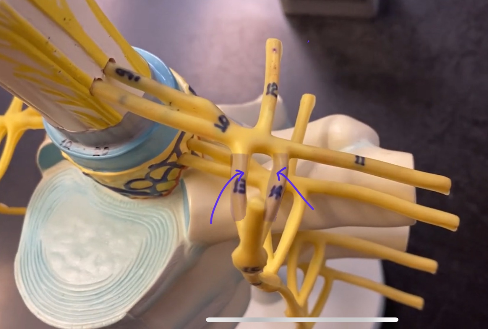
epidural space


dura mater


subdural space


arachnoid mater


subarachnoid space


pia mater


denticulate ligaments

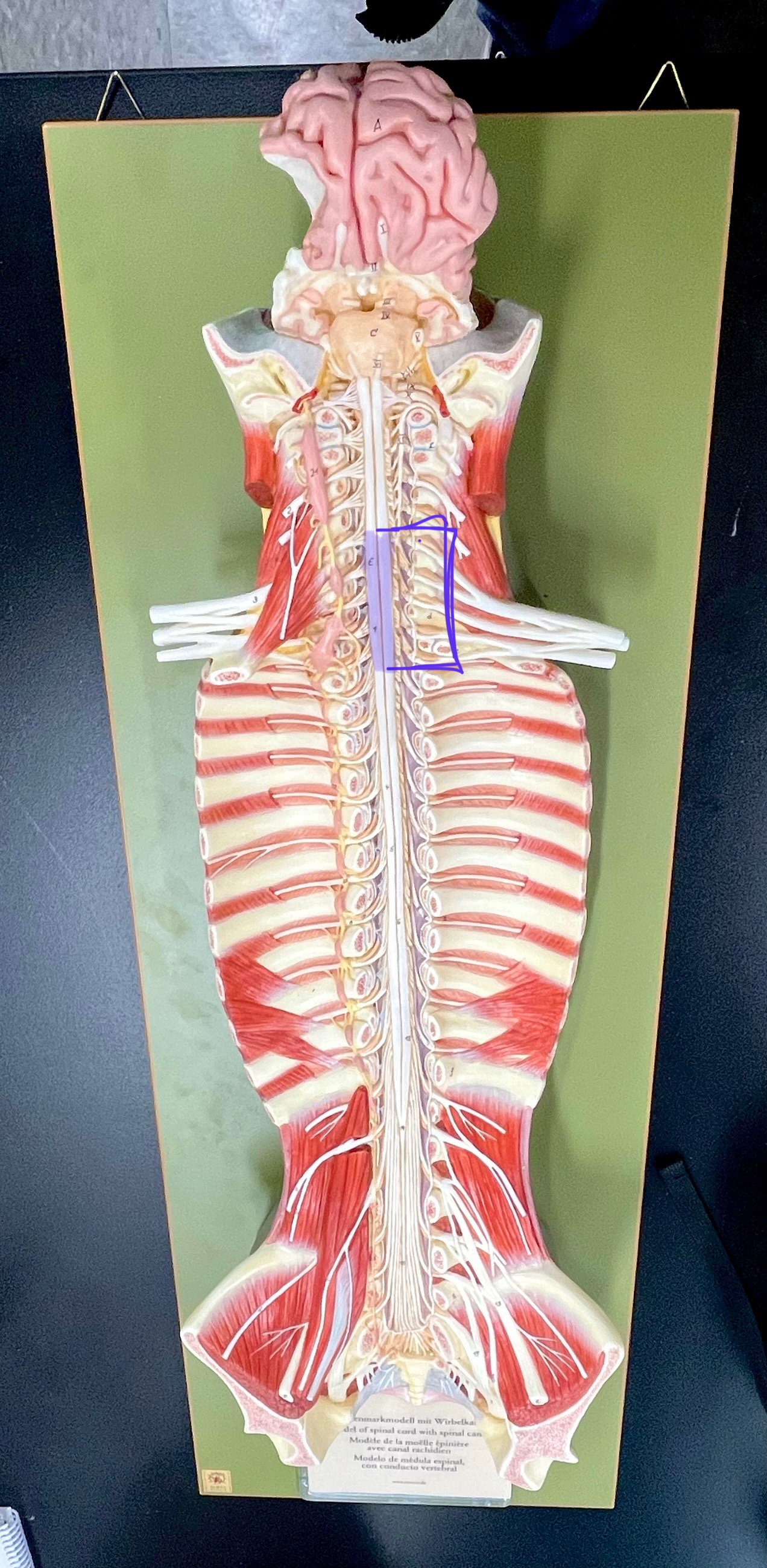
cervical enlargement
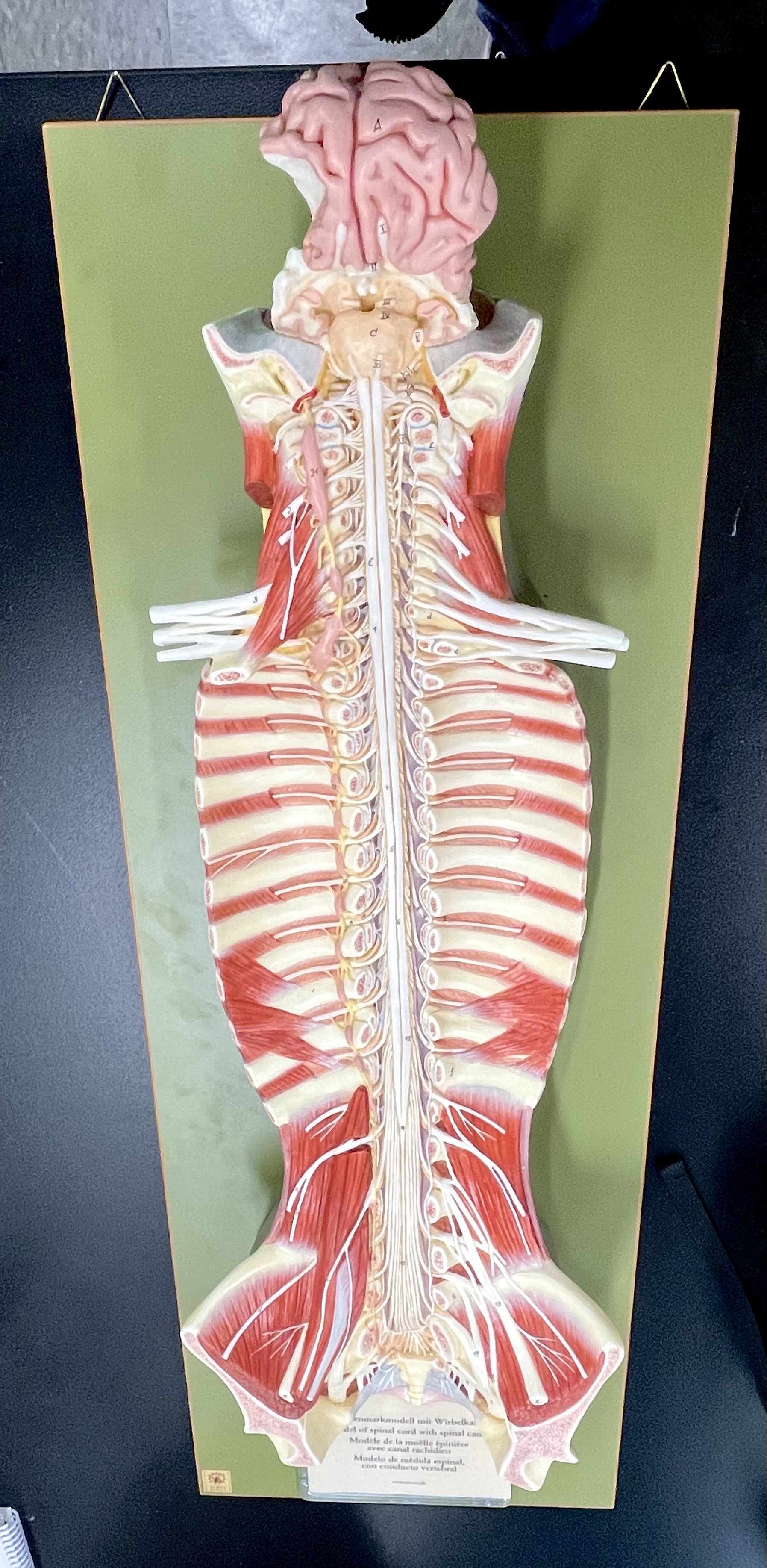

cervical enlargement

thoracic segment

lumbar enlargement

lumbar enlargement


conus medullaris


conus medullaris

filum terminale
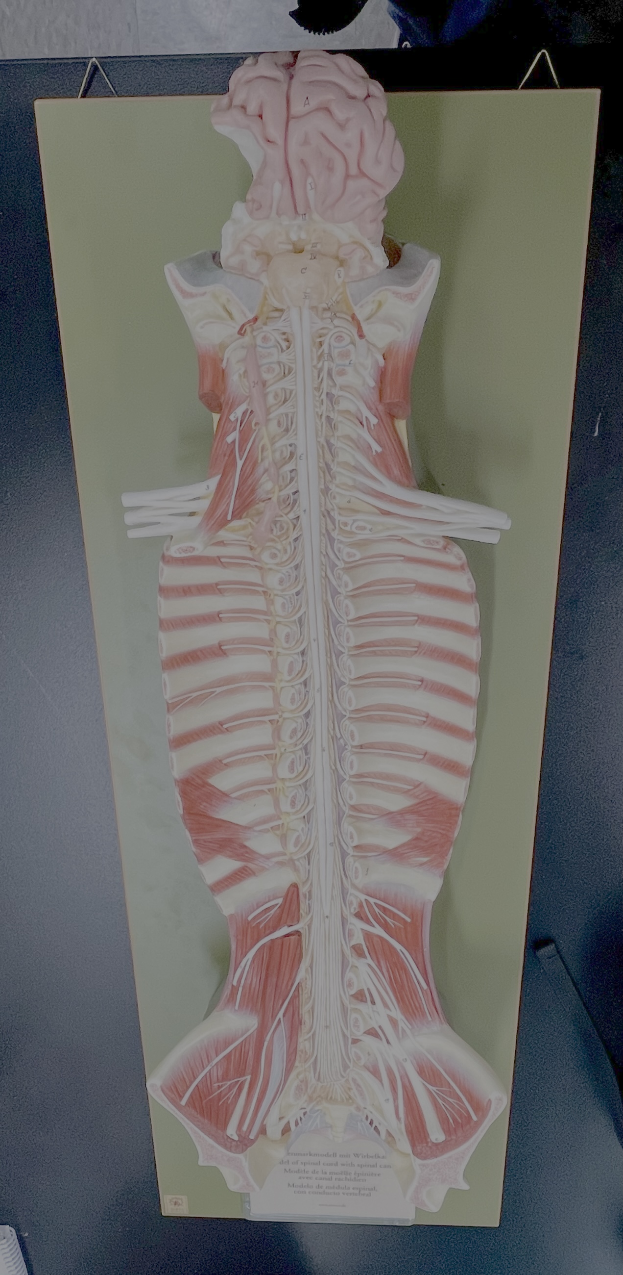

cauda equina


sympathetic chain ganglia


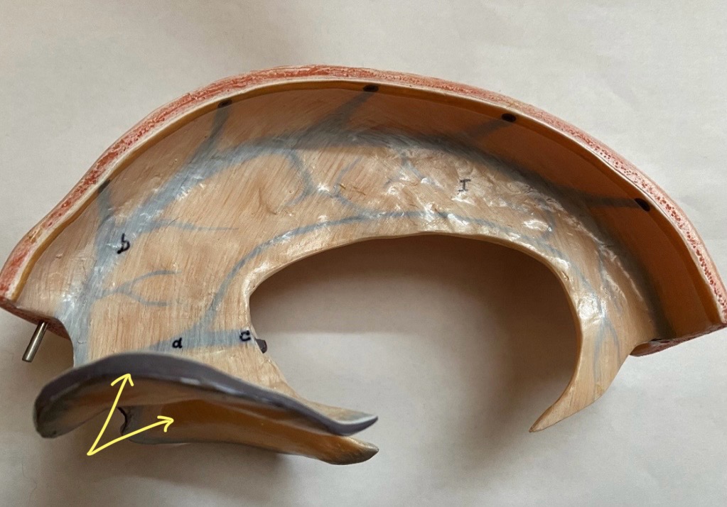
What is the groove here?
anterior median fissure


posterior median sulcus


posterior (dorsal) horn


posterior (dorsal) horn; R—>L


anterior (ventral) horn


lateral horn (selected models)


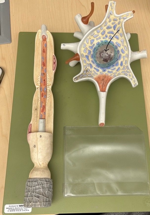
gray commissure


central canal


anterior column

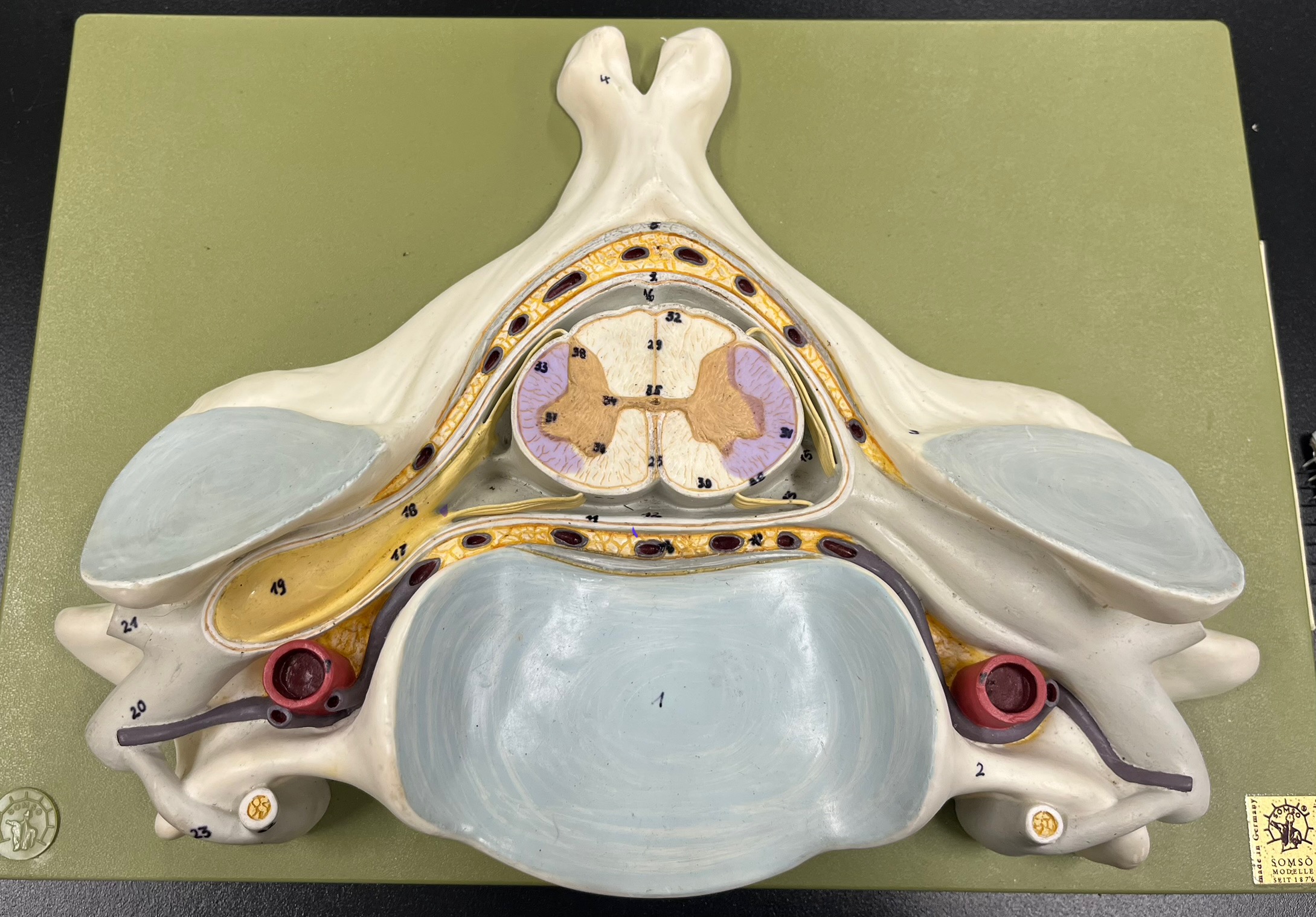
lateral column
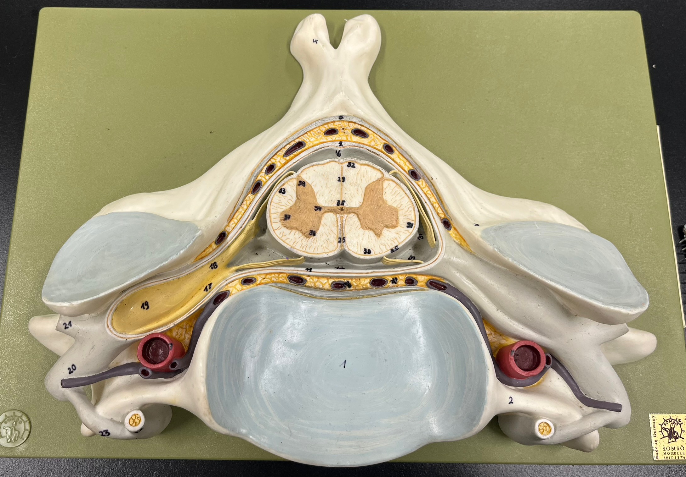

posterior column

white commissure

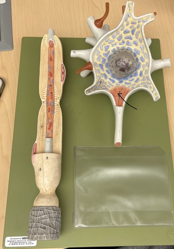
posterior (dorsal) root ganglion


What is the bulb here?
posterior (dorsal) root ganglion


posterior (dorsal) root

posterior (dorsal) root


anterior (ventral) root

anterior (ventral) root


dorsal ramus

dorsal ramus


ventral ramus


ventral ramus


rami communicantes

rami communicantes


sympathetic chain ganglia

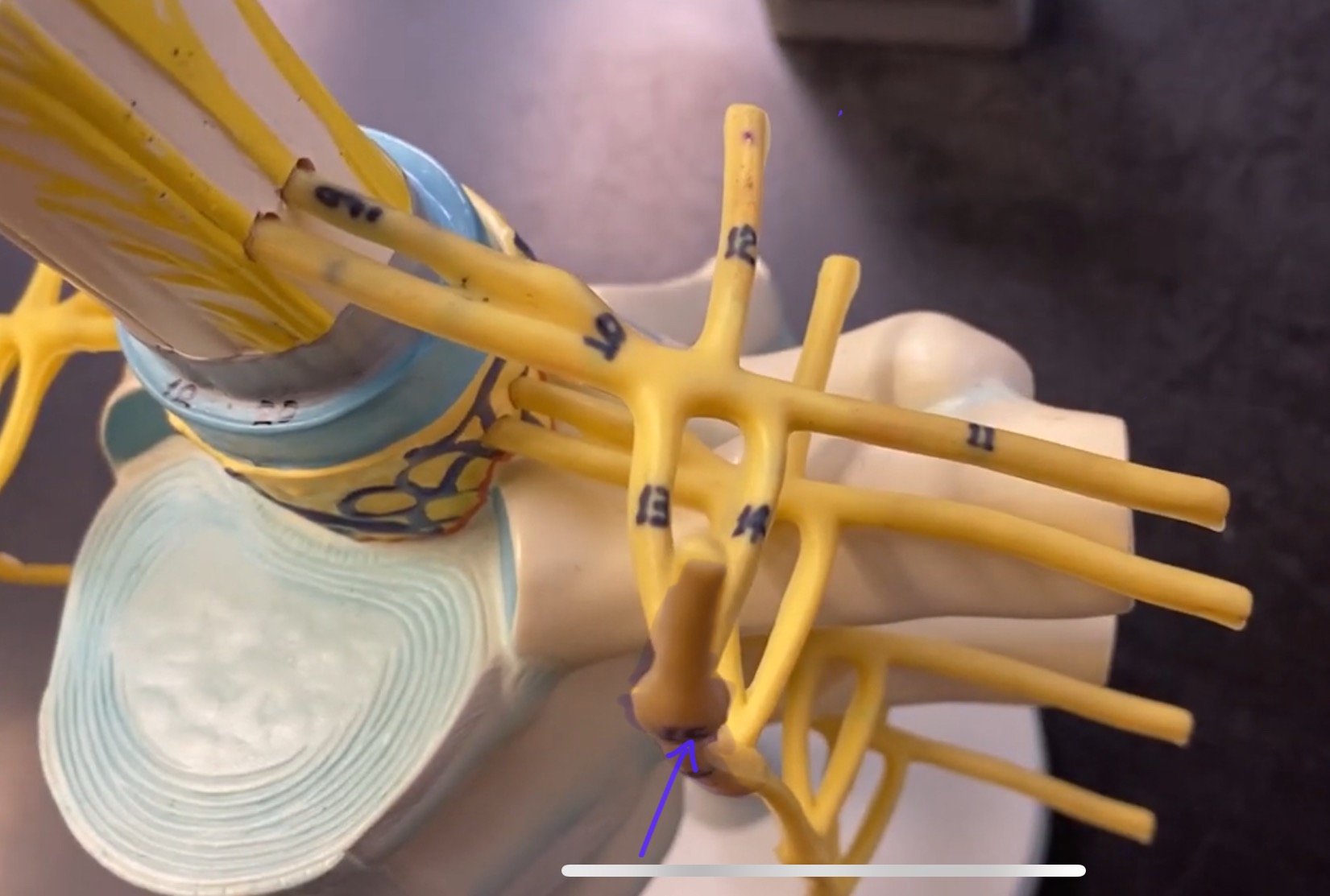
sympathetic chain ganglia

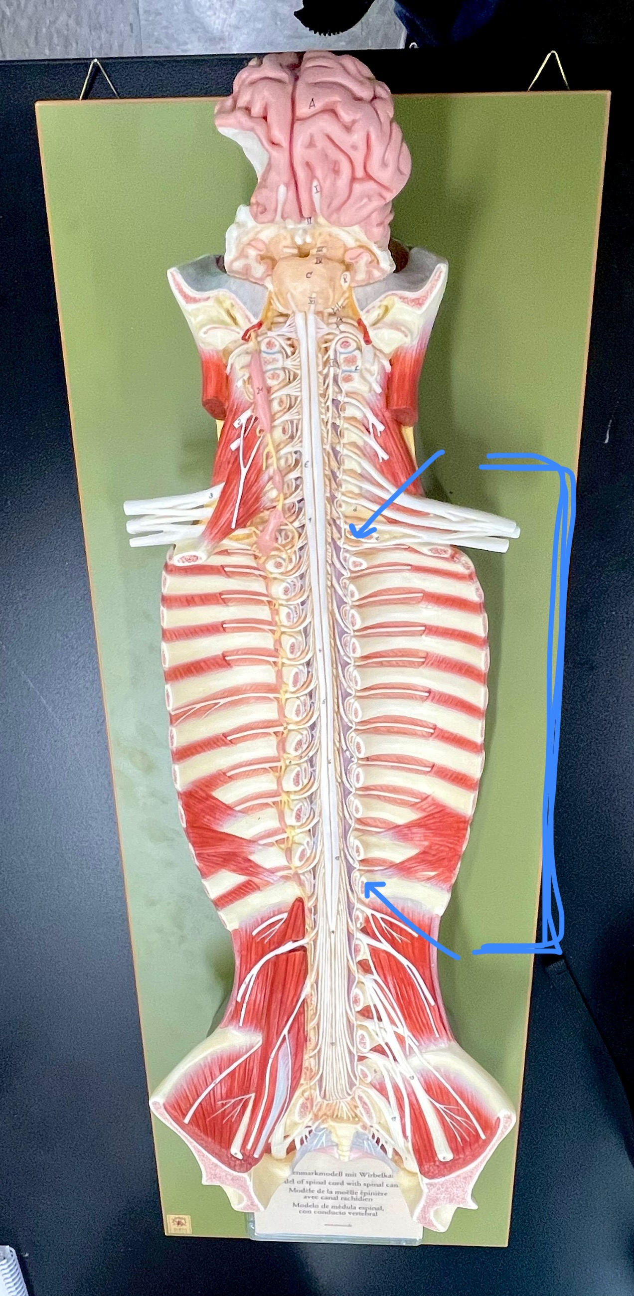
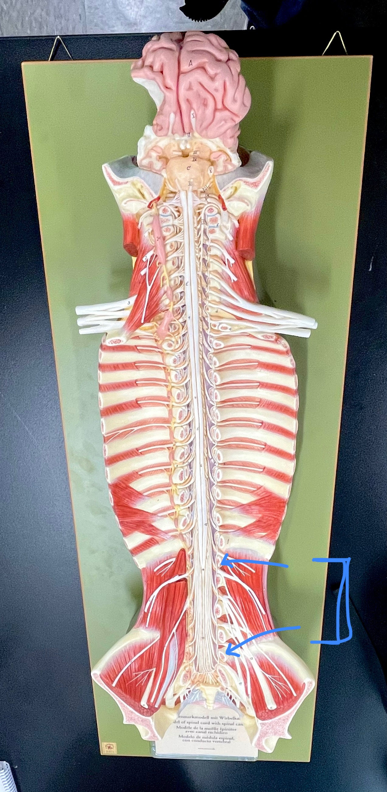
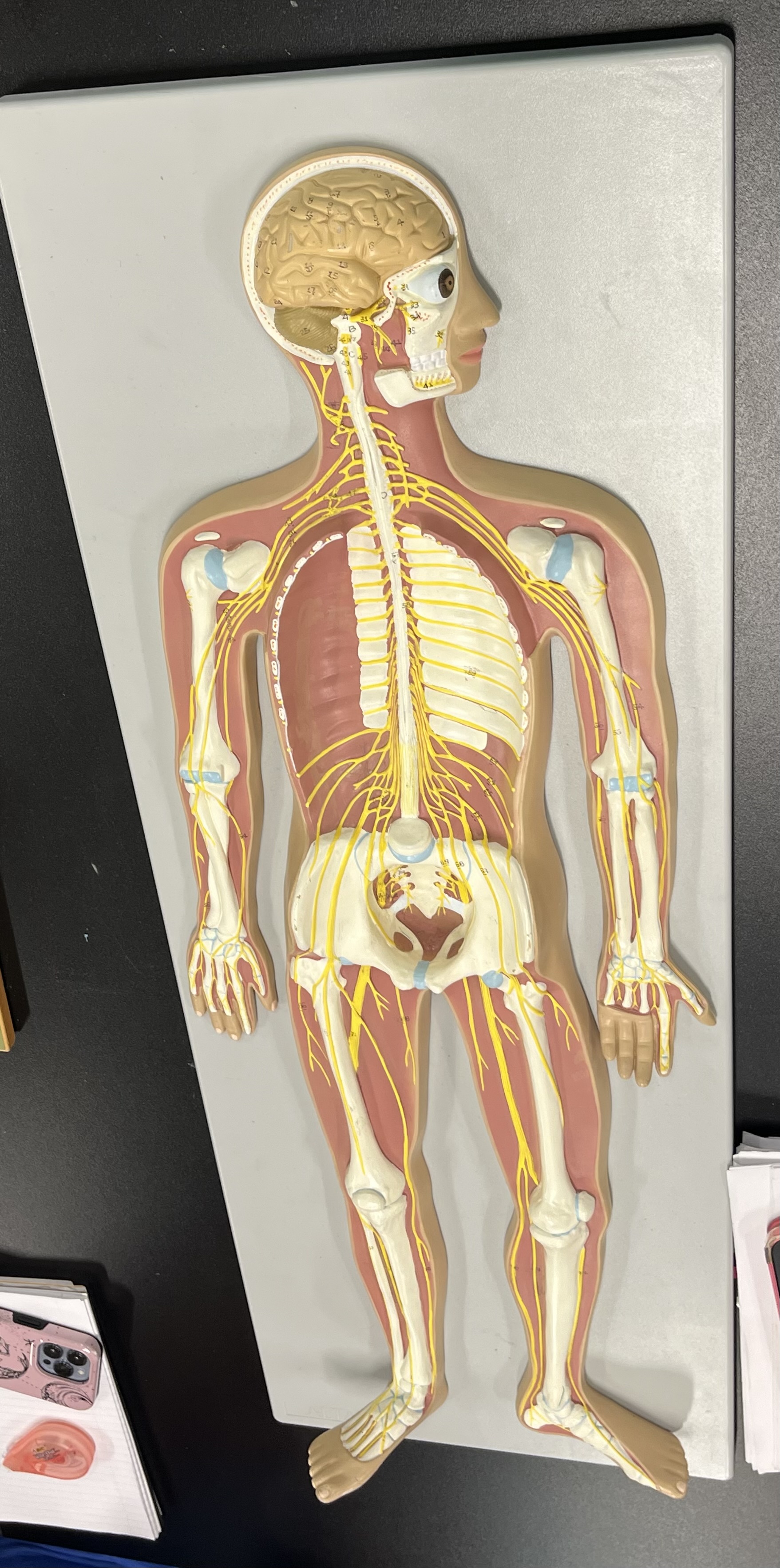
cervical plexus – phrenic nerve


cervical plexus – phrenic nerve


brachial plexus – ulnar nerve


brachial plexus – median nerve


brachial plexus – radial nerve


lumbar plexus – femoral nerve


lumbar plexus - femoral nerve

sacral plexus – sciatic nerve


sacral plexus – sciatic nerve

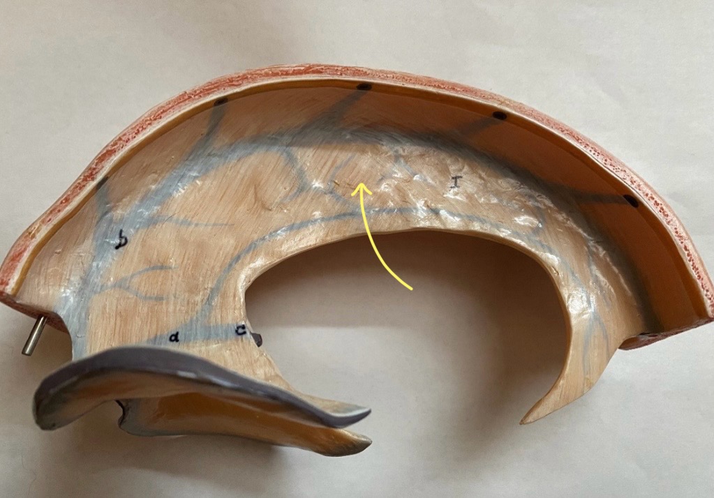
dura mater folds: falx cerebri
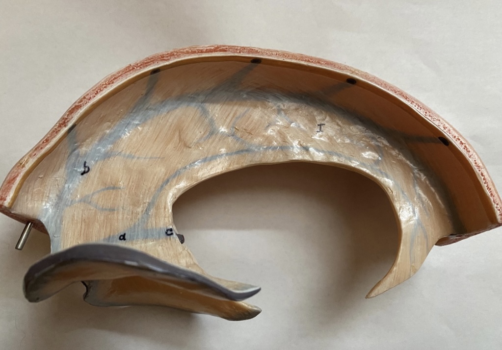
dura mater folds: falx cerebelli

dura mater folds: tentorium cerebelli


What is D?
lateral ventricle

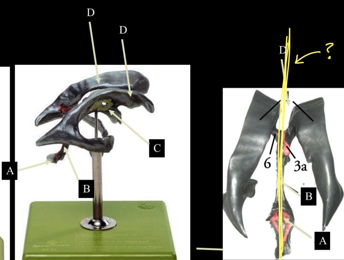
Imagine thin membrane separating the 2 lateral ventricles
septum pellucidum

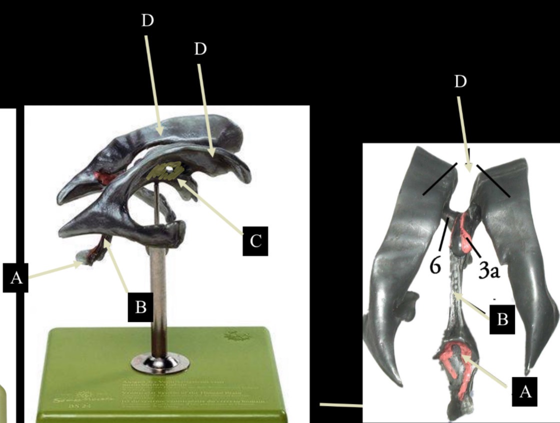
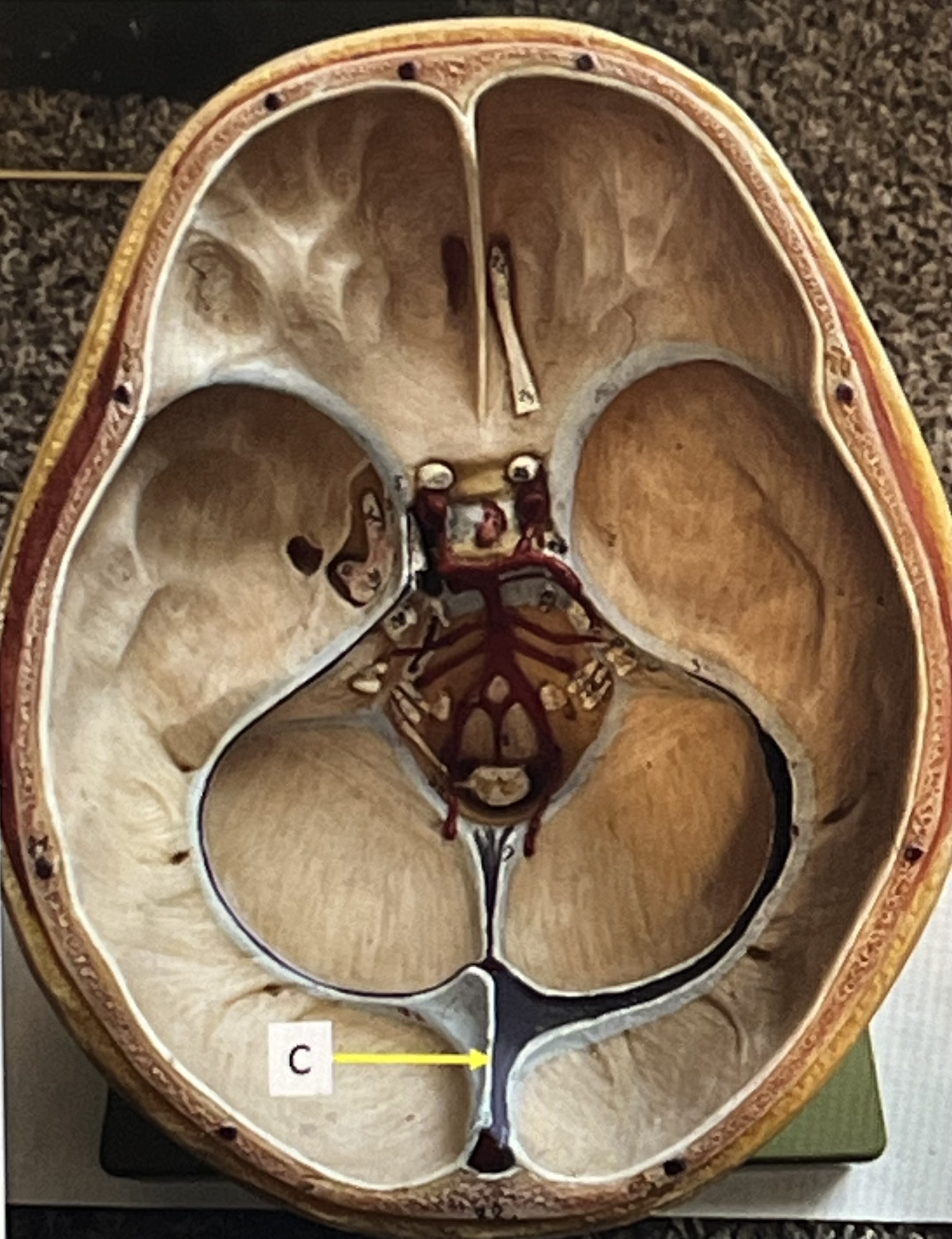
What is C?
3rd ventricle

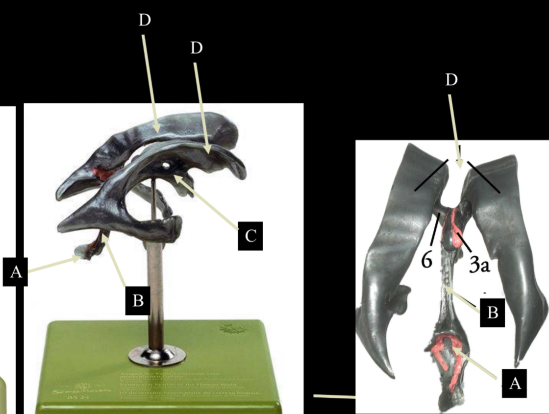
What is B?
cerebral aqueduct


What is A?
4th ventricle


What is below A at the very end?
central canal (spinal cord)


What is C?
midbrain


tectum (corpora quadrigemina): superior colliculus


tectum (corpora quadrigemina): inferior colliculus


What is a?
pons

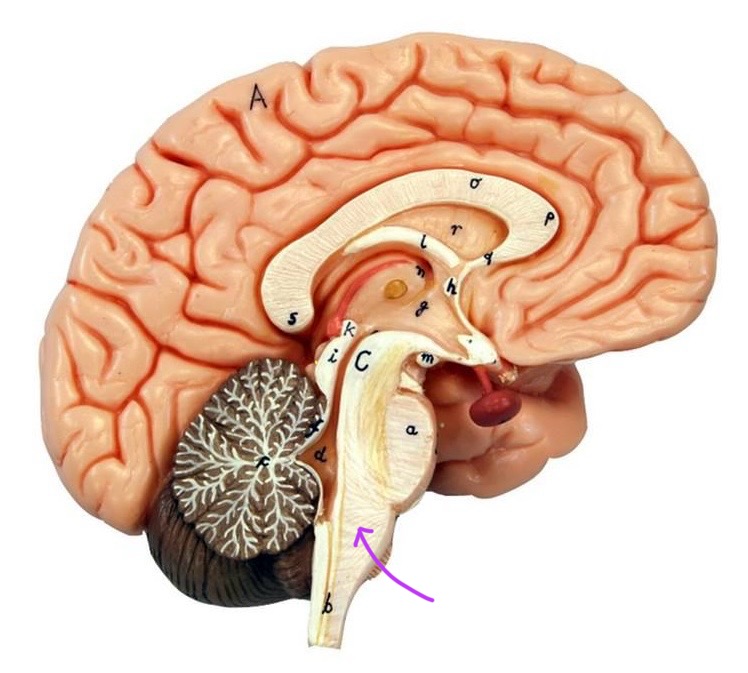
medulla oblongata


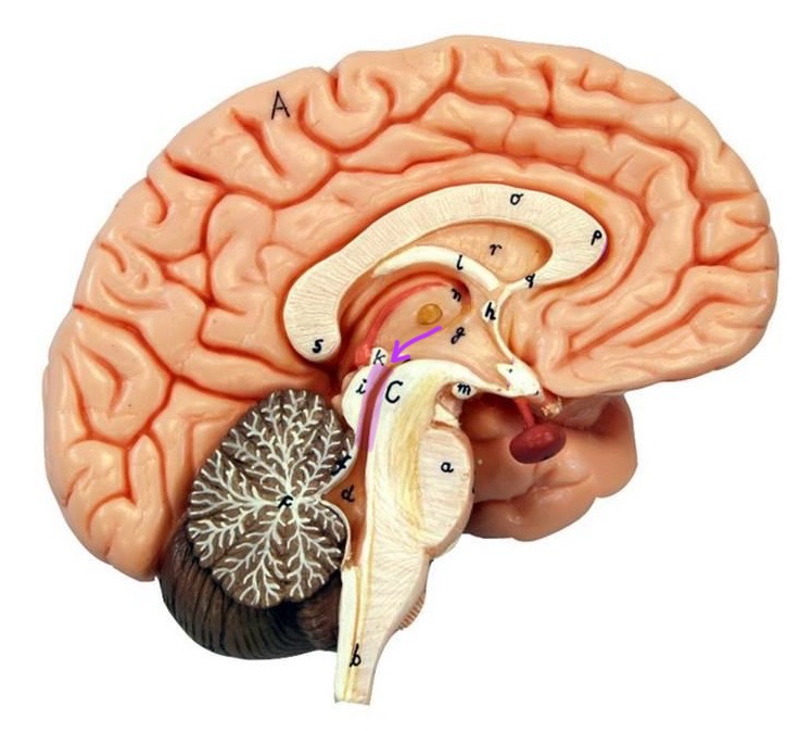
intermediate mass of thalamus

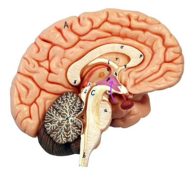
hypothalamus


What is the pink bulb?
pineal gland

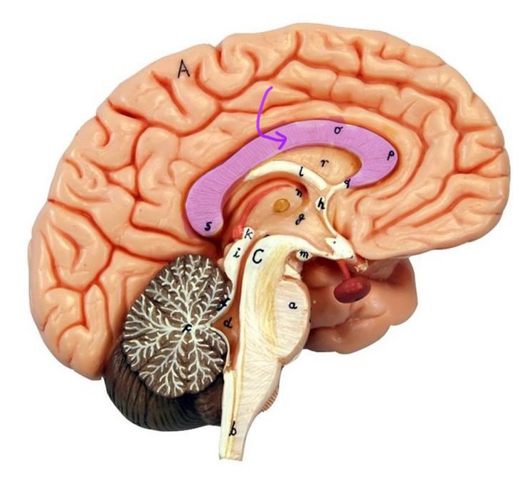
corpus callosum
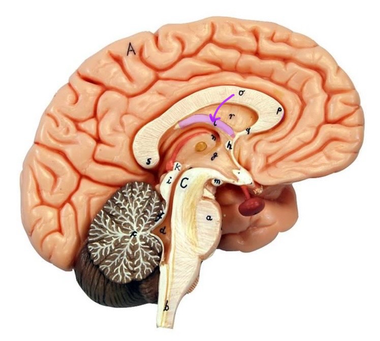
fornix


What is A?
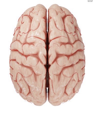
cerebral cortex

cerebral tracts

cerebral hemispheres

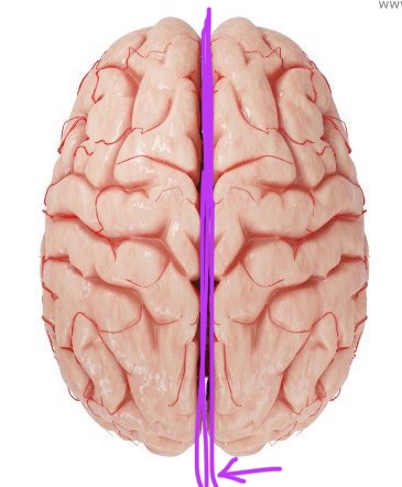
longitudinal fissure

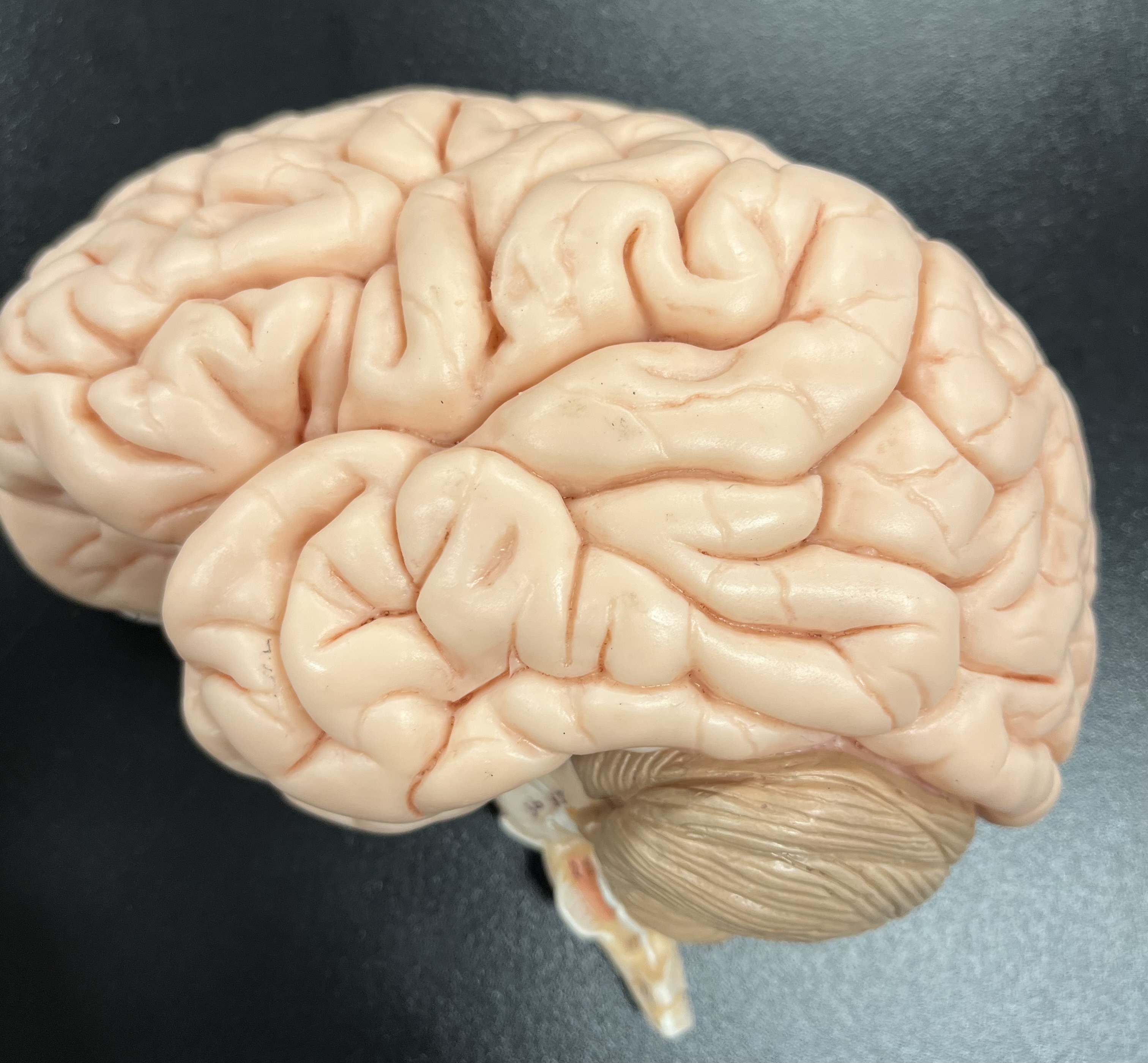
What are these ridges?
gyri


What are these shallow grooves?
sulci
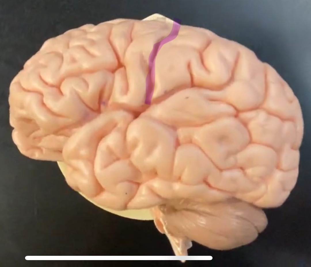
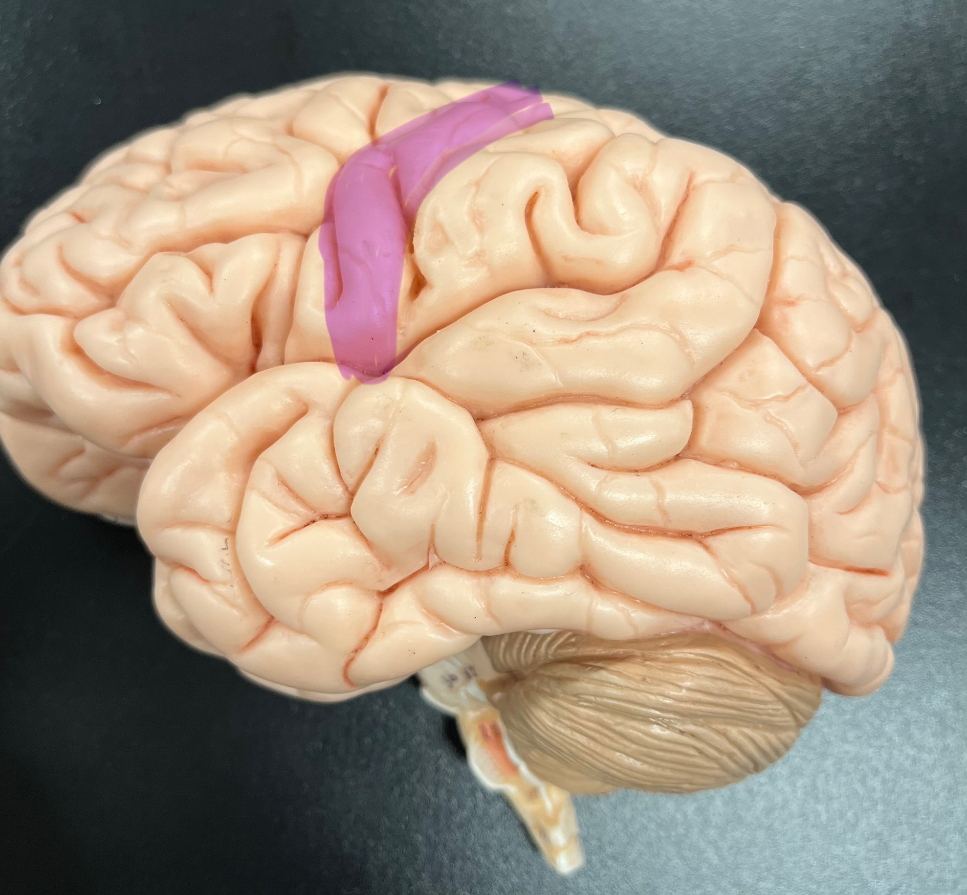
Divides parietal from frontal lobe
central sulcus

precentral gyrus
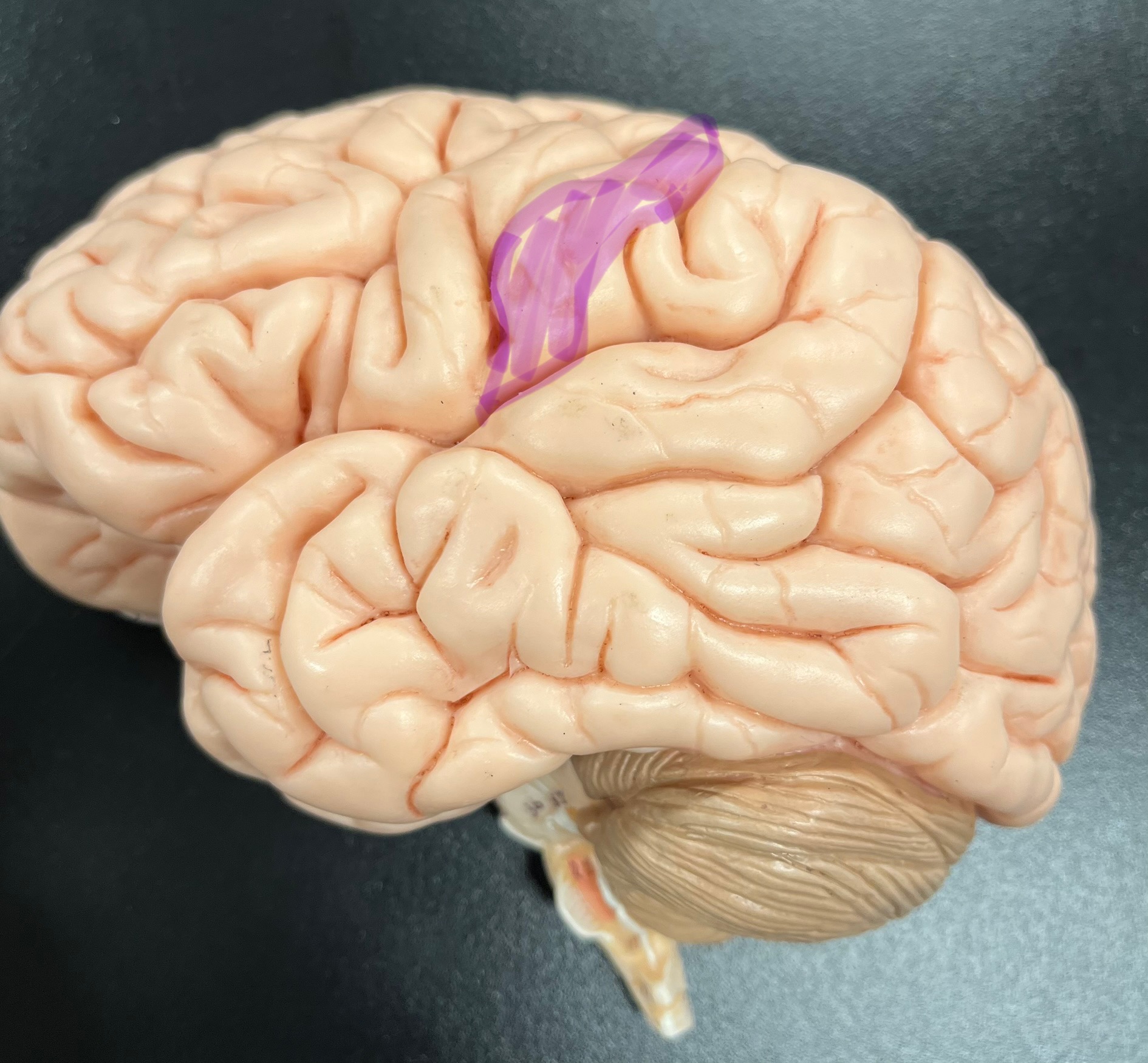
postcentral gyrus
