Cell Bio - Exam 1 study Aug 2026 - Baines
 Call Kai
 Learn
Learn Practice Test
 Spaced Repetition
 Match
Match Flashcards
 Knowt Play
1/185

There's no tags or description
Looks like no tags are added yet.
Name | Mastery | Learn | Test | Matching | Spaced | Call with Kai | Chat |
|---|
No analytics yet
Send a link to your students to track their progress
186 Terms
⭐ Q: What are the three major components of the cytoskeleton, and how does the main function of each compare?
The three major cytoskeletal structures are actin filaments, intermediate filaments, and microtubules.
Actin filaments: cell movement, contraction, and cell attachment
Intermediate filaments: mechanical strength and nuclear structure
Microtubules: organelle movement and cell division
💡 Memory trick:
Actin = Action
Intermediate = Infrastructure
Microtubules = Moving cargo + Mitosis

⭐ Q: How do actin filaments, intermediate filaments, and microtubules compare in size, composition, and physical structure?
From smallest to largest:
Actin filaments are the smallest and most flexible
- made of two-stranded rods made from monomeric actin
Intermediate filaments are middle-sized and rope-like
- made of rope-like filaments made from various cell-specific proteins
Microtubules are the largest and form rigid hollow tubes.
- made of hollow rods from α- and β-tubulin
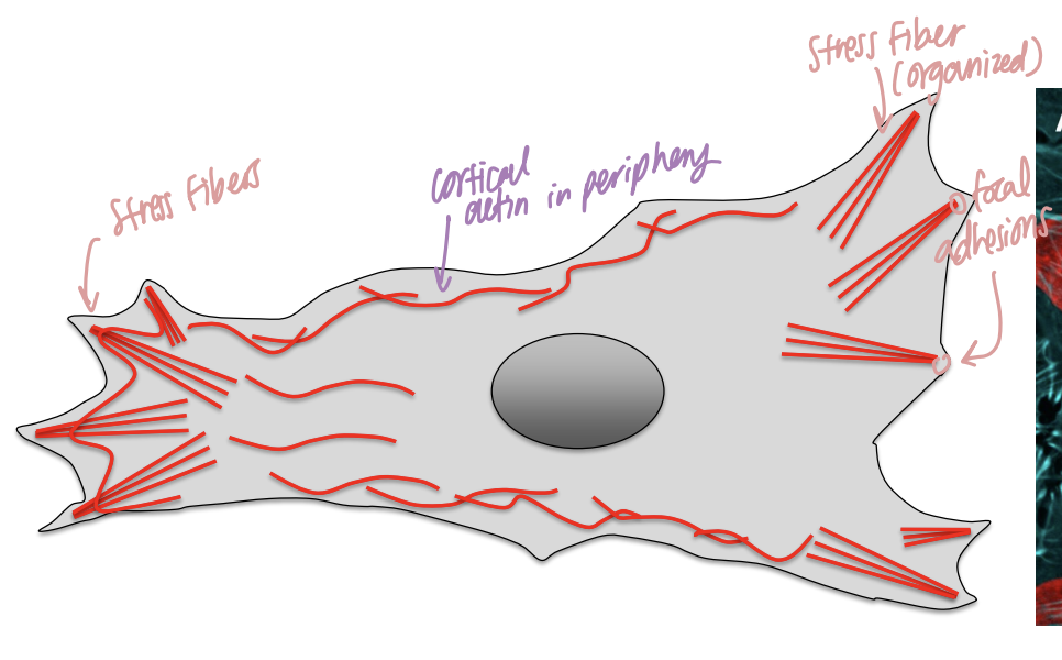
⭐ Q: What are actin filaments made of, what forms do they take in the cell, and what are their major functions?
Actin filaments are flexible, two-stranded rods made from actin monomers, and they occur primarily as cortical actin and stress fibers to support movement, contraction, and attachment.
Cortical actin: actin concentrated near the cell periphery
Stress fibers: bundles of actin extending through the cell
Major functions:
- cell locomotion
- contraction
- cell attachment
Cortical means near the outer region of the cell, just underneath the plasma membrane.
Stress fibers are larger actin bundles that span parts of the cell and can transmit force.
So actin is especially important anywhere the cell needs to change shape or generate force.
💡 Think: actin is the cell's muscle-and-movement system.
⭐ Q: How do actin filaments allow a cell to crawl across a surface?
Actin-driven cell locomotion occurs through a coordinated cycle of front-end protrusion, attachment to the surface, and contraction that pulls the rest of the cell forward.
The diagram in lecture shows:
1. Actin polymerizes at the front of the cell
→ pushes out the lamellipodium
2. The front attaches to the substrate
→ through focal contacts/adhesions
3. Actin and myosin generate contraction
→ pulls the cell body forward
🔎 What this means:
Polymerization means adding more protein subunits to a filament so it grows.
A lamellipodium is the broad protruding leading edge of a crawling cell.
So the cell basically:
reaches forward → grabs the ground → pulls itself forward.

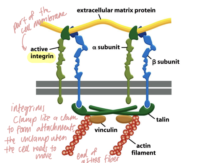
⭐ Q: How do actin filaments attach a cell to the extracellular matrix through focal adhesions?
Actin filaments attach cells to the extracellular matrix through focal adhesions, which physically connect intracellular actin to extracellular proteins through membrane proteins such as integrins.
The diagram in lecture shows:
extracellular matrix → integrin → talin/vinculin → actin filament
🔎 What this means:
The extracellular matrix (ECM) is the supportive material outside cells.
A focal adhesion is basically a strong attachment point between the inside of the cell and that outside matrix.
Integrins are membrane proteins that span the plasma membrane:
- one side interacts outside the cell
- the other side connects indirectly to actin inside the cell
That means the cell can both anchor itself and use actin-generated force against its surroundings.
💡 Analogy:
Focal adhesion = a wall anchor.
ECM 🧱 ← integrin/attachment proteins → actin rope 🪢
Q: In the example in lecture, how does disruption of actin contribute to Staphylococcus-associated mastitis in cattle?
In the example taught in the lecture, Staphylococcus aureus produces a toxin that causes actin depolymerization in mammary epithelial cells, disrupting the epithelial barrier and contributing to edema and inflammatory-cell migration.
🔎 What this means:
Depolymerization means breaking a filament apart into smaller components. It becomes “leaky” because it loses focal adhesion and cells come apart
The epithelial cells lining the mammary gland rely partly on their cytoskeleton to maintain their organization and barrier.
If their actin network falls apart:
actin disruption leads to
→ epithelial barrier disruption
→ fluid/inflammatory responses
→ edema and inflammatory-cell movement
This is an application of why actin matters structurally and functionally.

⭐ Q: What are intermediate filaments made of, and how are they different from actin filaments and microtubules?
Intermediate filaments are rope-like protein filaments whose size falls between actin filaments and microtubules, and unlike the other systems, their protein composition varies greatly depending on the cell type.
🔎 What this means:
There isn't one universal "intermediate filament protein" used identically in every tissue.
Different cells use different proteins to construct them.
The slide describes up to about 70 genes associated with intermediate-filament proteins.
That cell-specific composition is one of the key features distinguishing intermediate filaments from actin and microtubules.

⭐ Q: What is the primary mechanical function of intermediate filaments?
Intermediate filaments are the cytoskeletal system that provides the cell with its major mechanical stability, strength, and internal scaffolding.
⭐ Q: What are nuclear lamins, and what important functions do these intermediate filaments perform?
Nuclear lamins are intermediate-filament proteins that support nuclear structure and help connect the nucleus to important cellular processes.
Their functions include:
maintaining nuclear structure
attaching the nucleus to the cytoskeleton
helping anchor/move the nucleus
supporting chromatin attachment
helping position nuclear pores
Q: How can abnormal intermediate filaments contribute to Equine Motor Neuron Disease?
In the lecture's Equine Motor Neuron Disease example, neurofilaments accumulate in affected neurons, contributing to neuronal death and ultimately muscle wasting.
🔎 What this means:
Neurofilaments are intermediate-filament proteins associated with neurons.
The disease example shows that intermediate filaments must be properly organized and regulated.
Too much abnormal accumulation can damage the cells they are supposed to support.

⭐ Q: What are microtubules made of, how are they organized in the cell, and what are their major functions?
Microtubules are the largest cytoskeletal filaments and are rigid, hollow tubes made of α- and β-tubulin, usually extending outward from a microtubule-organizing center called the centrosome.
Their major functions are:
intracellular organelle/cargo movement
chromosome separation during mitosis

⭐ Q: How do microtubules and motor proteins move organelles through the cell?
Microtubules provide the tracks for intracellular transport, while kinesin and dynein are ATP-powered motor proteins that carry cargo along those tracks.
The lecture diagram shows:
Kinesin → generally toward the + end
Dynein → generally toward the − end
movement requires ATP
Cargo can include:
mitochondria
lysosomes
vesicles
🔎 What this means:
A motor protein is a protein capable of converting chemical energy into physical movement.
An ATPase is an enzyme that uses ATP.
So organelles don't simply float randomly until they reach the correct place.
They can be physically carried along microtubules.
The neuron example is especially useful:
cell body ↔ long axon ↔ synaptic terminal
Mitochondria may have to travel enormous distances relative to the size of the cell.
💡 Analogy:
Microtubule = train track
Kinesin/dynein = locomotives
Organelle = cargo 🚂📦

⭐ Q: Why are microtubules essential for mitosis?
Microtubules are essential for mitosis because they form and operate the mitotic spindle, which separates duplicated chromosomes into the two daughter cells.
🔎 What this means:
During division, the cell has to make sure each daughter cell receives the appropriate chromosome set.
Microtubules form the spindle machinery that organizes and separates those chromosomes.
So if microtubules are severely disrupted:
chromosome separation fails → normal cell division is disrupted.
⭐ Q: Which drugs in this lecture target microtubules, and what are their major clinical uses?
The microtubule-targeting drugs listed in the lecture are paclitaxel, vincristine/vinblastine, and benzimidazoles, and they are used particularly in cancer treatment and treatment of worm infections.
Paclitaxel/Taxol → cancer treatment by disrupting microtubules
Vincristine and vinblastine → cancer treatment by disrupting microtubules
Benzimidazoles → anti-helminthic by attacking their secretory vesicles - starving them
🔎 What this means:
Why would cancer drugs target microtubules?
Because cancer cells depend heavily on cell division, and mitosis depends on microtubules.
Why would worm drugs target them?
Because worms also depend on tubulin/microtubules for important cellular processes.
⭐ Q: How do benzimidazoles interfere with parasitic worms?
Benzimidazoles disrupt worm microtubule function and, in the mechanism specifically described in the lecture, prevent normal movement of secretory vesicles in the worm's gut, interfering with digestion and thus starving them to death
🔎 What this means:
Remember:
microtubules = railroad tracks for vesicle transport.
So if the drug damages those tracks:
→ secretory vesicles can't move normally
→ normal gut secretion/digestion fails
→ worm is unable to digest food and starves to death
⭐ Q: Why can microtubule-targeting drugs cause bone marrow suppression, and what side effects can be predicted from their mechanism?
Microtubule-targeting drugs can suppress bone marrow because blood-cell precursors must divide rapidly, and normal mitosis requires functional microtubules.
The case study showed:
leukopenia
lymphopenia
eosinopenia
thrombocytopenia
after fenbendazole treatment.
🔎 What this means:
Bone marrow hypoplasia means the marrow is producing fewer blood cells than normal.
The mechanism can be connected directly to what you already learned:
microtubules disrupted
→ mitotic spindle impaired
→ rapidly dividing marrow precursor cells cannot divide normally
→ fewer mature blood cells enter circulation.
This is exactly the kind of reasoning the learning objective means by “predict potential side effects.”
⭐ Q: What is the endoplasmic reticulum, and what major jobs does the ER perform overall?
The endoplasmic reticulum is a single-membrane organelle continuous with the outer nuclear membrane that performs major roles in protein production, metabolism, detoxification, and intracellular calcium storage.
Major ER functions include:
production of transmembrane and secretory proteins
lipid, steroid, and carbohydrate metabolism
drug/toxin detoxification in the liver
storage of intracellular Ca²⁺
🔎 What this means:
The ER is an extensive membrane network inside the cell.
It isn't one single-purpose organelle.
Different regions specialize in different jobs, which is why the lecture divides it into rough ER, smooth ER, and transitional ER.
The chemotherapeutic drug vincristine affects which cytoskeleton filament?
a. Actin Filaments
b. Intermediate Filaments
c. Microtubules
c. Microtubules
What is one of the primary functions of actin filaments?
a. Mitosis
b. Mechanical Strength
c. Cell Locomotion
d. Organellar Movement
c. Cell Locomotion
Which intermediate filament protein is present in all cells (except mature RBCs)?
a. Desmin
b. Lamin
c. Vimentin
d. Neurofilamin
b. Lamin
⭐ Q: How do rough ER, smooth ER, and transitional ER differ in structure and function?
The three ER regions specialize in different steps of cellular production: rough ER makes proteins, smooth ER performs metabolism/detoxification and stores Ca²⁺, and transitional ER packages molecules into vesicles for transport to the Golgi.
Rough ER
has ribosomes attached to membrane
major site of protein synthesis
Smooth ER
lacks ribosomes
lipid/steroid/carbohydrate metabolism
drug detoxification
Ca²⁺ storage
Transitional ER
specialized exit region
packages proteins and other molecules into transport vesicles
sends them toward the Golgi
🔎 What this means:
These are not three completely separate organelles.
They are specialized regions of the same ER network.
💡 Analogy:
Rough ER = factory 🏭
Smooth ER = chemistry lab + storage room 🧪
Transitional ER = shipping dock 📦
⭐ Q: What major functions does the smooth ER perform, and why is Ca²⁺ storage important?
Smooth ER performs drug detoxification, lipid/carbohydrate/steroid metabolism, and Ca²⁺ storage, with stored Ca²⁺ able to be released back into the cytosol through specific channels.
🔎 What this means:
Sequester means to take something out of the general cytosol and store it in a separate location.
So when the ER sequesters Ca²⁺, it keeps cytosolic calcium relatively controlled until the cell needs to release some.
The lecture particularly emphasizes Ca²⁺ storage in muscle cells.
Smooth ER also has a major metabolic role, especially in liver detoxification.
💡 Think:
Smooth ER = metobolism + calcium closet.
Q: How does West Nile virus demonstrate why normal ER structure and function matter?
West Nile virus uses the ER during viral replication, and accumulation of viral proteins and particle assembly can distort the ER, cause ER stress, trigger massive Ca²⁺ release, and contribute to cell death.
🔎 What this means:
The lecture uses West Nile virus as an application of ER biology.
Because the ER is also a major intracellular Ca²⁺ store:
ER disruption
→ abnormal Ca²⁺ release
→ severe cellular stress
→ cell death.
⭐ Q: What is the Golgi apparatus, and what is its overall function?
The Golgi apparatus is a single-membrane organelle made of multiple cisternae that receives proteins from the ER, modifies them, sorts them, and sends them to their correct cellular destinations.
🔎 What this means:
A cisterna is one of the flattened membrane compartments making up the Golgi stack.
The lecture calls the Golgi the cellular “Post Office.”
That's a very good analogy:
ER makes/sends package
→ Golgi receives package
→ modifies/address-labels it
→ sends it to the correct destination.
💡 Memory trick:
ER = makes it.
Golgi = modifies, sorts, mails it.

⭐ Q: What happens to proteins as they move from the cis side to the trans side of the Golgi?
Proteins are progressively modified as they travel from the cis toward the trans side of the Golgi, after which the trans-Golgi network sorts them to their final destinations.
Modifications named in the lecture include:
glycosylation
phosphorylation
sulfation
Possible destinations include:
lysosomes
secretory vesicles
plasma membrane
🔎 What this means:
The cis side is the receiving side closest to incoming ER cargo.
The trans side is closer to the shipping/sorting end.
At the simplest level:
glycosylation involves adding/modifying sugar groups
phosphorylation involves phosphate groups
sulfation involves sulfate groups
The lecture names these modifications but does not ask you to memorize detailed chemistry for each one.
Then the trans-Golgi network decides where the protein goes next.
⭐ Q: What are lysosomes, and what is their primary function?
Lysosomes are single-membrane organelles that serve as major intracellular digestion compartments, breaking down material delivered to them.
🔎 What this means:
The lecture emphasizes that lysosomes are heterogeneous in size, meaning they do not all have to be identical sizes.
Their digestive function depends strongly on their internal acidic environment.
💡 Analogy:
Lysosome = cellular recycling/demolition center. ♻
⭐ Q: Why is the acidic pH inside a lysosome essential for lysosomal function?
The acidic pH is essential because lysosomal digestion depends on acid hydrolases that function in the acidic lysosomal environment, which the slide depicts at about pH 5 compared with approximately pH 7.2 in the cytosol.
🔎 What this means:
A hydrolase is an enzyme that breaks chemical bonds using water.
The lysosomal enzymes shown include types that break down:
nucleic acids
proteins
sugars
lipids
phosphate-containing compounds
The slide calls them acid hydrolases because the lysosomal environment is acidic.
A membrane H⁺ pump uses ATP to move hydrogen ions into the lysosome and maintain that low pH.
So:
ATP-powered H⁺ pump
→ acidic lysosome
→ acid hydrolases can carry out digestion.
💡 Memory trick:
Lysosome = acidic stomach of the cell. 🧪
⭐ Q: Where do lysosomes get their proteins and membrane?
Lysosomes are formed using proteins supplied through the Golgi apparatus and membrane derived from the plasma-membrane/endosomal system.
🔎 What this means:
A lysosome isn't magically created as one complete bubble.
Different parts of the cellular trafficking system contribute components to it.
The lecture summarizes this as:
Golgi → proteins
plasma membrane → membrane
⭐ Q: What are the three major pathways that deliver material to lysosomes, and how does each pathway differ?
The three pathways are phagocytosis, endocytosis, and autophagy, and they differ mainly in what material they deliver to the lysosome.
Phagocytosis
brings in large extracellular particles such as bacteria
material enters a phagosome
phagosome ultimately delivers material toward lysosomal degradation
Endocytosis
internalizes extracellular material through membrane vesicles
cargo moves through early/late endosomes toward lysosomes
Autophagy
delivers the cell's own internal components, such as damaged organelles
material is enclosed in an autophagosome
autophagosome delivers it toward lysosomal degradation
🔎 What this means:
Phagocytosis: big thing from outside 🍽🦠
Endocytosis: smaller extracellular cargo from outside 📦
Autophagy: old/damaged stuff from inside the cell ♻
⭐ Q: How do lysosomal storage diseases develop?
Lysosomal storage diseases occur when genetic defects impair specific lysosomal acid hydrolases, so substrates that should normally be degraded instead accumulate inside lysosomes.
💡 Analogy:
Imagine a recycling center where the machine that crushes glass breaks.
Trash keeps arriving.
Everything else may still be processed, but the glass pile gets bigger and bigger until it interferes with the whole facility.
Q: How do neuronal ceroid lipofuscinoses illustrate the mechanism of lysosomal storage disease?
Neuronal ceroid lipofuscinoses are examples in which deficiencies in certain lysosomal enzymes or proteins lead to accumulation of material in neurons and progressive neurodegeneration.
The lecture associates NCLs with:
deficiencies such as TPP1
buildup of fluorescent material in neurons
neuronal degeneration
ataxia
impaired cognition
blindness
seizures
They occur in several veterinary species, including dogs, cats, sheep, goats, horses, and cattle.
⭐ Q: How do the ER, Golgi apparatus, and lysosomes work together as a cellular production, sorting, and degradation system?
The ER primarily produces and begins handling cellular products, the Golgi modifies and sorts those products, and lysosomes receive and digest material destined for degradation.
A simplified pathway is:
Rough ER
→ makes proteins
Transitional ER
→ packages cargo into vesicles
Golgi
→ receives, modifies, and sorts cargo
Trans-Golgi network
→ sends cargo to destinations such as:
plasma membrane
secretory vesicles
lysosomes
Lysosome
→ degrades material delivered through pathways such as endocytosis, phagocytosis, or autophagy
🔎 What this means:
ER = factory 🏭
Transitional ER = shipping dock 📦
Golgi = post office/distribution center 🏤
Lysosome = recycling and waste-processing center ♻
⭐ Q: What is the overall structure of a mitochondrion, and what are its major cellular functions?
A mitochondrion is a double-membrane organelle of bacterial origin whose major functions include ATP production, ion homeostasis, heme synthesis, lipid/steroid metabolism, and regulation of cell survival and death.
Its major compartments are:
Outer mitochondrial membrane
Intermembrane space (IMS) = space between the two membranes
Inner mitochondrial membrane
Matrix = innermost mitochondrial compartment
Major functions include:
generating an electrochemical proton gradient
oxidative phosphorylation and ATP production
Ca²⁺, Fe³⁺, and K⁺ homeostasis
heme synthesis
steroid and lipid metabolism
regulation of cell death and survival
🔎 What this means:
Their double-membrane structure is especially important for ATP production because the inner membrane separates the matrix from the intermembrane space, allowing the mitochondrion to create an H⁺ gradient between those compartments.
💡 Big picture:
The mitochondrion is simultaneously a:
⚡ power plant
🧪 metabolic center
🧲 ion regulator
☠ regulator of cell survival/death
⭐ Q: What are the major characteristics of the mitochondrial genome, and what does mitochondrial DNA encode?
Mitochondria have their own small, maternally inherited genome, which occurs in multiple copies and contains 37 genes needed for a small portion of mitochondrial protein synthesis.
The 37 genes encode:
13 oxidative-phosphorylation proteins
22 tRNAs (transfer RNA)
2 rRNAs (ribosomal RNA)
Mitochondria also contain basic machinery for:
replication
transcription
translation
🔎 What this means:
A tRNA helps deliver amino acids during protein synthesis.
An rRNA is part of the ribosome itself.
And OXPHOS, or oxidative phosphorylation, is the process mitochondria use to make ATP using the electron transport chain.
⭐ Q: If mitochondria have their own DNA, where do most mitochondrial proteins actually come from?
Although mitochondria have their own genome, 99.9% of mitochondrial proteins are encoded by nuclear genes, synthesized in the cytosol, and then imported into the mitochondria.
These proteins contain a mitochondrial localization signal (MLS) that directs them to the mitochondrion.
🔎 What this means:
This is an important distinction:
Mitochondria can make some proteins themselves.
But the overwhelming majority are encoded by DNA in the nucleus.
So the cell makes those proteins outside the mitochondrion and then has to deliver them to the correct organelle.
The MLS acts like an address label saying:
“This protein belongs in a mitochondrion.” 📦
⭐ Q: What is the general process of nuclear-encoded proteins being imported into mitochondria?
Nuclear-encoded mitochondrial proteins are synthesized in the cytosol as precursor proteins, recognized through their MLS (mitochondrial localization signal), transported across mitochondrial membranes, and then processed into mature mitochondrial proteins.
The general sequence shown is:
Nuclear-encoded mitochondrial proteins made in cytosol
→ recognize MLS
→ enters mitochondrion
→ transported through mitochondrial membrane
→ localization signal is cleaved
→ mature mitochondrial protein
⭐ Q: How does the mitochondrial electron transport chain ultimately generate ATP?
The electron transport chain generates ATP by using electron transfer to pump H⁺ across the inner mitochondrial membrane, creating an electrochemical proton gradient that drives ATP synthase.
The mechanism is:
1. NADH and FADH₂ donate electrons
↓
2. Electrons move through the electron transport chain
↓
3. ETC complexes pump H⁺ from the matrix into the intermembrane space
↓
4. An H⁺ gradient builds across the inner membrane
↓
5. Electrons ultimately reach O₂, contributing to water formation
↓
6. H⁺ flows back into the matrix through ATP synthase
↓
7. ATP synthase uses that energy to convert ADP → ATP
🔎 What this means:
There are really two stages to remember.
Stage 1: Store energy
The ETC uses electron energy to move H⁺ to one side of the inner membrane.
Now there is a difference in H⁺ concentration and electrical charge across that membrane.
That's the electrochemical proton gradient.
Stage 2: Use the stored energy
H⁺ wants to flow back toward the matrix.
ATP synthase gives it a controlled pathway through the membrane and uses that movement to make ATP.
💡 Dam analogy:
ETC = pumps water uphill
H⁺ gradient = reservoir behind a dam
ATP synthase = turbine
H⁺ flowing through ATP synthase = water spinning turbine
ATP = electricity ⚡
⭐ Q: What is the difference between the mitochondrial H⁺ gradient and mitochondrial Ca²⁺ transport?
The H⁺ gradient directly drives ATP synthesis, whereas mitochondrial Ca²⁺ transport helps regulate cellular calcium levels and can influence metabolism but does not create the proton gradient used by ATP synthase.
⭐ Q: How do mitochondria regulate Ca²⁺ movement, and why is mitochondrial Ca²⁺ handling important?
Mitochondria regulate calcium using a Ca²⁺ uniporter that moves Ca²⁺ into the matrix and a Na⁺/Ca²⁺ exchanger that moves Ca²⁺ back out, allowing mitochondria to influence ATP production and buffer excessive cytosolic calcium.
Ca²⁺ uniporter → Ca²⁺ enters mitochondrion
Na⁺/Ca²⁺ exchanger → Ca²⁺ exits mitochondrion
Ca²⁺ activates TCA-cycle enzymes
this can increase ATP synthesis
mitochondria buffer local Ca²⁺ concentrations
they can act as a “firewall” when cytosolic Ca²⁺ becomes pathologically high
🔎 What this means:
A uniporter transports one type of substance across a membrane.
An exchanger couples movement of one ion to movement of another.
To buffer Ca²⁺ means mitochondria can temporarily take some Ca²⁺ out of the surrounding cytosol, preventing the local concentration from rising as dramatically.
That's what the professor means by mitochondria acting as a “firewall.”
⭐ Q: How do mitochondria contribute to Fe³⁺ homeostasis and heme synthesis?
Mitochondria contribute to iron regulation and heme biosynthesis, including the final incorporation of iron into protoporphyrin by the enzyme ferrochelatase.
🔎 What this means:
Heme is an iron-containing molecule that becomes part of important proteins such as hemoglobin.
The pathway shown on the slide ends with:
protoporphyrin + Fe → heme
The enzyme performing the iron-insertion step is:
ferrochelatase
💡 Memory trick:
FERROchelatase → think iron.
Q: How does bovine protoporphyria demonstrate why ferrochelatase is important?
Bovine protoporphyria demonstrates that a ferrochelatase mutation can prevent normal heme synthesis and cause protoporphyrin to accumulate, producing serious disease.
The lecture associates it with:
high blood/urine protoporphyrin
photosensitization
photophobia
failure to thrive
ataxia
seizures
anemia
liver failure
🔎 What this means:
If ferrochelatase is defective:
protoporphyrin cannot be processed normally
→ protoporphyrin accumulates.
⚠ Lower priority: Know the ferrochelatase → iron → heme mechanism before memorizing the complete clinical-sign list.
⭐ Q: What is the basic structure of the nucleus?
The nucleus is a double-membrane organelle whose outer membrane is continuous with the ER and that contains the vast majority of the cell's DNA.
🔎 What this means:
The double membrane surrounding the nucleus is called the nuclear envelope.
Because most cellular DNA stays inside the nucleus, cells need a controlled way to move things such as proteins and RNA:
That is the job of nuclear pores.
⭐ Q: What role does the nuclear lamina play in nuclear structure and organization?
The nuclear lamina is a network of lamin intermediate filaments that supports the nucleus, connects it to the cytoskeleton, helps anchor chromatin, and positions nuclear pores.
🔎 What this means:
We studied lamins in your intermediate-filament lecture already.
This lecture brings them back because they are part of nuclear structure.
Main roles:
structural support
nucleus anchoring/movement
chromatin attachment
nuclear-pore positioning
⭐ Q: What are nuclear pores, and what kinds of material move through them?
Nuclear pores are protein complexes that span both nuclear membranes and allow regulated bidirectional traffic between the nucleus and cytosol.
Examples moving into the nucleus:
histones
replication machinery
transcription machinery
transcription factors
RNA-processing proteins
Examples moving out of the nucleus:
mRNA
rRNA
tRNA
some transcription factors
🔎 What this means:
Bidirectional means movement occurs both:
cytoplasm → nucleus
and
nucleus → cytoplasm
A nucleus cannot simply be completely sealed off.
DNA stays protected inside, but lots of proteins must enter to work on that DNA, while RNA made in the nucleus often needs to leave so it can perform its function elsewhere.
⭐ Q: What two components determine whether a protein is imported into or exported from the nucleus?
Nuclear protein transport requires an internal signal sequence on the protein and an external carrier protein that recognizes that signal.
For import:
NLS = Nuclear Localization Signal (think of it like an address)
Importin = carrier protein
For export:
NES = Nuclear Export Signal (think of it like an address)
Exportin = carrier protein
⭐ Q: What is the mechanism of protein import and export through nuclear pores?
During nuclear import, importin binds the NLS of a cytosolic protein, carries it through the nuclear pore, and releases it inside the nucleus; during export, exportin binds an NES-containing nuclear protein, carries it through the pore, and releases it into the cytosol.
Import:
protein with NLS
→ importin binds
→ transport through nuclear pore
→ release in nucleus
Export:
protein with NES
→ exportin binds
→ transport through nuclear pore
→ release in cytosol
⚠ Lower priority: Detailed Ran mechanism unless your professor emphasized it verbally.
Q: How is vesicular stomatitis virus related to nuclear transport?
Vesicular stomatitis virus interferes with nuclear transport because its VSV-M protein interacts with Nup98 and blocks nuclear export of mRNAs.
🔎 What this means:
Normally:
DNA is transcribed
→ mRNA is made in nucleus
→ mRNA exits through nuclear pore
→ cytoplasmic ribosome translates it
If mRNA cannot leave:
normal host protein production becomes disrupted.
⚠ Lower priority: Clinical example rather than a direct learning objective.
Mitochondrial DNA encodes proteins involved in which process?
a. Calcium homeostasis
b. TCA cycle
c. Oxidative phosphorylation
d. Heme biosynthesis
c. Oxidative phosphorylation
Many molecules are transported through the nuclear pores except for:
a. Histones
b. RNA
c. Transcription factors
d. Chromatin
d. Chromatin
Ferrochelatase inserts which ion into
protoporphryin?
a. Hydrogen
b. Copper
c. Calcium
d. Iron
d. Iron
Calcium ions are pumped out of the matrix to
generate the mitochondrial electrochemical
potential needed to drive ATP synthesis:
a. True
b. False
b. False
the answer is Hydrogen not calcium
Q: What is the central dogma relationship among DNA, RNA, and protein?
The central dogma describes genetic information flowing through DNA → RNA → protein, while DNA can also be copied through replication.
Replication: DNA → DNA
Transcription: DNA → RNA
Translation: RNA → amino-acid chain/protein
Protein then folds into its functional form
⭐ Q: What are DNA and RNA made of, and what three components make up every nucleotide?
DNA and RNA are chains of nucleotides, and each nucleotide consists of a nitrogenous base, a sugar, and a phosphate group.
The nitrogenous bases are divided into:
Purines: Adenine and Guanine (A,G)
Pyrimidines: Cytosine, Thymidine, and Uracil (C, T, U)
Sugars:
DNA → deoxyribose
RNA → ribose
🔎 What this means:
A nucleotide is the basic building block of DNA or RNA.
Every nucleotide can be thought of as:
BASE + SUGAR + PHOSPHATE
The sequence of bases carries the genetic information.
The sugar and phosphate form much of the structural backbone.
💡 Memory trick:
Nucleotide = BSP
Base
Sugar
Phosphate
⭐ Q: How do DNA and RNA compare in their structure and composition?
DNA and RNA are both nucleotide polymers, but DNA is generally double-stranded and contains deoxyribose and thymidine/T, while RNA is generally single-stranded and contains ribose and uracil/U.
DNA
double-stranded helix
deoxyribose sugar
uses T
strands are antiparallel
strands are complementary
A pairs with T
C pairs with G
RNA
generally single-stranded
ribose sugar
uses U instead of T
🔎 What this means:
Antiparallel means the two DNA strands run in opposite orientations.
⭐ Q: What structural features hold the DNA double helix together?
DNA contains two complementary, antiparallel strands with negatively charged sugar-phosphate backbones and specific base pairing between A–T and C–G.
🔎 What this means:
The sugar-phosphate backbone forms the outside of each DNA strand.
The bases point inward and interact with the complementary bases on the opposite strand.
The fact that the backbone is negatively charged becomes extremely important for understanding why DNA binds to histones.

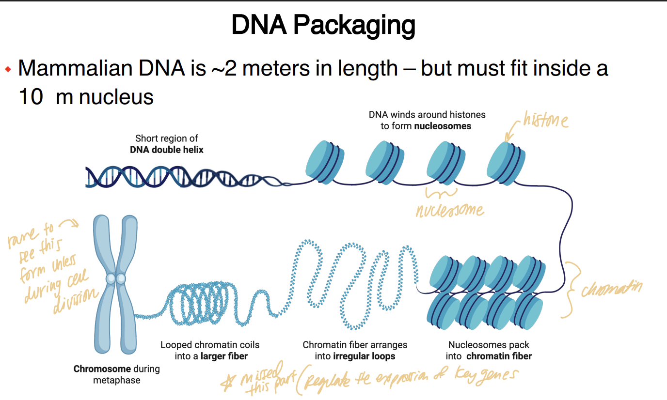
⭐ Q: Why does DNA need to be packaged, and how do histones help accomplish this?
DNA must be heavily packaged because the mammalian genome is extremely long relative to the size of the nucleus, and histone proteins allow DNA to wrap, condense, and ultimately form chromatin.
The general hierarchy shown is:
DNA double helix
→ DNA wraps around histones
→ nucleosomes
→ chromatin fibers/loops
→ highly condensed chromosome
🔎 What this means:
You can't just stuff a giant loose DNA strand into a tiny nucleus.
The cell solves this by progressively organizing and folding it.
A nucleosome is DNA wrapped around histone proteins.
Chromatin means DNA together with its associated proteins.
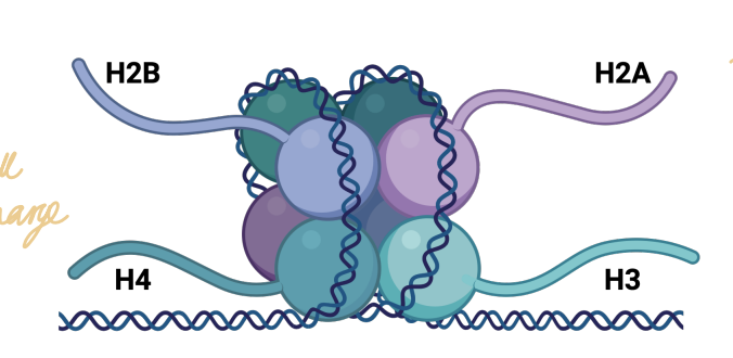
⭐ Q: What are histones structurally, and why does DNA bind strongly to them?
Histones are positively charged proteins that form an octamer around which negatively charged DNA wraps, allowing DNA to condense into chromatin.
Histones are especially rich in positively charged:
lysine ⭐️
arginine⭐
need to remember those two^
🔎 What this means:
An octamer means a complex containing eight protein subunits.
DNA is negatively charged because of its phosphate-containing backbone.
Histones contain lots of positively charged amino acids.
Opposite charges attract:
negative DNA ↔ positive histones
This is called an electrostatic interaction.
⭐ Q: Why does the tightness of DNA wrapping around histones affect gene expression?
DNA-histone packing affects gene expression because tightly condensed DNA is less accessible to cellular machinery, while more open DNA is more accessible, and histone modifications help regulate this packaging.
⚠ Lower priority
⭐ Q: Why must DNA replication occur before cell division, when does it occur, and why is proofreading important?
DNA must replicate before cell division so both daughter cells receive the same genetic information, and this replication occurs during the S phase (Synthesis) of the cell cycle while newly synthesized DNA is proofread and repaired.
🔎 What this means:
Before one cell becomes two, it has to produce a second copy of its genome.
S phase = DNA synthesis phase.
Proofreading is important because mistakes made while copying DNA can become permanent sequence changes if they aren't corrected.

⭐ Q: What does it mean that DNA replication is semi-conservative?
DNA replication is semi-conservative because each original DNA strand serves as a template for a new strand, leaving each daughter DNA molecule with one parental strand and one newly synthesized strand.
🔎 What this means:
Start with:
OLD strand 1
OLD strand 2
Result:
DNA molecule A: OLD + NEW
DNA molecule B: OLD + NEW
💡 Memory trick:
Each new DNA molecule conserves half of the original molecule.
That's why it's semi-conservative.
⭐ Q: What are the major proteins involved in DNA replication, and what does each one do?
The major DNA-replication proteins work together to unwind DNA, create a starting point, synthesize and proofread new DNA, remove temporary RNA primers, fill remaining gaps, and connect DNA fragments.
Helicase → unwinds/separates DNA strands
Primase → makes RNA primer: the starting point
DNA polymerase → synthesizes new DNA
DNA polymerase → also proofreads
RNase/RNA exonuclease → removes RNA primers
DNA polymerase → replaces primer regions with DNA
DNA ligase → joins Okazaki fragments
🔎 What this means:
An RNA primer is a short RNA sequence that gives DNA polymerase somewhere to begin.
An Okazaki fragment is one of the short DNA sections formed on the lagging strand.
Ligase seals those pieces together afterward.
💡 Story:
Helicase = unzip 🧬
Primase = makes the starting block RNA primer 🏁
Polymerase = build 🧱
RNase = remove temporary starter 🗑
Polymerase = fill hole
Ligase = glue 🩹
⭐ Q: What is the complete sequence of DNA replication taught in this lecture?
DNA replication occurs through DNA unwinding → RNA primer placement → DNA synthesis → primer removal → gap filling → ligation.
1. DNA unwinding
Replication begins at an origin of replication.
Helicase opens the strands, creating a replication fork.
2. RNA primers are added
Primase makes short RNA primers.
These provide a starting point for DNA polymerase.
3. DNA is synthesized
DNA polymerase uses each parental strand as a template.
It also proofreads the DNA as it goes.
4. RNA primers are removed
RNA exonuclease/RNase removes the temporary RNA primers.
5. DNA fills the gaps
Another DNA polymerase replaces those regions with DNA.
6. DNA fragments are connected
DNA ligase joins the Okazaki fragments on the lagging strand.
⭐ Q: How do the leading and lagging strands differ during DNA replication?
The leading strand is synthesized continuously using one RNA primer, whereas the lagging strand is synthesized discontinuously as multiple Okazaki fragments and therefore requires multiple RNA primers.
Lagging strand
discontinuous synthesis
multiple primers
produces Okazaki fragments
fragments later joined by DNA ligase
⭐ Q: What major classes of chemotherapy drugs in the lecture inhibit DNA replication, and what examples are given?
The lecture gives antimetabolites, crosslinking agents, alkylating agents, and anti-tumor antibiotics as major drug classes that interfere with DNA replication.
Examples:
Antimetabolite: 5-fluorouracil
Crosslinking agent: carboplatin
Alkylating agent: cyclophosphamide
Anti-tumor antibiotic: doxorubicin
🔎 What this means:
The learning objective specifically asks you to give examples of chemotherapeutic drugs that inhibit DNA replication, so I would prioritize remembering the category-example pairings.
The following slide also gives general mechanisms that all stop DNA replication:
Antimetabolites
→ messes with nucleotide bases → slows/stops DNA replication
Crosslinking agents
→ glues DNA strands together so they can’t be copied
Alkylating agents
→ damages DNA by adding alkyl group, it also works as a crosslinking agent
Anti-tumor antibiotics
→ the drug slips in between DNA base pairs to deform it and prevents proper DNA unwinding, causing the strand break
All of these make it harder for a cell to successfully replicate DNA.
💡 Why cancer cells are targeted conceptually:
Cancer treatment often targets processes required for cell proliferation, and a cell cannot divide successfully without replicating its DNA first.
How do microtubule-targeting mitotic inhibitors differ from the DNA-replication drugs in this lecture?
DNA-replication drugs interfere with copying DNA, whereas microtubule-targeting mitotic inhibitors interfere with chromosome separation during mitosis.
🔎 What this means:
These both interfere with cell division, but at different points.
DNA replication inhibitors
→ problem during genome copying
Microtubule inhibitors
→ problem later when chromosomes need to be separated
The final chemotherapy slide includes both groups, but the current learning objective specifically asks for drugs that inhibit DNA replication, so that should be your priority here.
Q: True or False: Acidification of the lysosome is energy-dependent.
True. Lysosomal acidification requires energy because an ATP-dependent H⁺ pump maintains the acidic interior.
Q: In which form of the ER is Ca²⁺ stored?
Ca²⁺ is stored in the smooth ER.
What is the default destination for proteins leaving the Golgi when they are not specially diverted elsewhere?
the plasma membrane
Which of the following pathways bring material to lysosomes: endocytosis, exocytosis, and/or autophagy?
Endocytosis and autophagy bring material toward lysosomes, whereas exocytosis sends material out of the cell.
Which is NOT a component of RNA?
a. Cytosine
b. Phosphates
c. Uracil
d. Deoxyribose sugar
d. Deoxyribose sugar
Deoxyribonucleic acid (DNA) is negatively
charged:
a. True
b. False
a. True
Which amino acid is involved in histone
binding to DNA?
a. Lysine
b. Glutamine
c. Alanine
d. Tyrosine
a. Lysine
the other one is Arginine
During DNA replication, helicase is responsible
for laying down the RNA primers needed as
“starting blocks” for DNA polymerase:
a. True
b. False
b. False
Primase is the one responsible
⭐ Q: How do replication, transcription, and translation fit together in the central dogma?
Replication copies DNA, transcription uses DNA to make RNA, and translation uses mRNA to make a protein.
Replication: DNA → DNA
Transcription: DNA → RNA
Translation: RNA → amino-acid chain
The amino-acid chain then folds into a functional protein.
⭐ Q: How do transcription and translation compare in what they make, what template they use, and where they occur?
Transcription uses DNA as a template to make RNA in the nucleus, whereas translation uses mRNA as a template to make protein in the cytosol.
Transcription
template = DNA
product = RNA
occurs in nucleus
mediated by RNA polymerase
Translation
template = mRNA
product = amino-acid chain/protein
occurs in cytosol
mediated by ribosomes and tRNAs
💡 Memory trick:
Transcription = write a transcript (RNA). 📝
Translation = translate the message (mRNA) into another “language,” (protein).
⭐ Q: What major types of RNA are identified in this lecture, and how do their functions compare?
mRNA
tRNA
rRNA
snRNA
miRNA
lncRNA
The lecture divides RNA into protein-coding mRNA and several non-coding RNAs that perform other roles in RNA processing, translation, or gene regulation.
mRNA = messenger RNA
coding RNA
carries information that is translated into protein
tRNA = transfer RNA
non-coding
links mRNA codons to amino acids during translation
rRNA = ribosomal RNA
non-coding
helps form the ribosome that reads mRNA the build amino acid chains
snRNA = small nuclear RNA
non-coding
Help splice pre-mRNA into mature mRNA that can actually be exported and translated
miRNA = microRNA
non-coding
recognizes particular mRNAs and decreases the expression of a specific gene
lncRNA = long non-coding RNA
non-coding
regulates gene expression
🔎 What this means:
“Non-coding” does not mean useless.
It means the RNA itself is not translated into a protein.
Some RNAs instead help the cell:
translate other RNAs
process RNA
regulate gene expression
The slide identifies mRNA as the coding RNA and lists the other five as non-coding types.
Most of these RNAs exist either to help turn the information in DNA into protein, or to control whether/how that protein gets made.
A codon is a group of 3 nucleotides on mRNA that tells the translation machinery which amino acid to add next.
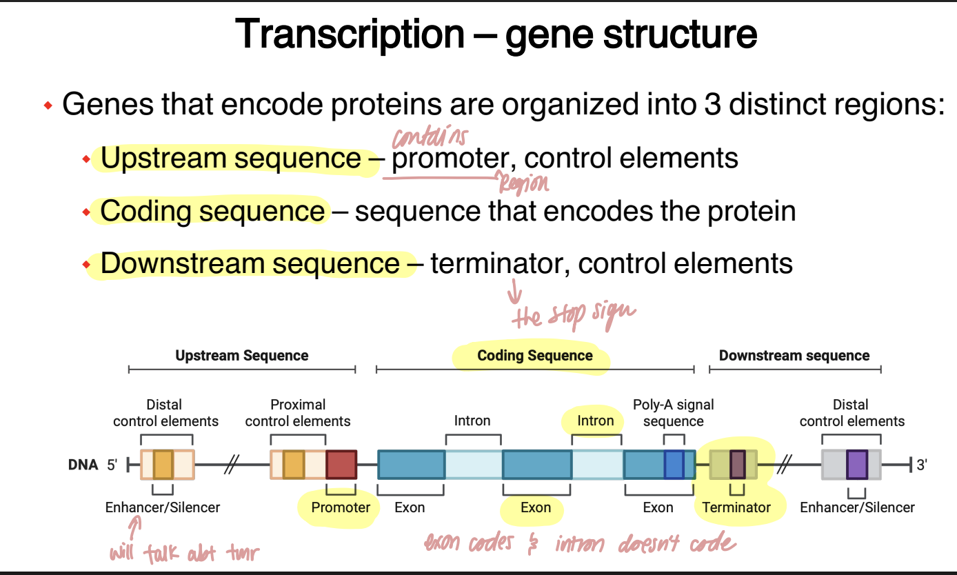
⭐ Q: What are the three major regions of a protein-coding gene, and what does each region do?
A protein-coding gene contains an upstream region that helps control transcription, a coding region that contains the protein-coding information, and a downstream region containing termination and additional control elements.
Upstream sequence: contains promoter & control elements
Coding sequence: contains the sequence that ultimately encodes the protein & includes exons and introns
Downstream sequence: contains the terminator & additional control elements
🔎 What this means:
Genes contains instructions controlling:
where transcription starts → what gets copied → where transcription ends
💡 Analogy:
Upstream = start/control signs 🚦
Coding region = actual message 📝
Downstream = stop/control signs 🛑
⭐ Q: What is transcription, what enzyme performs it, and what are its three stages?
Transcription is the production of RNA using genomic DNA as a template, occurs in the nucleus, is mediated by RNA polymerase, and consists of initiation, elongation, and termination.
The stages are:
1. Initiation
→ transcription machinery assembles
2. Elongation
→ RNA is actively synthesized
3. Termination
→ RNA synthesis stops and the transcript is released
🔎 What this means:
RNA polymerase is the enzyme that performs the central copying step.
It reads DNA information and builds a complementary RNA molecule.
⭐ Q: What happens during transcription initiation?
Transcription initiation begins at the promoter, where general transcription factors bind sequentially and recruit RNA polymerase.
Sequence:
promoter
→ general transcription factors bind
→ RNA polymerase is recruited
→ transcription can begin
🔎 What this means:
RNA polymerase doesn't just randomly land anywhere on DNA and start copying.
The promoter tells the transcription machinery where the gene begins.
General transcription factors are proteins that help assemble the transcription machinery at that promoter.
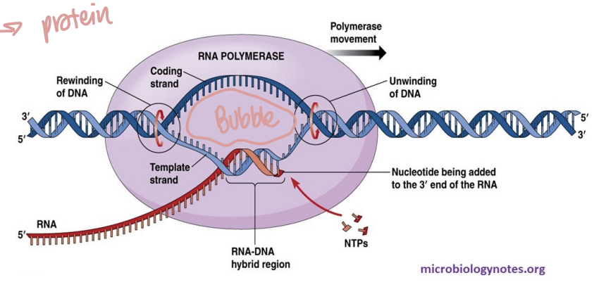
⭐ Q: What happens during transcription elongation, and what is RNA polymerase doing?
During elongation, RNA polymerase locally unwinds DNA, reads the template DNA strand, synthesizes complementary RNA, and rewinds the DNA behind itself.
RNA polymerase:
has helicase-like activity to open the strands
reads the template/antisense strand
synthesizes complementary sense mRNA
does not proofread
creates a temporary transcription bubble
🔎 What this means:
A transcription bubble is the small region where DNA is temporarily opened so RNA polymerase can access the template strand.
Unlike DNA replication, the whole chromosome isn't being copied.
RNA polymerase opens only the local region it needs, makes RNA, and closes the DNA again behind itself.
💡 Analogy:
Imagine opening a zipper just a few inches at a time:
open ahead → read/copy → zip back up behind.
⭐ Q: What happens during transcription termination?
Transcription terminates when RNA polymerase reaches a termination sequence, stops transcription, the RNA strand is released, and RNA polymerase detaches from the DNA.
⭐ Q: Why isn't the RNA produced immediately by transcription ready to function as mature mRNA?
Newly transcribed RNA is not yet mature because it still contains non-coding introns that must be removed through RNA splicing.
So transcription initially makes a pre-mRNA, and RNA processing turns that into mature mRNA.
⭐ Q: What is RNA splicing, and what does the spliceosome do?
RNA splicing is the process in which a spliceosome removes introns from pre-mRNA and joins the exons together to produce mature mRNA.
🔎 What this means:
A spliceosome is an RNA-protein complex that carries out splicing.
⭐ Q: What happens to mature mRNA after RNA processing?
Once mature mRNA has been produced, it is exported through nuclear pores into the cytosol, where it can undergo translation.
⭐ Q: What is translation, what molecules carry it out, and what determines the amino-acid sequence of the protein?
Translation is the synthesis of a protein using mRNA as a template, and it is carried out in the cytosol by ribosomes and tRNAs.
mRNA provides the nucleotide sequence
tRNAs connect that sequence to specific amino acids
ribosomes read the mRNA and join the amino acids together
🔎 What this means:
Translation converts information written in the language of nucleotides into the language of amino acids.
The order of nucleotides in the mRNA determines the order of amino acids in the resulting protein.
⭐ Q: What is a codon, and how does the genetic code connect mRNA to amino acids?
A codon is a sequence of three mRNA nucleotides that specifies one amino acid or a translation signal.
🔎 What this means:
The ribosome doesn't read one nucleotide at a time independently.
It reads mRNA in groups of three.
Example from the lecture:
AUG → methionine
AUG also serves as the start codon
💡 Memory trick:
3 RNA letters = 1 codon.
⭐ Q: What is the role of tRNA during translation?
transfer RNA acts as an adapter between an mRNA codon and its corresponding amino acid, allowing the nucleotide sequence to be converted into an amino-acid sequence.
Each tRNA has:
an attached amino acid
an anticodon
the anticodon corresponds to an mRNA codon
🔎 What this means:
An anticodon is the sequence on the tRNA that recognizes the corresponding codon on mRNA.
The lecture also states that tRNAs are transcribed in the nucleus and exported into the cytosol.
💡 Analogy:
tRNA = delivery driver 🚚
Codon = delivery address
Amino acid = package

What is the one codon that Baines wants us to remember and why?
AUG = Methionine
Because translation always starts with methionine
⭐ Q: What is the role and basic structure of a ribosome during translation?
Ribosomes read mRNA and join the amino acids delivered by tRNAs into a growing protein, and they consist of rRNAs and proteins organized into large and small subunits.
Ribosomes are made of 2 separate subunits joined together: the 60S large subunit and the 40S small subunit
These are assembled separately in the nucleus and then exported.
🔎 What this means:
Ribosomes are not made only of protein.
They contain ribosomal RNA, or rRNA, plus many proteins.
That is why rRNA is one of the non-coding RNA types essential for translation.

⭐ Q: What happens during translation initiation with ribosomes?
Translation initiation begins when the 40S ribosomal subunit and a methionine (AUG) -carrying tRNA identify the AUG start codon, after which the 60S subunit joins to form the translation complex.
🔎 What this means:
AUG tells the ribosome where protein synthesis should begin.
AUG encodes methionine, so methionine is the amino acid associated with translation initiation in this lecture.
💡 Memory trick:
AUG = “Ayo, Start!” 😭

⭐ Q: What happens during translation elongation with ribosomes?
During translation elongation, new tRNAs repeatedly enter with amino acids, the growing amino-acid chain is transferred, the ribosome moves to the next codon, and empty tRNAs leave.
The cycle is:
1. New tRNA enters with amino acid
→ 2. growing chain is transferred to the new amino acid
→ 3. ribosome moves forward one codon
→ 4. empty tRNA exits
→ 5. cycle repeats
🔎 What this means:
Each time the ribosome moves to a new codon, another amino acid can be added.

⭐ Q: What happens during translation termination with ribosomes?
Translation terminates when the ribosome reaches a stop codon, a release factor triggers release of the amino-acid chain, and the ribosome disassembles.
🔎 What this means:
A release factor is the factor shown in the lecture that recognizes the termination situation and causes the completed chain to be released.
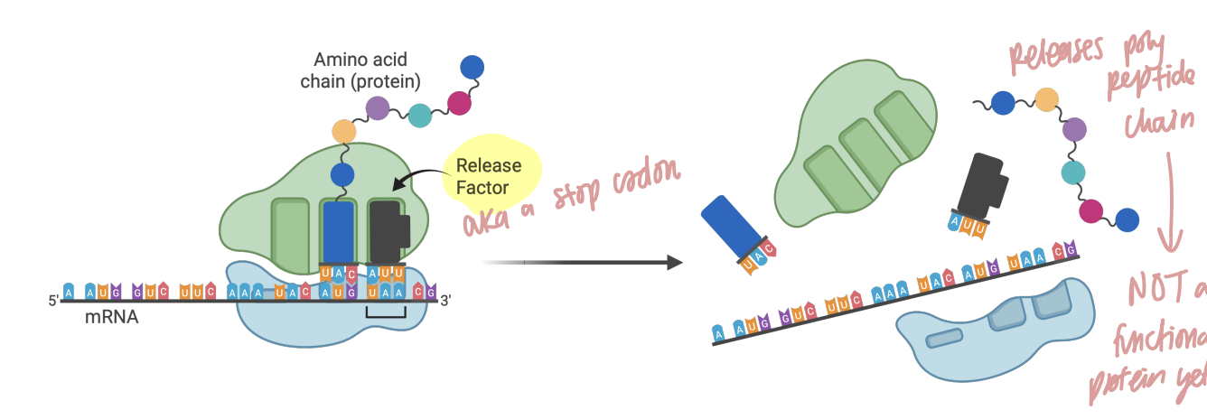
Q: Is the amino-acid chain released from the ribosome already a fully functional protein?
No. After translation, the amino-acid chain must fold into its final three-dimensional form to become a functional protein.
Q: How did α-amanitin poisoning cause severe disease in the Dachshund case?
α-Amanitin from death cap mushrooms inhibited RNA polymerase, thereby disrupting transcription and especially damaging cells with high protein-synthesis demands such as hepatocytes
Leading to acute liver failure
DNA polymerase is required for transcription
of DNA into RNA:
a. True
b. False
b. False
It is RNA polymerase that is needed instead, not DNA polymerase
In which of the 3 steps of transcription is the
RNA actively synthesized?
a. Initiation
b. Elongation
c. Termination
b. Elongation
Removal of introns from the pre-RNA to
produce the mature mRNA is known as:
a. RNA Editing
b. RNA Splicing
c. RNA Interference
b. RNA Splicing
Which other types of RNA are necessary for
the translation of mRNA into protein (select
all that apply)?
a. Transfer RNA (tRNA)
b. Micro RNA (miRNA)
c. Ribosomal RNA (rRNA)
d. Small nuclear RNA (snRNA)
a. Transfer RNA (tRNA)
c. Ribosomal RNA (rRNA)
⭐ Q: At what stages can gene expression be regulated?
Gene expression can be regulated at multiple stages from transcription through RNA processing and translation, allowing the cell to control how much final protein is produced.
This lecture focuses on:
Regulation of transcription:
histone modifications
DNA methylation
enhancers
silencers
Regulation of RNA processing:
alternative splicing
Regulation of translation:
RNA interference, especially miRNA
🔎 What this means:
Gene expression means using a gene to ultimately produce its functional product, which this lecture frames primarily as production of protein.
The cell doesn't only have one ON/OFF switch.
It can regulate the process at several checkpoints.
💡 Analogy:
Imagine a factory line.
You can control production by:
preventing the order from being written
changing how the order is edited
preventing the order from being translated into a product
⭐ Q: What is epigenetics according to this lecture, and what are the “Big 3” epigenetic mechanisms it identifies?
Epigenetics refers to heritable changes in gene expression that occur without changing the gene's DNA sequence, and the lecture identifies histone modifications, DNA methylation, and RNA interference (miRNA) as its “Big 3.”
🔎 What this means:
The DNA sequence itself does not have to change for the amount of gene expression to change.
Instead, the cell can alter:
how accessible the DNA is
whether the promoter is available
whether an mRNA is allowed to produce protein