term 1 pos
 Call Kai
 Learn
Learn Practice Test
 Spaced Repetition
 Match
Match Flashcards
 Knowt Play
1/49

There's no tags or description
Looks like no tags are added yet.
Name | Mastery | Learn | Test | Matching | Spaced | Call with Kai | Chat |
|---|
No analytics yet
Send a link to your students to track their progress
50 Terms
basic structure of chromosomes-
centromere at centre
telomeres at ends- protective, repetitive sequence of DNA
there are replication origins
genes spread around
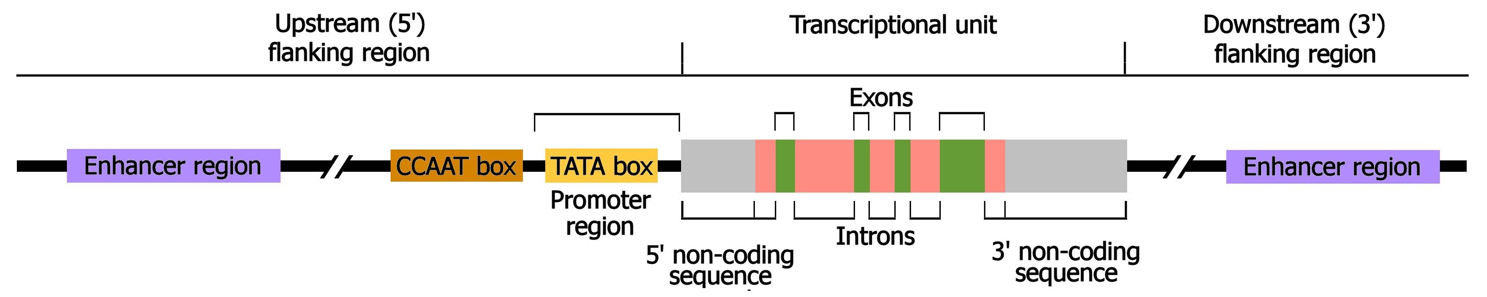
structure of a eukaryotic gene
promotor region
exons and introns
TATA box in promotor- initiation of transcription
transcription initiation site
translation initiaton site in exon 1 (ATG)
translation termination site
transcription termination site
what is the central dogma
DNA to RNA to protein
(transcription, splicing, export, translation, folding)
what are RNA polymerases and what are the 3 types
protein complexes made up of 8-14 subunits
Pol 1 - ribosomal RNA
Pol 2- mRNA for translation into protein
Pol 3- small RNA (tRNA)
transcription initiation
transcription factor binds to eukaryotic promotor
formation of preinitation complex
RNA pOL 2 begins elongation
what do transcription factors bind to
gene specific transcription factors bind to regulatory sequences of the gene
gene definition
A locus (or region) of DNA, which is made up of nucleotides and is the molecular unit of heredity
chromosome definition
A thread-like structure of nucleic acids and protein found in the nucleus of most living cells, carrying genetic information in the form of genes.
genome definition
The complete set of genes or genetic material present in a cell or organism
initiation
general transcription factors bind , eg TBP to TATA box
pre initiation complex formed
rna polymerase holoenzyme binds to the promotor region
where do transcription factors bind to?
regulatory sequences
what is a transcription factor homodimer
2 protein monomers that bind together to regulate gene expression
trans activating domain
regions of transcription factors which bind to coactivator complexes to activate transcription
helix turn helix motif
two alpha helices joined by a short strand of amino acid
recognition and binding of DNA is done by the 2 helices, one at n terminus and one at c terminus
c terminal binds to major groove, n terminal helps to position the complex
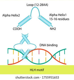
other dna binding motifs
zinc finger
leucine zipper
helix loop helix
elongation
what does rna polymerase do
what helps maintain the correct length
what does the zipper do
RNA polymerase travels along from the 5’ to 3’ end (upstream to downstream)
rudder in the upstream end of the RNA-DNA hybrid helps maintain the correct length, etc
the zipper is another protein loop within RNA polymerase and maintains boundaries of the transcription bubble especially at the downstream end
what are topoisomerases
regulate dna overwinding or underwinding
5’ cap addition
7 methyl guanosine triphosphate cap
this is a modified guanine cap to the 5’ end of mRNA
protection from degradation
initiation of protein synthesis
3’ polyadenylation
example of the sequence
what recognises it
what happens then to the mRNA
what then happens at the 3 end
what binds
why does this happen
sequences of the pre mRNA such as the AAUAAA sequence are recognised by the cleavage and polyadenylation specifity factor (CPSF)
then an endonuclease cleaves mRNA at specific site
enzyme poly-a-polymerase joins multiple adenine nucleotides to the 3’ end
poly a binding proteins bind to the newly formed tail
provides stability and facilitates exit of mRNA from the nucleus
xm2
nuclease that degrades remaining nacsent transcript and leads to termination of transcription
splicing
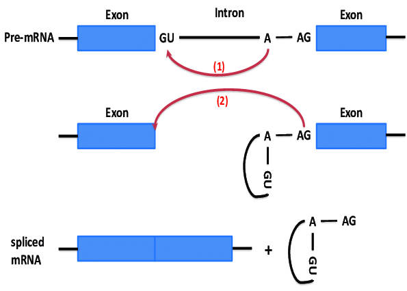
introns always begin with the residues GU and end with AG and an A somewhere towards the middle
phosphodiester bond is attacked between GU and A
3 prime end attacks phosphodiester bond between A and AG joining exons and removing the introns
how can proteins be altered for a specific function?
alternative splicing
what is the genetic code
20 amino acids
4 diff nucleotides
if code is x nucleotides , number of possiblity is 4 to the power of x
codons
charging of tRNAs with amino acids
what does amino acyl tRNA synthetase do
what does it then attach and what is released as a result
what is created
what does the enzyme bind this to
what then happens and what kind of bond is formed
what molecule is released
amino acyl tRNA synthetase binds an amino acid and ATP
the enzyme catalyses a reaction that attaches AMP to amino acid releasing pyrophosphate
this creates a high energy aminoacyl-adenylate intermediate
The same enzyme, aminoacyl-tRNA synthetase, binds the activated aminoacyl-adenylate and its corresponding tRNA molecule.
amino acid is transferred from AMP to the 3 end of tRNA which forms an ester bond
AMP is released, as the amino acid is now covalently linked to the tRNA, creating a "charged" aminoacyl-tRNA.
what are the site of the ribosome
p site holds trna with growing polypeptide chain attached
e site holdsthe trna which will exit
a site holds aminoacyl trna
elongation cycle
what is it
what is delivered to the A site by what. what does this consume
what catalyses formation of a peptide bond
what happens and what 2 things does this need
process of adding amino acids to a polypeptide chain
aminoacyl trna is delivered to the A site. elongation factor escorts the trna and binds it to the ribosome consuming GTP (to GDP)
peptidyl transferase catalyses formation of peptide bond
ribosome shifts one codon along in the 3 direction. facilitated by elongation factor 2, this requires GTP.This action moves the tRNAs: the now uncharged tRNA from the P-site moves to the E-site, and the tRNA carrying the polypeptide chain moves from the A-site to the P-site, freeing up the A-site for the next cycle.
translation initiation
what do the eukaryotic initation factors bind to
what does initiator tRNA carry and what does this do
what does the small ribosomal subunit do
what happens to the GTP on the tRNA and what does this cause and allow
what sits on the p site
eukaryotic initiation factors bound on the 5’ end join to the (poly a binding protein) bound on the poly a tail of the 3’ end
initiator tRNA carries methionine, GTP and initiation factor 2, and the 40s ribosome both recognise the initiation factors bound to the 5 and 3 ends. They all interact
small ribosomal subunit finds the start codon, using protein initiation factors. but some are easier to find than others (purine 3 residues upstream of A -Kozak sequence) 40s subunits slides down
GTPase activity converts the GTP on the tRNA to GDP
causes conformational change, and GDP is release
this allows the large ribosomal subunit to bind
methionine sites on the p site of the ribosomal complex
ready to begin elongation
termination
stop codon do not have a corresponding trna to recognise
instead when a ribosome reaches the stop codon the a site accepts a protein called release factor instead of tnra
release factor hydrolyses the bond between trna in the p site and the amino acid in the polypeptide chain, freeing it
two ribosomal subunits and other structures dissociate
antibiotics targeting translation
chloramphenicol
erythryomycin
tetracycline
streptomycin
chloramphenicol binds to 50s rrna inhibiting formation of a peptide bond
erythromycin binds to 50s of rrna and prevents movement along mrna
tetracycline inhibits trna anticodon reading of mrna codon
streptomycin changes shape of 30s rrna and causes mrna to be read incorrectly
what is meant by a protein structure hierarchy
-primary, secondary, tertiary, quaternary(eg 2 beta and 2 alpha globin polypeptides with a heme group)
what type of amino acids are mammalian proteins
how many amino acids
what is the peptide bond classified by
how many amino acids makes a protein
shape of peptide bond
mammalian proteins are alpha amino acids
20 amino acids
carboxyl group reacts with amino group to form a peptide bond and lose a water molecule
catalysed by peptidyl transferase (28s ribozyme-ribonucleic acid enzymes)
within a polypeptide chain, individual amino acids = residues
when >50 amino acids are in a polypeptide is a protein
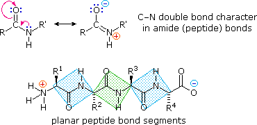
peptide bond is planar (partial double bond between carbonyl O and N)
bonding determining protein structure
covalent primary
hydrogen secondary
hydrophobic+VDW tertiary
electrostatic within tertiary helps maintain
VDW/ electrostatic quaternary
alpha helix
what runs parallel to the helix axis
what winds around the axis
what does htis lead to
how many amino acid residues per complete turn
beta sheets
what orientation do H bonds run to the chain direction
alpha helix- H bonds run parallel to helix axis. alpha carbon backbone winds around an axis so that each carbonyl O atom is H bonded to each amino N of the amino acid located 4 residues closer to C terminus
The standard alpha helix has approximately 3.6 amino acid residues per complete turn
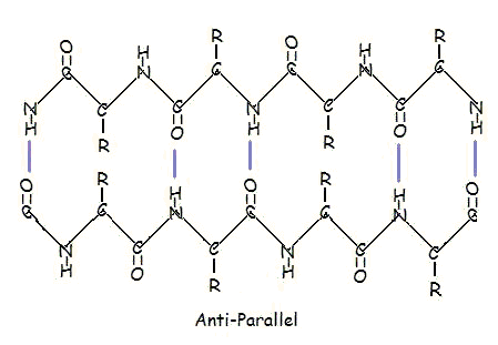
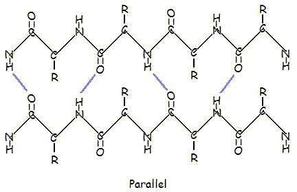
beta sheets- H bonds run perpendicular to chain direction
parallel and antiparallel
types of r groups
nonpolar, polar, electrically charged
cysteine
can form disulphide bonds as it has an SH group
examples of tertiary proteins
-catalase
-triose phosphate isomerase
-actin
tertiary domain
the smallest stable unit of a tertiary structure. a domain is defined as that region of a polypeptide chain that can fold into an autonomous stable tertiary structure
domain shuffling- when domains have been switched around between proteins through evolution
haemoglobin- quaternary potein
what are the two states
how many units
haemoglobin- needs to deliver oxygen tissues. needs high enough affinity to pick up oxygen but low enough to release it.
t state- oxygen unbound
r state- oxygen bound. so that the 3 other units have higher affinity
small changes in oxygen concentration can dramatically affect binding to haemoglobin giving a sigmoidal curve of oxygen binding
collagen
what type of protein
what is the general structure
extracellular fibrous protein but not an alpha helix
3 helical chains that wind around a central axis
general structure is Gly-X-Y where X can be any amino acid especially proline lysine or hydroxyproline
Glycine is small so glycines from each chain fit at the centre of each helix
H bonds between the chains
functions of chromatin
package and condense dna
prevent dna damage
control dna expression
what are nucelosomes
how many base pairs
what is the core comprised of
what are the cores separated by
what brings nucleosomes together and what does this do
what do multiple stacked rings form
what is formed when they are coiled further
‘beads on a string’
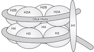
around 146 base pairs of dna wrapped around 1.7 times around a core
core is comprised of an octamer of histone proteins
nucleosome core proteins are separated by linker proteins
histone H1 brings nucelosomes together to condense chromatin further
multiple stacked rings form the solenoid fibre/30nm fibres
these are coiled further to form chromosomes
pcr
heat to 94 to denature (separate) dna strands
cool to 50-60 to allow primers to anneal
72 to allow elongation of new strands using taq polymerase
so at the end of the first cycle 2 new strands
begin cycle two by denaturing and annealing again
elongation generates strands of different sizes depending on the template strands
sanger sequencing
mix small amount dideoxy with deoxy nucleotides
cycle through multiple rounds of pcr
each ddNTP contains fluorescent tags
separate by gel electrophoresis
detect by laser
(chain synthesis terminates when ddNTP is incorporated)
technical applications of PCR that could be used to an advantage in veterinary medicine
VNTR
RAPD
AFLP
they are all used to determine breed, pedigree and/or parentage
what components are essential for basic PCR?
DNA polymerase
all 4 deoxyribonucleotides
dsDNA template
a pair of primers which bind to opposite polynucleotide strands and have their 3 ends pointing towards each other