Upper GI - Esophagus
 Call Kai
 Learn
Learn Practice Test
 Spaced Repetition
 Match
Match Flashcards
 Knowt Play
1/131

There's no tags or description
Looks like no tags are added yet.
Name | Mastery | Learn | Test | Matching | Spaced | Call with Kai | Chat |
|---|
No analytics yet
Send a link to your students to track their progress
132 Terms
What type of epithelium lines the normal esophagus?
Nonkeratinized stratified squamous epithelium
What are the major layers of the esophageal wall?
Mucosa → submucosa → muscularis propria → adventitia
What layers make up the mucosa of the esophagus?
Nonkeratinized squamous epithelium
Lamina propria
Muscularis mucosae
What are the two layers of the muscularis propria?
Circular
Longitudinal
Where does the esophagus extend to?
Epiglottis → Gastroesophageal junction
What is unusual about the esophagus compared with most of the GI tract?
It does not have a serosa
It has an adventitia instead
What type of muscle is found predominantly in the upper 2/3 of the esophagus?
Straited skeletal muscle
Stratified squamous epithelium
What type of muscle is found predominantly in the lower 1/3 of the esophagus?
Smooth muscle
Columnar epithelium
How does lymphatic drainage differ with the upper esophagus?
Neck and mediastinal lymph nodes
How does lymphatic drainage differ with the lower esophagus?
Gastric and para-aortic lymph nodes
How does innervation differ with the upper esophagus?
Somatic sensation
Allowing relatively better localization of pathology
How does innervation differ with the lower esophagus?
Visceral innervation
Resulting in poorer localization of pathology
What type of carcinoma can arise from the upper esophageal epithelium?
Squamous cell carcinoma
What type of carcinoma commonly arises in the lower esophagus?
Adenocarcinoma
What are examples of mechanical obstructions?
Atresia
Fistulas
Duplications
What is an example of a functional obstruction?
Achalasia
What is esophageal atresia?
Thin, noncanalized cord
A congenital interruption of the esophagus in which a segment is replaced by
Where does esophageal atresia most commonly occur?
At or near the tracheal bifurcation
What congenital abnormality is commonly associated with esophageal atresia?
A tracheoesophageal fistula connecting an esophageal pouch to the trachea or bronchus
What are major complications of esophageal atresia/tracheoesophageal fistula?
Aspiration
Suffocation
Pneumonitis/Pneumonia
Severe fluid/electrolyte abnormalities
How does esophageal atresia typically present?
Shortly after birth
Commonly with regurgitation during feeding
How is esophageal atresia treated?
Prompt surgical repair
What is an important long-term complication of repaired esophageal atresia?
Increased risk of:
Esophageal dysmotility
Aspiration
Squamous cell carcinoma
Which abnormality is the most common atresia/fistula?
Proximal atresia w/ distal fistula

What is esophageal stenosis?
Narrowing of the esophageal lumen
1-99%
Is esophageal stenosis more commonly congenital or acquired?
Acquired
Strong bases heavily contribute
What causes acquired esophageal stenosis?
Fibrous thickening of the submucosa and atrophy of the muscularis propria
What conditions can cause acquired esophageal stenosis?
Chronic GERD/inflammation and scarring
Radiation
Ingestion of caustic substances
What is the classic progression of dysphagia in esophageal stenosis?
Difficulty swallowing solids first
Followed by difficulty with liquids
What is achalasia?
Discoordinated contraction or spasm of the muscularis leading to esophageal dysmotility
Waves of peristaltic contraction
failure of progressive peristalsis
Incomplete relaxation of the lower esophageal sphincter (LES)
What complication is associated with achalasia?
Diverticula
What are the three classic features of achalasia?
Incomplete LES relaxation
Increased LES tone
Esophageal aperistalsis — Muscles are unable to squeeze to move food downward
What is the underlying pathologic process in primary achalasia?
Progressive inflammatory destruction of ganglion cells in the distal esophagus
What happens to the esophagus proximal to the LES in achalasia?
It becomes progressively dilated
What is the classic radiographic appearance of achalasia?
Dilated proximal esophagus with distal narrowing producing a “bird-beak” appearance
What histologic findings can be seen in achalasia?
Chronic ganglionitis or aganglionosis
Wallerian degeneration
Hypertrophy of the circular layer of the muscularis propria
What are complications of achalasia?
Regurgitation
Aspiration pneumonia
Diverticula
Markedly increased risk of esophageal squamous cell carcinoma
How much is the risk of esophageal SCC increased in achalasia according to the lecture?
Approximately 33-50x
What infections can cause an achalasia-like disorder?
Chagas disease, caused by Trypanosoma cruzi, through destruction of the myenteric plexus
What is pseudoachalasia?
An achalasia-like syndrome caused by another process
Such as tumor invasion of the myenteric plexuses
What is an esophageal “inlet patch”?
A focus of ectopic gastric mucosa within the esophagus
Ectopia
Where is an inlet patch most commonly located?
The upper third of the esophagus
Mostly asymptomatic
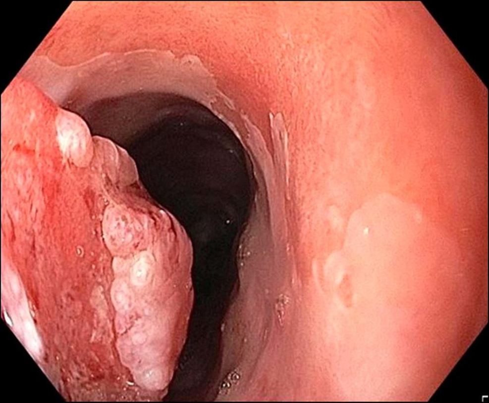
Why can an inlet patch cause symptoms?
Gastric mucosa can secrete acid into the esophagus
What complications can result from an ectopia?
Dysphagia
Esophagitis
Barrett esophagus
Rarely adenocarcinoma
What are esophageal varices?
Engorged, tortuous, dilated veins caused by portal hypertension
Can cause massive and life-threating bleeding (medical emergency)

What is the most common cause of esophageal varices according to the lecture?
Alcoholic liver disease
What other cause of esophageal varices is listed?
Hepatic schistosomiasis
Why do esophageal varices develop in portal hypertension?
Increased portal venous pressure causes blood to be diverted through portosystemic collateral vessels
Including veins in the distal esophagus
What is the classic clinical presentation of bleeding esophageal varices?
Hematemesis
What is the approximate mortality associated with the first variceal bleeding episode according to the lecture?
~50%
What is the normal venous blood flow for the GI tract>
GI tract venous blood flow → portal vein → liver → inferior vena cava (First-pass effect)
What is a Mallory-Weiss tear?
A mucosal laceration at/near the gastroesophageal junction caused by severe retching or vomiting
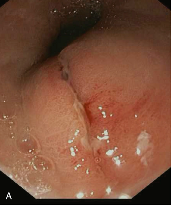
What is the most common cause of esophageal laceration?
Mallory-Weiss tear
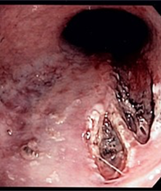
Where does a Mallory-Weiss tear typically occur?
At the gastroesophageal junction
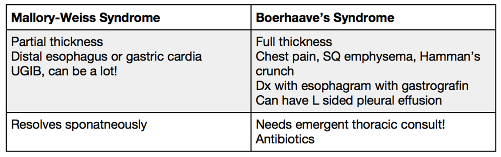
What is Boerhaave syndrome?
A transmural esophageal rupture/perforation
Classically following severe vomiting
Which is more severe: Mallory-Weiss tear or Boerhaave syndrome?
Boerhaave syndrome
It involves full-thickness perforation
In what patient population does infectious esophagitis primarily occur?
Immunocompromised patients
What are the major causes of infectious esophagitis?
Candida (fungal organism)
HSV
CMV
Which organism is the most common cause of infectious esophagitis?
Candida
What is the characteristic gross/endoscopic appearance of esophageal candidiasis?
Adherent gray-white pseudomembranes/plaques covering the mucosa

What is the classic description of Candida plaques?
“Cottage cheese” plaques

What makes up the pseudomembranes in esophageal candidiasis?
Densely matted fungal hyphae and inflammatory cells
What is the characteristic endoscopic appearance of HSV esophagitis?
Punched-out ulcers
Where are HSV inclusions found histologically?
Nuclear viral inclusions within a rim of degenerating epithelial cells at the ulcer edge

What are the classic HSV nuclear changes (Triple M)?
Multinucleation
Margination of chromatin
Molding of nuclei
How does CMV esophagitis typically appear endoscopically?
Relatively shallow ulcerations
Compared with HSV
Which cells are characteristically infected by CMV in esophageal biopsies?
Endothelial cells
Stromal cells
What type of inclusions are characteristic of CMV?
Both nuclear and cytoplasmic inclusions
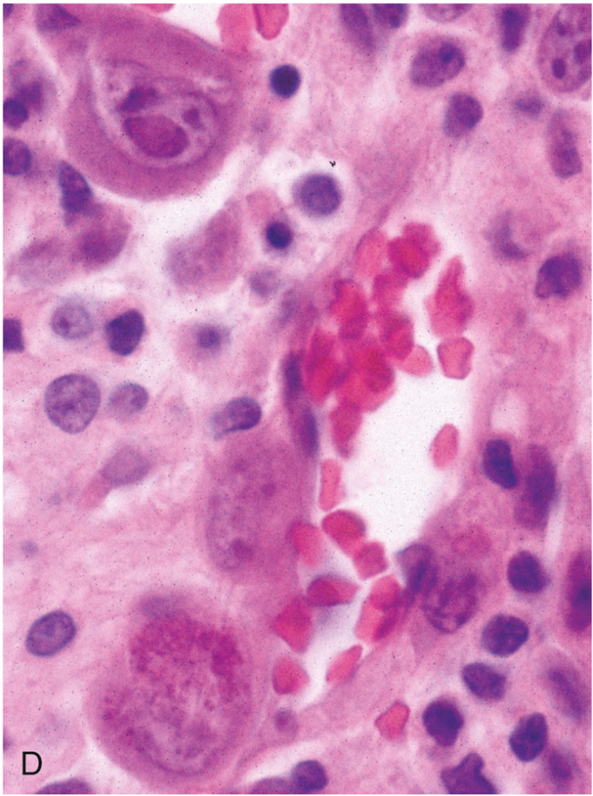
What is the classic appearance of the CMV nuclear inclusion?
“Owl-eye” inclusion
Compare the classic findings of the three infectious esophagitis causes
Infection | Classic finding |
|---|---|
Candida | Cottage-cheese plaques |
HSV | Punched-out ulcers + Triple M |
CMV | Shallow ulcers + owl-eye inclusions |
What are the three types of chemical esophagitis causes?
Pill esophagitis (bisphosphanates, doxycycline)
Cytotoxic chemotherapy
Radiation therapy
What is the most common cause of esophagitis?
Gastroesophageal reflux disease (GERD)/reflux esophagitis.
What causes reflux esophagitis?
Reflux of gastric contents into the esophagus → Produce mucosal injury
What mechanisms can promote GERD?
Decreased lower esophageal sphincter tone and/or increased abdominal pressure
In what age group is reflux esophagitis commonly seen according to the lecture?
Patients >40 years old
What are the characteristic histologic findings of reflux esophagitis?
Basal cell hyperplasia >15% of epithelial thickness
Elongation of lamina propria papillae to >2/3 of epithelial thickness
Scattered intraepithelial eosinophils
Neutrophils in more severe disease
What is eosinophilic esophagitis?
An immunologically mediated chronic esophageal disorder
What are the classic symptoms of eosinophilic esophagitis?
Dysphagia and food impaction
What type of patients are commonly associated with eosinophilic esophagitis?
Patients with other allergic/atopic conditions
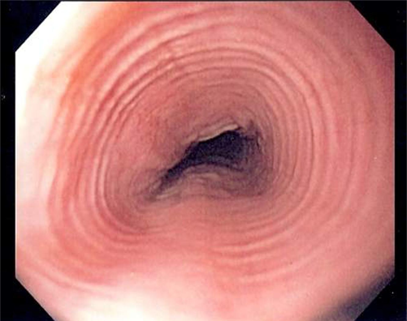
What is the characteristic endoscopic appearance of eosinophilic esophagitis?
Multiple concentric rings producing “trachealization” or “feline esophagus”
What is the histologic diagnostic feature of eosinophilic esophagitis emphasized in the lecture?
>15 eosinophils/high-power field, particularly superficially
(Notice the eosinophilic abscess)

Where should the eosinophils be located to support eosinophilic esophagitis?
They can be present at sites far from the gastroesophageal junction
What is Barrett esophagus?
Intestinal metaplasia of the esophageal squamous mucosa
Usually resulting from chronic GERD
What is the major clinical significance of Barrett esophagus?
It increases the risk of esophageal adenocarcinoma
What is the classic endoscopic appearance of Barrett esophagus?
Salmon-colored mucosa

What is seen microscopically in Barrett esophagus?
Metaplastic intestinal-type mucosa containing goblet cells

How is Barrett esophagus diagnosed?
Endoscopic findings PLUS biopsy demonstrating intestinal metaplasia
What is the common progression of adenocarcinoma due to GERD?
Chronic GERD → Barrett metaplasia → dysplasia → adenocarcinoma
What are the two esophageal tumors?
Adenocarcinoma
Squamous cell carcinoma
What are major risk factors for esophageal adenocarcinoma?
Barrett esophagus (Chronic GERD)
Obesity
Alcohol
Radiation therapy
What demographic pattern is associated with esophageal adenocarcinoma?
Much more common in males
Approximately 7:1 male-to-female predominance
Where does esophageal adenocarcinoma most commonly arise?
Distal third of the esophagus
Why does adenocarcinoma favor the distal esophagus?
Most arise from Barrett esophagus
Which develops in the distal esophagus in the setting of chronic reflux
How can one reduce the risks of esophageal adenocarcinoma?
Fresh fruits and vegetables
H. pylori → Gastric atrophy → Reduced acid secretion and reflux → Decreased incidence of Barrett esophagus
How can the location of H. pylori affect effects?
H pylori + esophagus → Good
H pylori + stomach → Bad (carcinoma & lymphoma)
What is the metaplasia-dysplasia-carcinoma sequence for esophageal adenocarcinoma?
Normal squamous epithelium → inflammation/hyperplasia (esophagitis) → Barrett intestinal metaplasia → low-grade dysplasia → high-grade dysplasia → adenocarcinoma
(Likelihood of developing into ECA increases with overall sequence)

What are the common symptoms of esophageal adenocarcinoma?
Pain or difficulty swallowing
Progressive weight loss
Hematemesis
Chest pain
Vomiting
What molecular abnormalities increase as Barrett esophagus progresses toward adenocarcinoma according to the lecture?
Abnormal ploidy
Increasing expression of p53 and GLUT-1
What is the prognosis of esophageal adenocarcinoma when diagnosed at an advanced stage?
A 5-year survival of <25%
What is the approximate 5-year survival when adenocarcinoma is limited to the mucosa and submucosa?
~80%