Human Anatomy- Lab exam 1
 Call Kai
 Learn
Learn Practice Test
 Spaced Repetition
 Match
Match Flashcards
 Knowt Play
1/103

There's no tags or description
Looks like no tags are added yet.
Name | Mastery | Learn | Test | Matching | Spaced | Call with Kai | Chat |
|---|
No analytics yet
Send a link to your students to track their progress
104 Terms
ocular lens
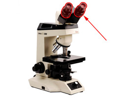
revolving nosepiece
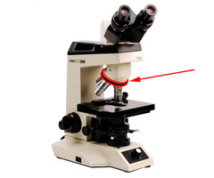
objective lens

Mechanical stage

Diaphragm adjustment lever

Base

Power switch

Mechanical stage adjustment knob

Variable light control
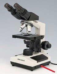
stage aperture

Condenser

Condenser heigh adjustment knob
allows the condenser to move up & down. It also is location on the left side of our microscopes.
Light source
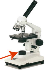
coarse focus knob

fine focus knob

Arm

Field of view- what do you see when you look trough the ocular lens

Nucleus

cytoplasm (histology)
The part of a cell between the plasma membrane and the nucleus; contains many organelles.
Total magnification

Plasma membrane histology
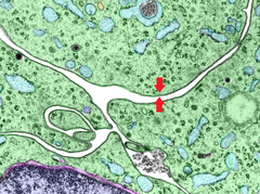
lumen
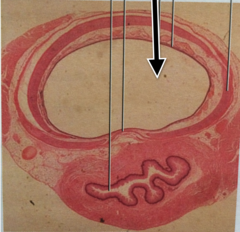
apical surface (free surface)
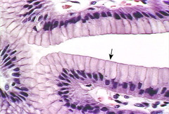
basal surface (attached surface)

basement membrane
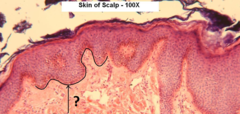
lateral view of simple squamous epithelium
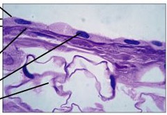
Top view of Simple Squamous Epithelium

simple cuboidal epithelium
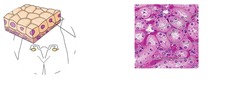
simple columnar epithelium

villus

brush boarder
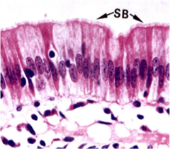
microvilli

goblet cell

pseudostratified ciliated columnar

transitional epithelium

areolar connective tissue

Fibroblasts

extracellular matrix
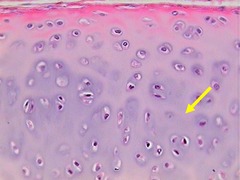
ground substance of connective tissue (the blue sections)

collagen fiber connective tissue

adipose tissue

adipocyte

fat vacuole

dense regular connective tissue

dense irregular connective tissue
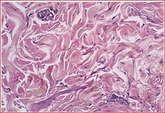
hyaline cartilage

chondrocyte
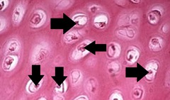
lacuna

perichondrium in hyaline cartilage

extracellular matrix hyaline cartilage (highlighted blue)
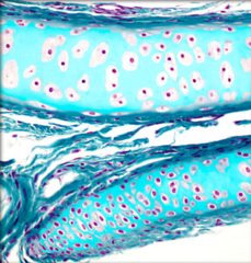
elastic cartilage

Fibrocartilage

osteocyte

Canaliculus (highlight blue)
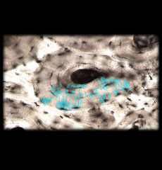
Osteon

central canal
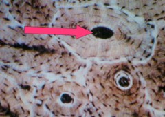
lamella
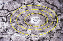
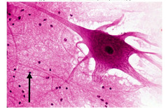
nervous tissue

neuron
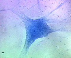
neuron process
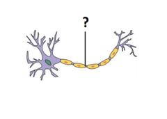
cell body of neuron

nucelus of neuron

glial (neuroglial) cell
elastic cartilage

hyaline cartilage

Thrombocyte (blood slide)

Erythrocytes

leukocyte

Skeletal muscle

Sarcolemma (plasma membrane of muscle)
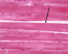
Sarcoplasm (cytoplasm of muscle)

striations (RED ARROWS)

cardiac muscle

sarcolemma of cardiac muscle

Sarcoplasm of cardiac muscle
the cytoplasm of the cardiac muscle
intercalated discs of cardiac muscle

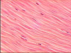
Smooth muscle
Epidermis

Stratum corneum (letter A)
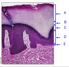
Stratum lucidum

Stratum granulosum (letter C)

Stratum spinosum
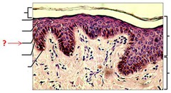
stratum basale (letter E)

Dermis

Papillary layer (areolar CT) (outlined in blue)
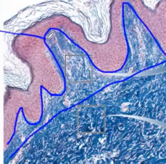
Reticular layer (dense irregular CT)

Hypodermis (superficial fascia or subcutaneous layer)

Trachea

Lung
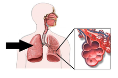
Parietal pleura (RED ARROWS)

Visceral pleura (RED ARROW)
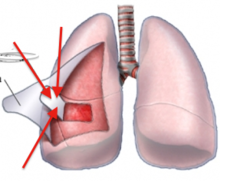
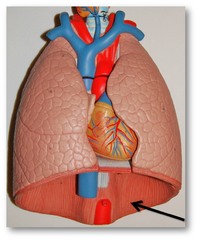
Diaphragm
Esophagus

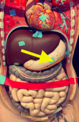
Stomach
Small intestine

Large intestine (grey organ)

Liver

heart

aorta artery

Kidney
