microbio quiz 1/test 1 pt 2
 Call Kai
 Learn
Learn Practice Test
 Spaced Repetition
 Match
Match Flashcards
 Knowt Play
1/203

There's no tags or description
Looks like no tags are added yet.
Name | Mastery | Learn | Test | Matching | Spaced | Call with Kai | Chat |
|---|
No analytics yet
Send a link to your students to track their progress
204 Terms
trematodes: general characteristics
phylum Platyhelminthes; class Trematoda (aka flukes)
adults = dorsoventrally flattened + may infect intestine, liver, lungs, lymphatics, or blood vessels
most = hermaphroditic (have both male + female reproductive organs)
EXCEPT BLOOD FLUKES
eggs are usually operculated
EXCEPT BLOOD FLUKES
most require a SNAIL as the first intermediate host; many require a second host (e.g. fish or crab)
miracidium
larval stage that hatches from eggs in water and infects SNAIL
cercaria
larval stage that leaves the snail
in schistosomes, this is infective to humans by skin penetration
PENETRATION

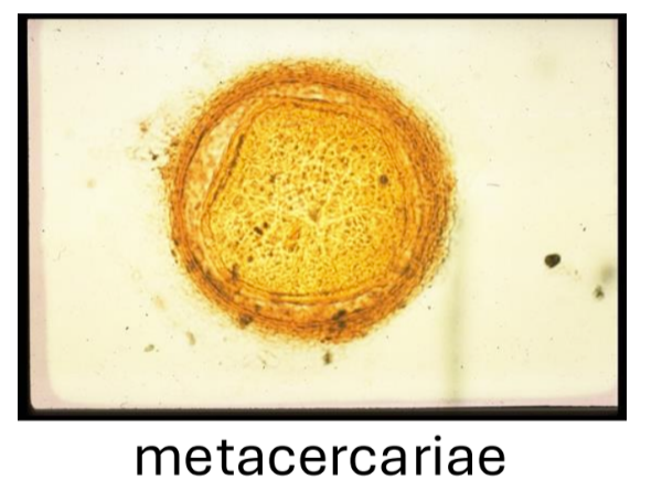
metacercaria
encysted larval stage on vegetation or in fish/crab
this is INFECTIVE for most non-blood flukes
INGESTION
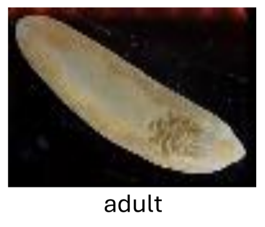
adult
lives in target organ/site and produces eggs
adult site helps predict specimen source
intestinal flukes
organisms
Fasciolopsis buski
Metagonimus yokogawai
Heterophyes heterophyes
classic exposure
water plants or raw/undercooked fish
likely specimen
stool
liver flukes
organisms
Fasciola hepatica
Clonorchis sinensis
classic exposure
watercress/vegetation
raw/pickled fish
likely specimen
stool and/or bile
lung fluke
organisms
Paragonimus westermani
classic exposure
raw/undercooked crab or crayfish
likely specimen
SPUTUM
stool
blood flukes
organisms
Schistosoma mansoni
S. haematobium
S. japonicum
classic exposure
freshwater skin exposure
likely specimen
stool
urine
look alikes
F. buski and F. hepatica EGGS
Metagonimus/Heterophyes/Clonorchis EGGS
Faciolopsis buski is also known as?
GIANT intestinal fluke
F. buski - region
Far east regions
china
vietnam
india
F. buski - reservoir hosts
dogs
pigs
F. buski - transmission stage and source
ingestion of metacercariae on raw or undercooked water plants
F. buski - life cycle
eggs reach water and mature
SNAIL is required 1st intermediate host
METACERCARIAE encyst on aquatic vegetation
humans ingest contaminated water plants
adults mature in the intestine and EGGS pass in STOOL

F. buski - infective vs diagnostic stages
infective = metacercariae on water plants
diagnostic = eggs in stool
F. buski - clinical infection + diagnosis
often asymptomatic (light worm burden)
heavy infections:
mechanical + toxic damage of small intestine
persistent diarrhea, anorexia, edema, ascites, intestinal obstruction, eosinophilia
pt diagnosis
travel history, food history, symptoms, lab findings
F. buski - laboratory finding + sample type
adult or egg
EGG IN FECES is more common
CANNOT BE DIFFERENTIATED FROM FASCIOLA HEPATICA EGGS
if only egg: report as “F. buski/ F. hepatica”
F. buski - egg
yellow-brown
elliptical
approximately 130-140 × 80-85 microns
small operculum (small lid that can open/close) may not always be visible
important!!!!
CANNOT BE DIFFERENTIATED FROM FASCIOLA HEPATICA EGGS
if only egg: report as “F. buski/ F. hepatica”

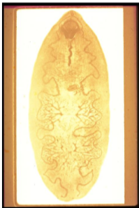
F. buski - adult
flattened fluke
approx. 2-7 cm

metagonimus/heterophyes - region
Far east
middle east
Metagonimus yokogawi and Heterophyes heterophyes
metagonimus/heterophyes - transmission stage + source
ingestion of metacercariae in raw/undercooked fish
metagonimus/heterophyes - clinical infection + diagnosis
often asymptomatic
heavy burden:
diarrhea
colic
LOOSE STOOLS W MUCUS
metagonimus/heterophyes - lab diagnosis
eggs = diagnostic and found in stool
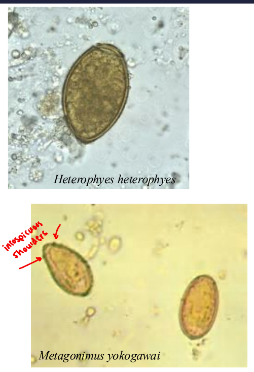
metagonimus/heterophyes - eggs
approx. 28-30 microns long
may have vase or flask shape
embryonated and operculated
inconspicuous shoulders
important!!!!!
EGGS RESEMBLE ONE ANOTHER + CLORONCHIS SINENSIS
metagonimus/heterophyes - adult
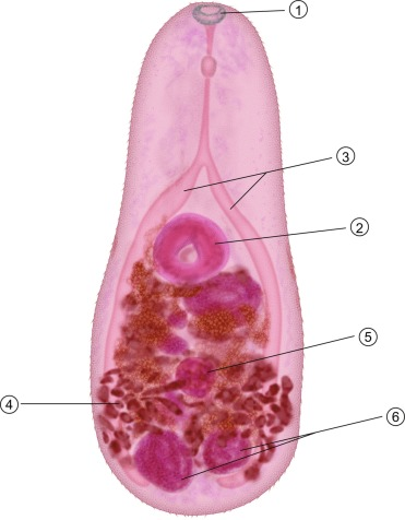
intestinal flukes comparison chart

Fasciola hepatica is also known as?
sheep liver fluke
infection in sheep is called liver rot
F. hepatica - region
found in cattle & sheep raising areas
parts of southwestern United States
F. hepatica - transmission stage + source
ingestion of metacercariae on raw water vegetation
esp watercress

F. hepatica - life cycle
eggs pass in feces and MUST reach WATER
SNAIL is required as FIRST intermediate host
cercariae leave snail and encyst as metacercariae on aquiatic vegetation
humans ingest metacercariae → larvae migrate to the liver and biliary tract

F. hepatica - infective vs diagnostic
infective = metacercariae on aquatic vegetation
diagnostic = egg in feces and/or bile
F. hepatica - clinical infection + diagnosis
tissue damage may occur during metacercarial migration thru liver tissue
adults may obstruct the biliary ducts or gallbladder
symptoms: OBSTRUCTIVE JAUNDICE, vomiting, fever, ENLARGED LIVER, CIRRHOSIS, diarrhea, eosinophilia, inflammation, and secondary bacterial infection
F. hepatica - lab diagnosis
recovery of eggs in feces and/or bile fluid
adult recovery is possible
F. hepatica - egg
yellow-brown
small operculum
approx. 130-150 × 60-90 microns
important!!!!
EGGS INDISTINGUISHABLE FROM F. BUSKI
report as “F. buski/F. hepatica egg”
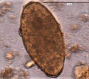
F. hepatica - adult
approx 3 cm long
conical anterior projection
broadly pointed posterior end

Clonorchis sinensis is also known as ?
chinese liver fluke
C. sinensis - region
mainly far east
china, japan, korea, vietnam
C. sinensis - reservoir hosts
dogs
cats
C. sinensis - transmission stage and source
ingestion of metacercariae in raw, undercooked, or pickled FISH
adults live in bile ducts
C. sinensis - life cycle
eggs pass in feces and reach water
SNAIL is the first intermediate host
FISH serve as the second intermediate host
humans ingest METACERCARIAE in raw, undercooked, or pickled FISH
adults live in the bile ducts and eggs may be found in feces or biliary contents

C. sinensis - clinical infection + diagnosis
light infections = few/no symptoms
heavy infections = inflammation, diarrhea, pain, fibrosis, obstruction of bile duct
complications = obstructive jaundice, cirrhosis, neoplasm of bile ducts
C. sinensis - lab diagnosis
recovery of eggs in feces and/or biliary contents
C. sinensis - egg
eggs are diagnostic
broad, ovoid egg w thick brown shell
approx. 29-35 microns long
operculum has clearly demarcated shoulders
KNOB at the opposite end, may show COMMA SHAPED POSTERIOR APPENDAGE

C. sinensis - adult
adults may be recovered from bile duct aspiration
adults are approx 1-2.5 cm

liver flukes comparison chart

Pragonimus westermani is also known as ?
the oriental lung fluke
P. westermani - region
primarily in southeast asia, latin america, africa
P. westermani - transmission stage + source
ingestion of raw or undercooked crabs or crayfish (shellfish) containing metacercariae
metacercariae excyst in the small intestine, then burrows into the diaphragm into the lungs
adults live within capsules in the bronchioles
P. westermani - life cycle
eggs leave the body in sputum or feces
larvae develop through SNAIL and CRUSTACEAN hosts
humans ingest METACERCARIAE in raw/undercooked CRAB or CRAYFISH
adults mature in the lungs

P. westermani - clinical infection + diagnosis
light infections = asymptomatic
heavy infections = persistent cough, chest pain, and hemoptysis
lung inflammation = infiltration of eosinophils and neutrophils and fibrotic capsule formation around the worm
P. westermani - lab diagnosis
recovery of eggs in sputum and/or feces
SPUTUM IS BLOOD TINGED W RUSTY BROWN FLECKS
complement fixation test may also help w extrapulmonary infections
P. westermani - egg
eggs are diagnostic (sputum = primary diagnostic specimen)
ovoid egg
approx. 80-118 × 48-60 microns
operculum has VERY SMALL SHOULDER + THICKENED SHELL AT OPPOSITE END
may also be found in feces if swallowed

P. westermani - adult
live within capsules in bronchioles

schistosoma spp - how they differ
male and female adult worms are SEPARATE forms
female lives INSIDE THE MALE within the gynecophoral canal
eggs are UNOPERCULATED
adults are CYLINDRICAL rather than dorsoventrally flattened
humans infected when CERCARIAE PENETRATE SKIN in contaminated freshwater
S. mansoni
geography: Africa, South America, West Indes, Puerto Rico
adults found in: venules of LARGE INTESTINE
egg specimen: FECES
S. japonicum
geography: far east
adults found in: venules of SMALL INTESTINE
egg specimen: FECES
S. haematobium
geography: east africa and middle east
adults found in: veins surrounding BLADDER
egg specimen: URINE
Schistosomiasis: clinical infection
skin entry
swimmer’s itch: dermatitis from cercariae w small papules, edema, erythema, intense itching
larval migration
urticaria, fever, malaise
egg production/migration
major acute damage occurs as eggs move through tissues
Schistosoma - egg production/migration leads to:
S. mansoni: stool w blood and mucus
S. japonicum: stool w blood and mucus; KAYETAMA FEVER
S. haematobium: hematuria and dysuria
Schistosoma - chronic infections
granuloma formulation, venous scarring, ascites, pain, anemia, hypertension, hepatomegaly, splenomegaly
S. haematobium chronic complications: urine retention, UTIs
Schistosoma - lab diagnosis
S. mansoni/japonicum: eggs in feces
S. haematobium: eggs in urine
Schistosoma eggs
oval w species specific spine location
NO SHOULDERS , NO LIDS
peak excretion time for eggs is 12 PM - 2 PM
S. mansoni
lateral spine
feces specimen

S. haematobium
terminal spine
urine specimen

S. japonicum
small curved rudimentary spine
feces specimen

Foodborne flukes vs Blood flukes chart

large operculated eggs consider:
Fasciola/Fasciolopsis or Paragonimus depending on specimen and type
small operculated eggs compare:
Clonorchis, Metagonimus, and Heterophyes
stage and specimen review chart

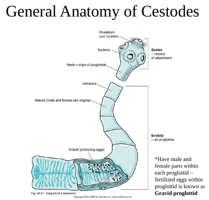
cestode anatomy vocab
strobila = adult body
scolex = “head” attachment organ
rostellum = tip of scolex; may be armed with hooks
proglottids = body segments added during growth
gravid proglottid = fertilized egg within proglottid
eggs contain an oncosphere = embryo w 3 pairs of hooks
Diphyllobothrium latum - transmission stage + source
pleroceroid larvae in pickled/raw/undercooked freshwater fish
adults live in the jejunum ; usually just 1 worm
D.latum - life cycle
eggs passed in feces reach water
coracidium is ingested by cyclops/copepod
fish ingests copepod; pleroceroid develops in fish tissue
humans ingest raw or undercooked freshwater fish
adult matures in intestine ; eggs are passed in stool

D. latum - infective vs diagnostic stage
diagnostic: egg in stool
infective: pleroceroid larva
D. latum - clinical infection + diagnosis
usually asymptomatic
vague GI symptoms - nausea, vomiting, etc
VITAMIN B12 DEFICIENCY + MEGALOBLASTIC ANEMIA
lab finding + sample type
egg in stool
can also find scolex and proglottid
diagnostic structures: scolex, egg, proglottid
D. latum - egg
yellow - brown
distinct OPERCULUM
NO SHOULDERS
small KNOB at the opposite end
UNEMBRYONATED when passed

D. latum - scolex + proglottids
scolex:
2 sucking grooves
elongated spoon shaped
proglottids:
wider rather than long
CENTRAL rosette shaped uterus

Taenia saginata - transmission stage + source
BEEF tapeworm
ingestion of raw/undercooked meat containing cysticerus larvae
adults attach to mucosa of small intestine
Taenia solium - transmission stage + source
PORK tapeworm
ingestion of raw/undercooked meat containing cysticerus larvae
or EGGS → cysticercosis
adults attach to the small intestinal mucosa
Taenia spp - taeniasis symptoms + lab findings
human ingests CYSTICERUS LARVA in beef or pork
adult worms develop in small intestine
eggs/proglottids may be seen in stool
pig/cow = intermediate host
BOTH T. sginata and T. solium
Taenia spp - cysticercosis symptoms + lab findings
human ingests infective eggs of T. solium
human becomes accidental intermediate host
eggs = NOT diagnostic focus ; TISSUE LARVAE require imaging/biopsy/serology
larvae dodge in TISSUES (muscle, eye, brain)

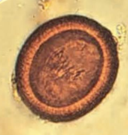
Taenia spp - egg
diagnostic but DO NOT SPECIATE
spherical
walnut - brown
smooth thick shell w RADIAL STRIATIONS
6-hooked oncosphere may not be visible
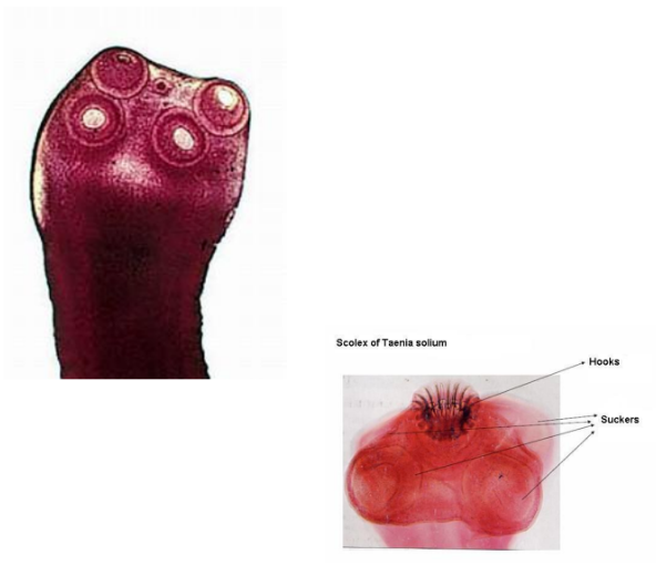
Tenia spp - scolex
allows speciation
T. saginata
NO HOOKS
< 5mm
T. solium
4 SUCKERS
double row of 25-30 HOOKLETS
evil one causes worse infection (cysticercosis) - has sharp teeth (hooks)
Taenia spp - proglottids
allow speciation
T. saginata
15-20 uterine branches
cow = bigger = MORE branches
T. solium
7-13 uterine branches
pig = smaller = LESS branches

Taeniasis treatment + prevention
SCOLEX EXPULSION = essential
worm will regrow otherwise
adequately cook meat
freeze meat below 10 C

Hymenolepis spp - transmission stage + source
H. nana = dwarf tapeworm
eggs ingested by humans in contaminated soil/ f/o route - autoinfective common in children
NO INT. HOST REQUIRED
H. diminuta = RAT tapeworm
eggs/arthropod-flea ingested by human in contaminated soil/ f/o route
FLEA = intermediate host
Hymenolepis spp. life cycle
eggs ingested by humans in contaminated soil or f/o route
H. nana can complete cycle WITHOUT intermediate host
AUTOINFECTION can maintain/increase worm burden
H. diminuta typically involves ingestion of an infected arthropod/ flea

Hymenolepis spp - clinical infection + diagnosis
light infection = asymptomatic
MANY worms = headache, irritability, dizziness, GI symptoms
Hymenolepis spp - lab diagnosis
EGG identification in feces
adults + proglottids = RARELY SEEN IN STOOL
H. nana vs H. diminuta egg
H. nana
gray color
hollow lemon oncosphere
4-8 POLAR FILAMENTS outside oncosphere
H. diminuta
gray/straw colored
hollow lemon oncosphere
NO POLAR FILAMENTS

H. nana - adult + scolex
40 mm long
small colex w 4 SUCKERS
ROSTELLUM W SPINES
egg ID = still most common lab approach

Diplylidum caninum - transmission stage + source
tapeworm of DOGS and CATS
ingestion of FLEAS containing the LARVAL CYSTICERCOID stage
human = accidental hosts
mainly children due to close pet exposure
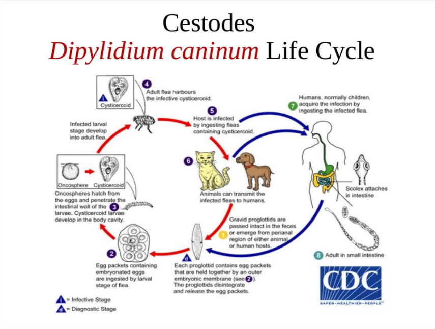
D. caninum - life cycle
egg packets/ proglottids pass from infected dog or cat
flea larvae ingests eggs
cysticercoid larva develops in flea
dog/cat or human ingests infected flea
adult tapeworm develops in intestine
D. caninum - infective vs diagnostic stages
infective: cysticercoid larva inside flea
diagnostic: proglottids (eggs)
D. caninum - clinical infection + diagnosis
pets: behavior relieve anal itching “butt scooting”
humans = usually asymptomatic + mild GI symptoms
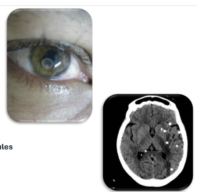
SPARGANOSIS
most common in SE asia
often seen in the eye
internal abscesses, cysts, subq nodules
associated w: ingestion of procercoid in copepod (water)/ plerocercoid larvae in other animals
D. caninum - lab diagnosis
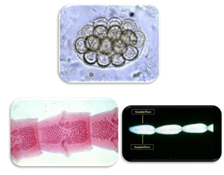
macroscopic (with eyes) ID of PROGLOTTIDS IN STOOL
may be motile when freshly passed
microscopic ID of EGGS in STOOL
large packets containing 15-25 eggs
D. caninum - proglottids and scolex
proglottids
2 GENITAL PORES
resemble PUMPKIN SEEDS
scolex
4 suckers
ARMED ROSTELLUM

Echinococcus granulosus - transmission stage + source
HYDATID = just E. granulosus → HYDATID tapeworm
INGESTION of EGGS containing HEXACANTH EMBRYO from food/water/fomites contaminated w DOG FECES
E. granulosus - hosts
human = accidental hosts
DOG = definitve host
SHEEP/ other herbivores = intermediate host
found worldwide, common in SHEEP raising areas