Ch. 25 LEC - Urinary System
 Call Kai
 Learn
Learn Practice Test
 Spaced Repetition
 Match
Match Flashcards
 Knowt Play
1/34

Earn XP
Description and Tags
Lecture Exam 4
Name | Mastery | Learn | Test | Matching | Spaced | Call with Kai | Chat |
|---|
No analytics yet
Send a link to your students to track their progress
35 Terms
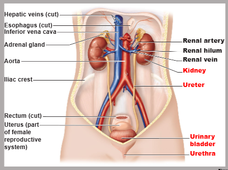
identify the major organs of the urinary system
kidneys, ureters, urinary bladder, urethra
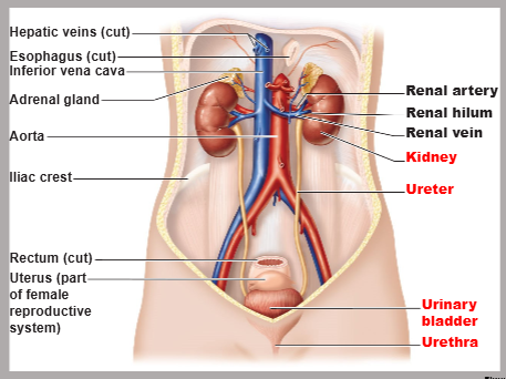
summarize ALL functions of the kidney
remove toxins and metabolic wastes
remove excess ions from blood
regulate blood volume, chemical composition, pH
activate vitamin D (crucial for bone strength)
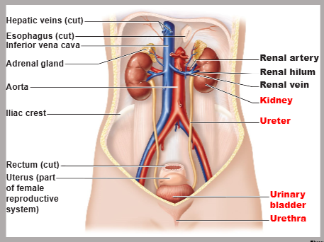
summarize the role of the urinary bladder
temporary storage reservoir for urine
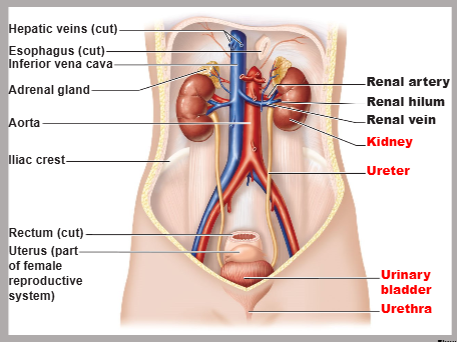
summarize the function of the ureters
transport urine from the kidneys to the bladder
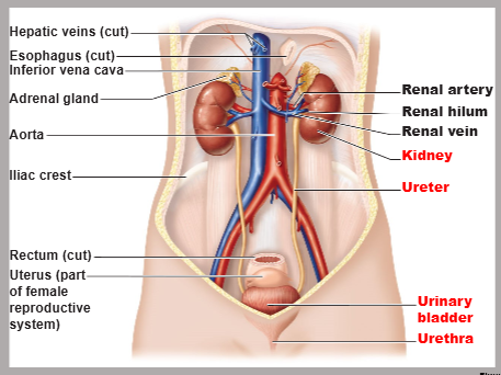
summarize the function of the urethra
transport urine out of the body
the kidneys perform gluconeogenesis during prolonged fasting. define gluconeogenesis
the metabolic process of creating new glucose from non-carbohydrate sources (e.g., lactate, amino acids, glycerol); ensures a stable blood sugar supply and energy
list the kidney’s endocrine duties
secrete renin for blood pressure control
secrete erythropoietin to stimulate RBC production
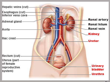
locate the kidneys
retroperitoneal (i.e., behind the peritoneum) in the superior lumbar region
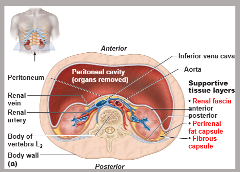
describe the kidney’s layers of supportive tissue
renal fascia (anchoring outer layer of dense connective tissue)
perirenal fat capsule (cushion)
fibrous capsule (infection shield)
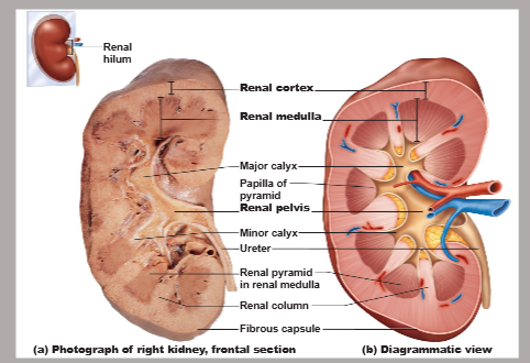
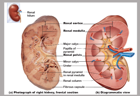
trace the journey of urine starting from the renal papilla to the ureter
renal papilla → minor calyx → major calyx → renal pelvis → ureter
![<p>the renal arteries deliver [FRACTION] ([NUMBER] mL) of cardiac output to the kidneys each minute</p>](https://assets.knowt.com/user-attachments/0858560e-1a4a-4b8b-958d-e6b7d1475dd0.png)
the renal arteries deliver [FRACTION] ([NUMBER] mL) of cardiac output to the kidneys each minute
1/4, 1200
the kidney’s nerve supply is via [PARASYMPATHETIC/SYMPATHETIC] fibers from the renal plexus
sympathetic
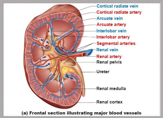
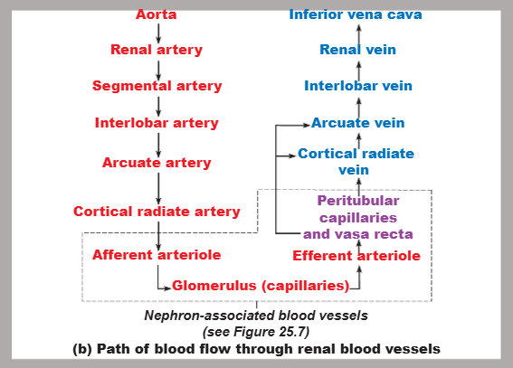
trace the path of blood flow through renal blood vessels (starting with the aorta and ending with the inferior vena cava)
aorta → renal artery → segmental artery → interlobar artery → arcuate artery → radiate artery → afferent arteriole → glomerulus → efferent arteriole → peritubular capillaries and vasa recta → radiate vein → arcuate vein → interlobar vein → renal vein → inferior vena cava
define the nephron
the structural and functional unit that forms urine (1 million per kidney) consisting of a renal corpuscle and a renal tubule
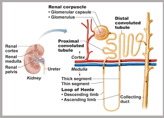
trace the path of filtrate in the nephron (starting with renal corpuscle and ending with collecting duct)
renal corpuscle → proximal convoluted tubule → descending loop of Henle → ascending loop of Henle → distal convoluted tubule → collecting duct
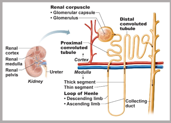
name the structures that make up the renal corpuscle of the kidney’s nephron
glomerulus (capillaries) and its glomerular capsule
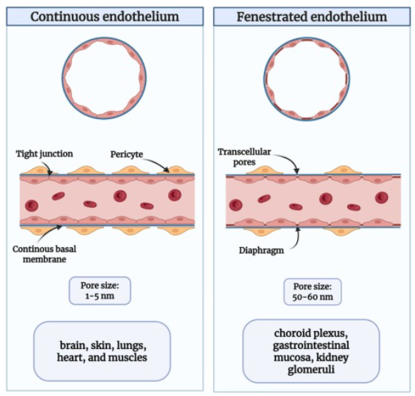
identify the histology of the glomerulus and its function
fenestrated glomerular endothelium allows filtrate from plasma to enter the glomerular capsule while ensuring RBCs stay in the blood vessel
name the different types of nephrons
cortical and juxtamedullary
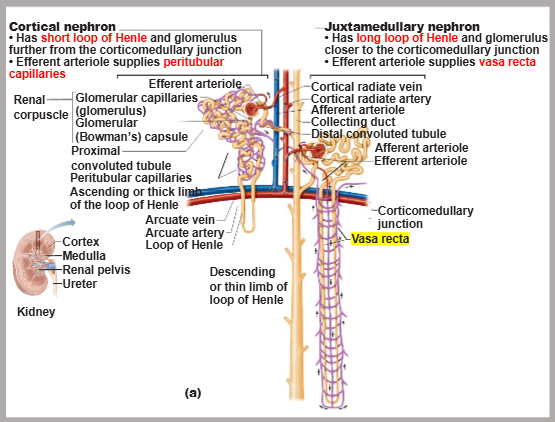
true/false: juxtamedullary nephrons make up 85% of nephrons in the kidney
false — cortical nephrons make up 85% of nephrons in the kidney
contrast the location of cortical and juxtamedullary nephrons
cortical nephrons: renal corpuscle sits high in the renal cortex AND loop of Henle barely dips into the medulla
juxtamedullary nephrons: renal corpuscle sits close to the corticomedullary junction AND loop of Henle reaches deep into medulla and towards the renal papilla
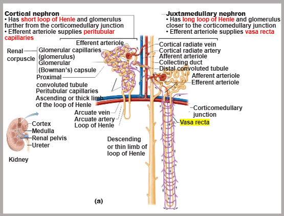
contrast the function of cortical and juxtamedullary nephrons
cortical nephrons: standard blood filtration, waste removal, and general fluid-electrolyte regulation
juxtamedullary nephrons: create concentrated urine to save water during dehydration
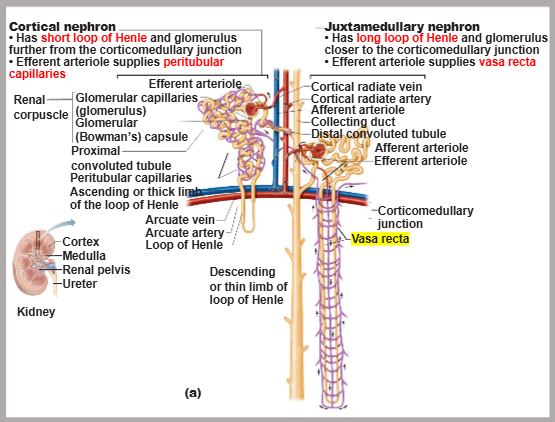
contrast the length of Loops of Henle between the cortical and juxtamedullary nephrons
cortical nephrons have SHORT while juxtamedullary nephrons have LONG loops of Henle
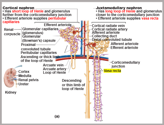
contrast the blood supply between cortical and juxtamedullary nephrons
cortical nephrons: surrounded by peritubular capillaries
juxtamedullary nephrons: surrounded by vasa recta
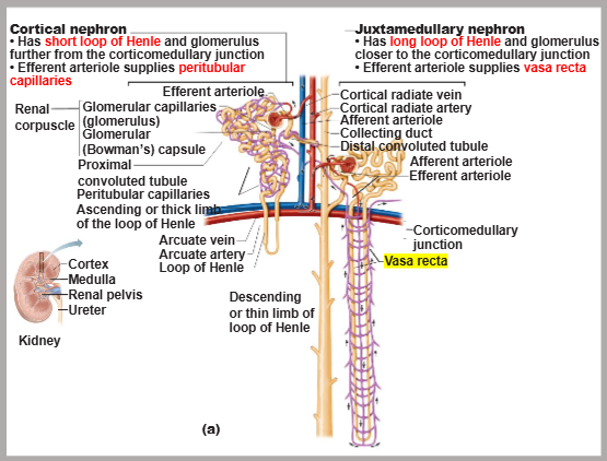
trace the blood flow through the renal corpuscle
afferent arteriole → glomerulus → efferent arteriole
the glomerulus is specialized for _
filtration
identify the reason why the glomerulus has high blood pressure
the efferent (away) arterioles are smaller in diameter than afferent (toward) arterioles
arterioles are high-resistance vessels in general
![<p><strong>peritubular capillaries:</strong></p><ul><li><p>[LOW/HIGH] pressure, porous capillaries adapted for [FILTRATION/ABSORPTION]</p></li><li><p>arise from [AFFERENT/EFFERENT] arterioles</p></li><li><p>cling to adjacent renal tubules in [CORTEX/MEDULLA]</p></li><li><p>empty into [ARTERIOLES/VENULES]</p></li></ul><p></p>](https://assets.knowt.com/user-attachments/16d408bc-81a9-49f0-8c49-1b7fe5f38c6a.jpg)
peritubular capillaries:
[LOW/HIGH] pressure, porous capillaries adapted for [FILTRATION/ABSORPTION]
arise from [AFFERENT/EFFERENT] arterioles
cling to adjacent renal tubules in [CORTEX/MEDULLA]
empty into [ARTERIOLES/VENULES]
low, absorption, efferent, cortex, venules
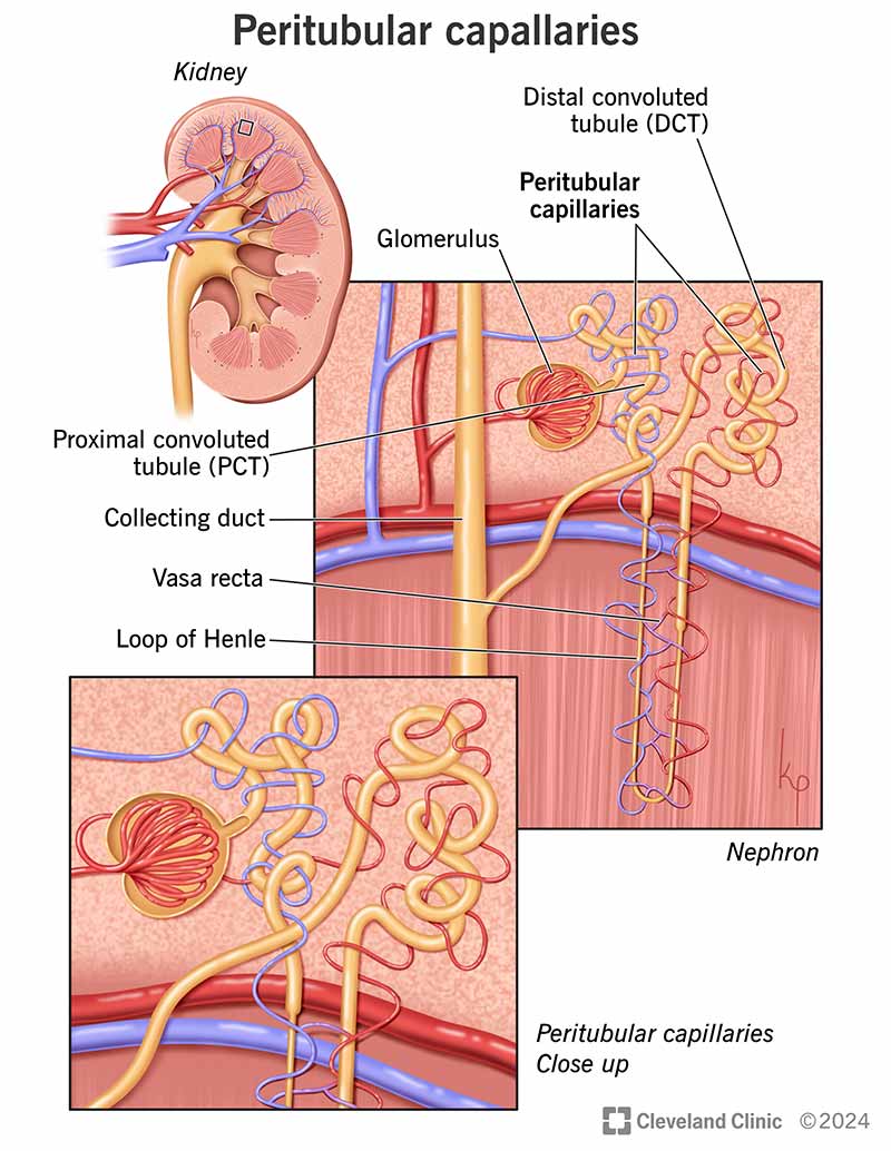
vasa recta:
[SHORT/LONG] vessels parallel to [SHORT/LONG] loops of Henle
arise from [AFFERENT/EFFERENT] arterioles of [CORTICAL/JUXTAMEDULLARY] nephrons
function: assist in the formation of concentrated urine
long, long, efferent, juxtamedullary
how does the high-resistance nature of the afferent and efferent arterioles affect the kidneys?
causes blood pressure to decline from 95 mmHg to 8 mmHg in kidneys
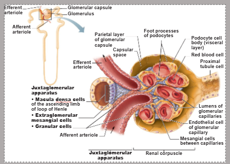
define juxtaglomerular apparatus (JGA)
a specialized structure in each nephron that is critical for regulating filtrate formation and blood pressure; located where the distal portion of the ascending Loop of Henle meets the afferent arteriole
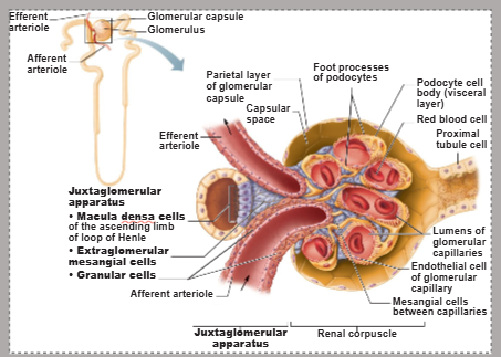
name the cells involved in the juxtaglomerular apparatus (JGA)
granular cells (JG cells), macula densa cells, and extraglomerular mesangial cells
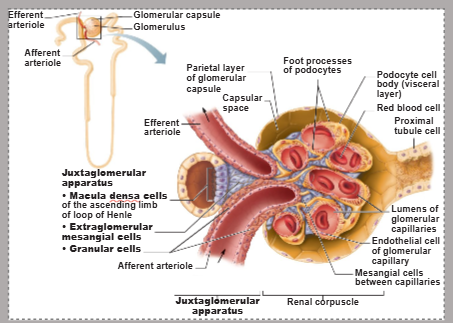
a part of the juxtaglomerular apparatus (JGA), [CELL TYPE] acts as mechanoreceptors that sense blood pressure
granular cells (JG cells)
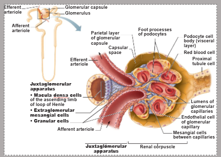
identify the histology and secretion of granular (JG) cells of the JGA
smooth muscle that secretes renin
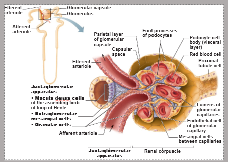
a part of the juxtaglomerular apparatus (JGA), [CELL TYPE] acts as chemoreceptors that sense NaCl content of filtrate
macula densa
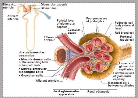
extraglomerular mesangial cells are interconnected with gap junctions. what is the function of extraglomerular mesangial cells’ function in the JGA?
to pass signals between macula densa and granular (JG) cells