CH. 20: Cardiovascular System
 Call Kai
 Learn
Learn Practice Test
 Spaced Repetition
 Match
Match Flashcards
 Knowt Play
1/133

There's no tags or description
Looks like no tags are added yet.
Name | Mastery | Learn | Test | Matching | Spaced | Call with Kai | Chat |
|---|
No analytics yet
Send a link to your students to track their progress
134 Terms
What are the two main functions of the heart?
Pumps blood through pulmonary circulation to lungs and systemic circulation to remaining tissues.
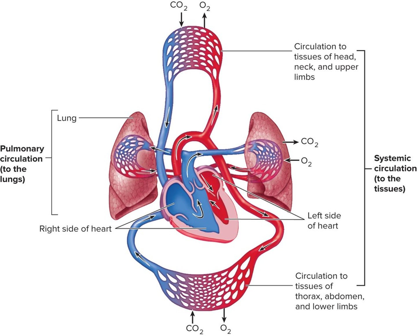
What does the right side of the heart do?
Pumps blood through pulmonary circulation to the lungs.
What does the left side of the heart do?
Pumps blood through systemic circulation to the body's tissues.
How does the heart generate blood pressure?
Through heart contractions.
What is the role of heart valves?
Ensure one-way blood flow.
What regulates blood supply in the heart?
Changes in heart rate and force of contraction based on tissue metabolic needs.
What is the sinoatrial (SA) node?
A specialized group of cardiac muscle cells that act as the pacemaker of the heart.
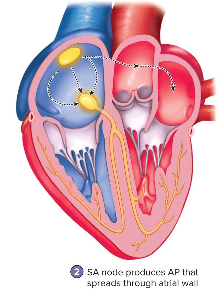
What is the function of the atrioventricular (AV) node?
Conducts action potentials more slowly to ensure ventricles contract after atria.
What does the AV bundle do?
Passes action potentials through the interventricular septum to the ventricles.
What are Purkinje fibers?
Large diameter cardiac muscle cells that conduct action potentials to ventricular muscle cells.
What initiates the pacemaker potential?
Sodium moves into cells via nongated sodium channels, causing depolarization.
What happens during repolarization in pacemaker cells?
Calcium channels close and potassium channels open, allowing potassium to leave the cell.
What does an electrocardiogram (ECG) record?
Electrical events in the myocardium correlated with mechanical events.
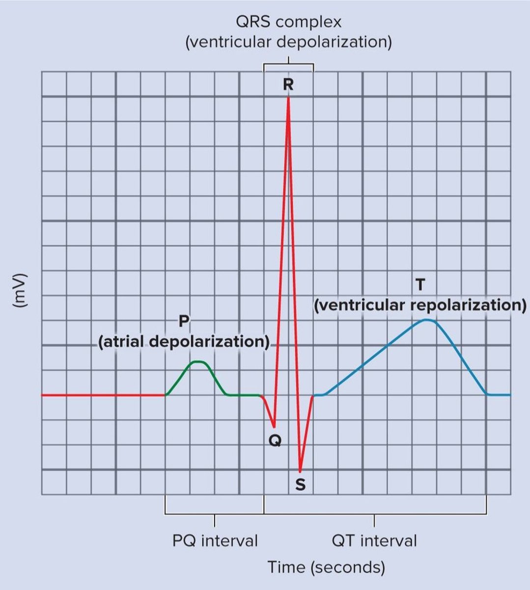
What does the P wave in an ECG represent?
Depolarization of atrial myocardium and the onset of atrial contraction.
What does the QRS complex in an ECG indicate?
Ventricular depolarization and the onset of ventricular contraction.
What does the T wave in an ECG signify?
Repolarization of the ventricles.
What is systole?
The contraction phase of the heart chambers.
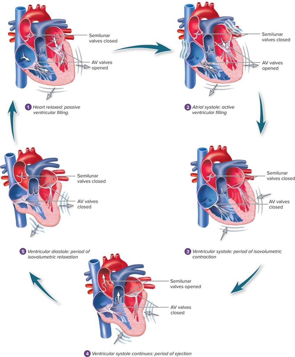
What is diastole?
The relaxation phase of the heart chambers.
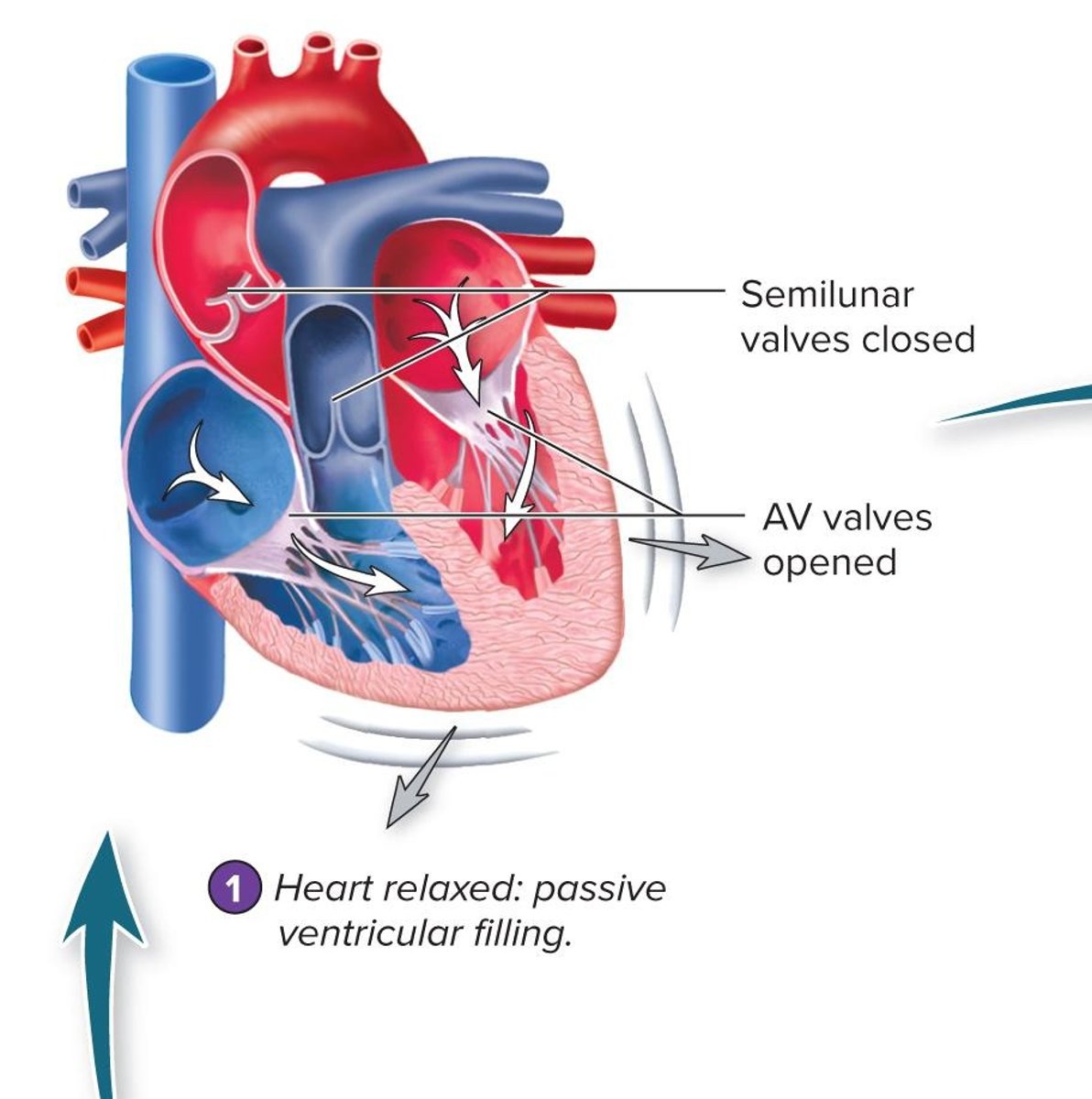
What occurs during passive ventricular filling?
Blood moves into the ventricles due to higher pressure in the veins.
What is the end diastolic volume (EDV)?
The volume of blood in the ventricles at the end of diastole.
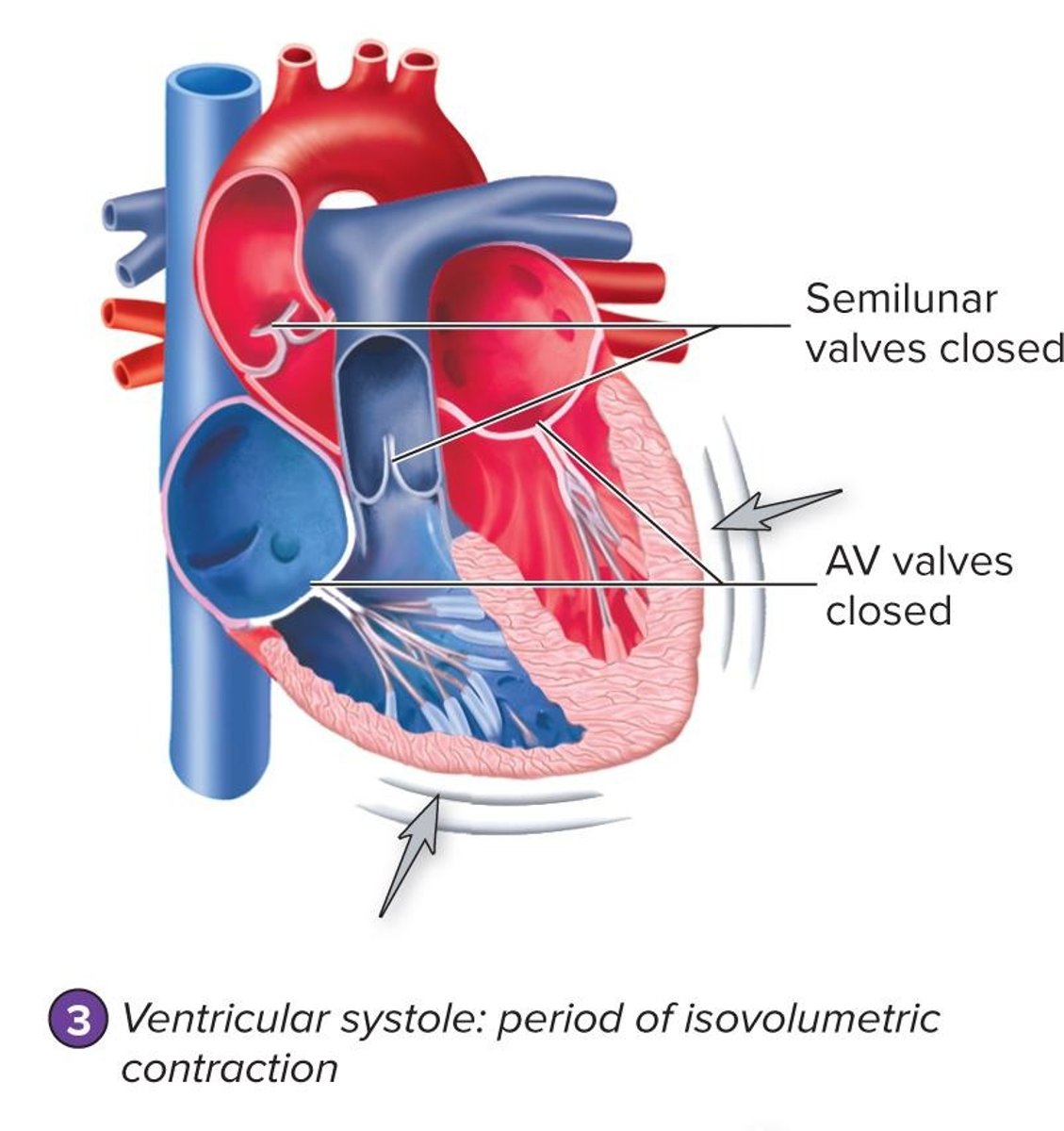
What happens during ventricular systole?
Pressure in the ventricles increases, causing AV valves to close and blood to be ejected.
What is the end-systolic volume (ESV)?
The volume of blood remaining in the ventricles after contraction.
What causes the first heart sound ('lubb')?
Closure of the atrioventricular valves at the beginning of ventricular systole.
What causes the second heart sound ('dupp')?
Closure of the aortic and pulmonary semilunar valves at the beginning of ventricular diastole.
What are heart murmurs?
Abnormal heart sounds caused by turbulent blood flow.
What is the cardiac cycle?
The repetitive pumping process that begins with contraction and ends with the next contraction.
What is the role of the conducting system in the heart?
Relays action potentials through the heart to coordinate contractions.
What is the significance of the PR interval in an ECG?
It measures the time between atrial contraction and the beginning of ventricular contraction.
What is the QT interval in an ECG?
The time from the beginning of ventricular depolarization to the end of ventricular repolarization.
What is an incompetent valve?
A valve that allows blood to flow backward when closed, causing regurgitation and turbulence.
What sound is produced by an incompetent AV valve?
A sound heard after the first heart sound.
What sound is produced by an incompetent SL valve?
A sound heard after the second heart sound.
What is a stenosed valve?
A valve with an abnormally narrow opening that produces turbulent blood flow and a rushing sound.
How is Mean Arterial Pressure (MAP) calculated?
MAP = Cardiac Output (CO) × Peripheral Resistance (PR).
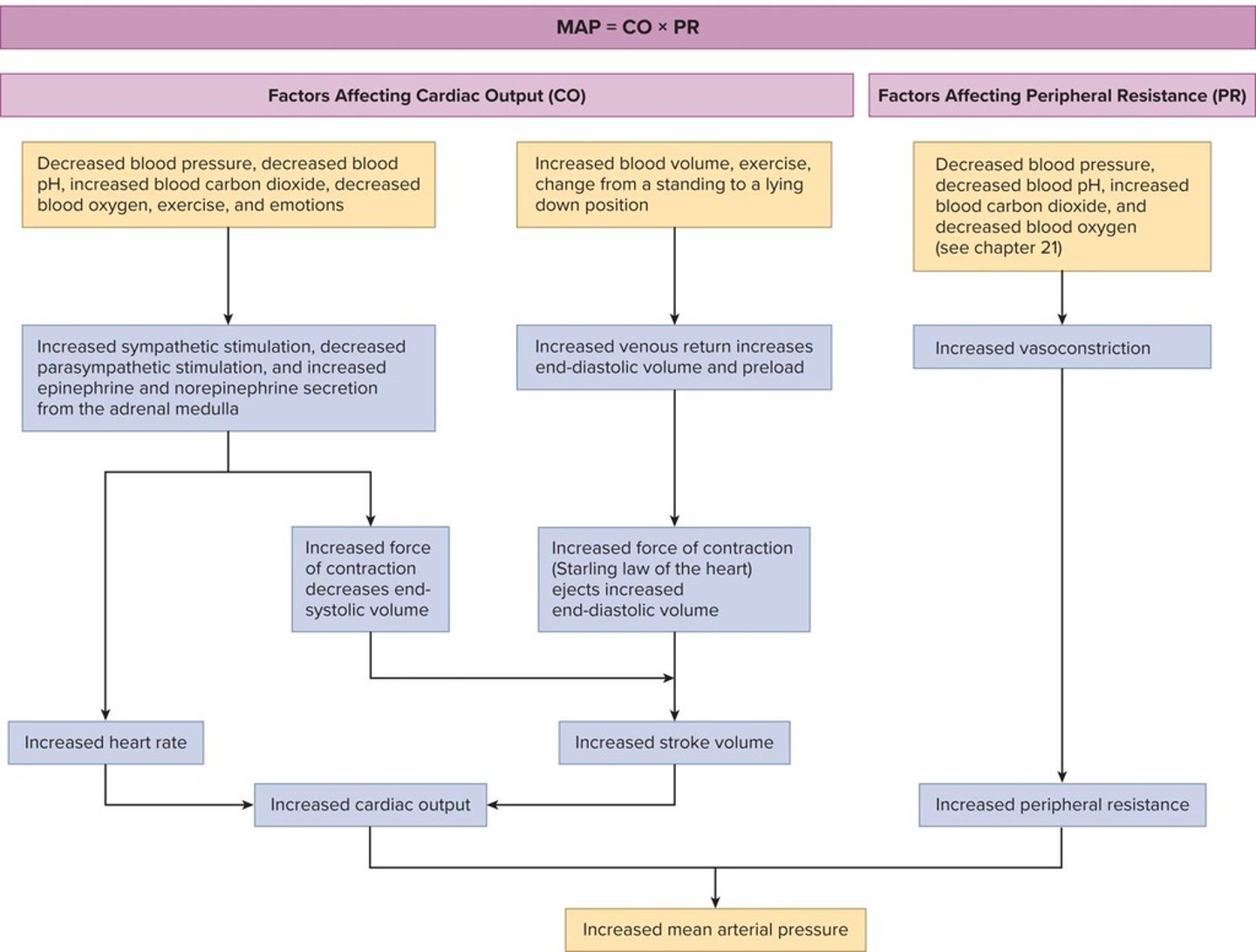
What does cardiac output (CO) represent?
The amount of blood pumped by the heart per minute.
How is cardiac output (CO) calculated?
CO = Stroke Volume (SV) × Heart Rate (HR).
What is stroke volume (SV)?
The amount of blood pumped during each heartbeat, calculated as EDV - ESV.
What does HR stand for in cardiovascular physiology?
Heart Rate, the number of beats per minute.
What factors can increase stroke volume?
Increasing End-Diastolic Volume (EDV) or decreasing End-Systolic Volume (ESV).
What is cardiac reserve?
The difference between cardiac output at rest and maximum cardiac output.
What happens to stroke volume in well-trained athletes?
It can increase to over 200 mL/beat, resulting in a cardiac output of 40 L/min or more.
What is the role of parasympathetic stimulation in heart regulation?
It decreases heart rate through the secretion of acetylcholine.
What is the effect of sympathetic stimulation on the heart?
It increases heart rate and force of contraction through epinephrine and norepinephrine release.
Where are baroreceptors located and what do they monitor?
In the walls of internal carotids and aorta; they monitor blood pressure.
What is the function of chemoreceptors in the cardiovascular system?
They regulate heart activity to maintain homeostasis of pH and blood gas values.
What happens when pH drops in the blood?
It increases sympathetic stimulation of the heart, leading to increased cardiac output.
What are some age-related changes in the heart?
Hypertrophy of the left ventricle, increased aortic pressure, decline in maximum heart rate, and less flexible valves.
What are common diseases associated with aging in the cardiovascular system?
Congestive heart disease and coronary artery disease.
What is the primary function of the heart?
To pump blood through the blood vessels of the body.
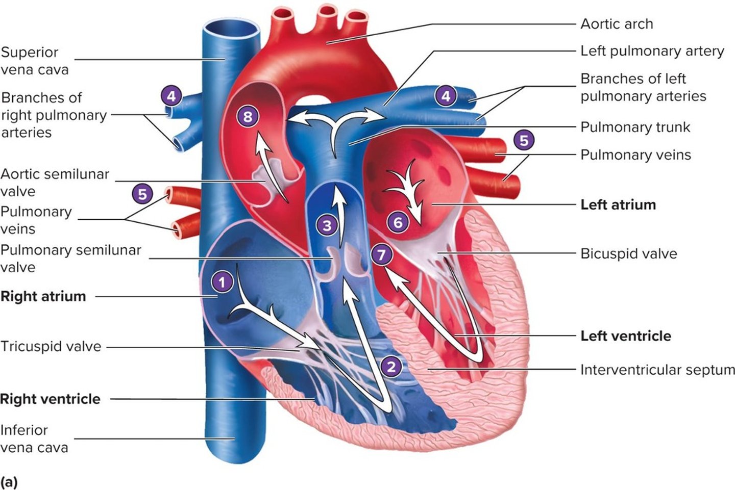
What are the three layers of the heart wall?
Epicardium, myocardium, and endocardium.
What is the pericardium?
The tissue that surrounds the heart.
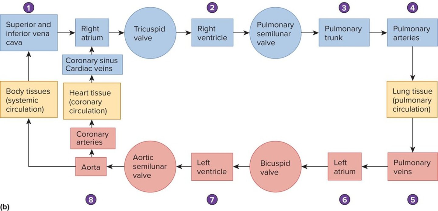
What is the difference between systemic and pulmonary circulation?
Systemic circulation carries oxygenated blood from the heart to the body, while pulmonary circulation moves deoxygenated blood from the heart to the lungs.
How does the diaphragm relate to the thoracic cavity?
It forms the inferior boundary of the thoracic cavity, separating it from the abdominal cavity.
What is the apex of the heart?
The pointed end of the heart that points downward and to the left.
Where is the heart located in the thorax?
Medially, anterior to the lungs, with the base near the 2nd intercostal space and apex at the 5th intercostal space.
What surrounds the heart?
The pericardium, which includes the visceral pericardium, pericardial cavity, parietal pericardium, and outer fibrous pericardium.
What is the function of the pericardial cavity?
It contains pericardial fluid that reduces friction between the heart and surrounding structures.
What are the main components of the heart wall?
The epicardium (visceral pericardium), myocardium, and endocardium.
What is the myocardium?
The thick muscular layer that forms the main component of the heart wall.
What are trabeculae carneae?
Ridges projecting into the ventricular cavity of the heart.
What major vessels enter the right atrium?
The superior vena cava and inferior vena cava.
What separates the atria from the ventricles?
The coronary sulcus.
What arteries supply blood to the heart?
The left coronary artery and right coronary artery.
What are the branches of the left coronary artery?
The circumflex artery, left marginal artery, and anterior interventricular artery.
What veins drain into the coronary sinus?
The great cardiac vein, middle cardiac vein, and small cardiac vein.
What is the role of the tricuspid valve?
It regulates blood flow from the right atrium to the right ventricle.
What is the bicuspid (mitral) valve?
The valve that regulates blood flow from the left atrium to the left ventricle.
What is the aortic semilunar valve's function?
It controls blood flow from the left ventricle into the aorta.
What is the pulmonary semilunar valve's function?
It controls blood flow from the right ventricle into the pulmonary trunk.
What is the role of the chordae tendineae?
They anchor the heart valves to the papillary muscles and prevent backflow of blood.
What is the mediastinum?
The tissue that surrounds and supports the thoracic organs, including the heart.
What is the significance of the anterior interventricular sulcus?
It separates the right and left ventricles and contains the anterior interventricular artery.
What is the function of the pulmonary trunk?
It carries deoxygenated blood from the right ventricle to the lungs.
What do the pulmonary veins do?
They return oxygenated blood from the lungs to the left atrium.
What does the coronary circulation include?
The arteries and veins that supply and drain blood to and from the heart muscle.
What is the location of the left ventricle?
It occupies much of the left posterior surface of the heart.
What is the function of the right atrioventricular canal?
It contains the tricuspid valve and connects the right atrium to the right ventricle.
What does the left atrioventricular canal contain?
The bicuspid (mitral) valve.
What is the role of the great cardiac vein?
It drains blood from the anterior part of the heart.
What is the significance of the coronary sulcus?
It contains the right coronary artery and separates the atria from the ventricles.
What anatomical structures are visible in the anterior view of the heart?
The right atrium, superior vena cava, inferior vena cava, and ascending aorta.
What structure delivers blood into the left atrium?
The pulmonary veins
What valve is open when blood flows into the left ventricle?
The bicuspid valve
What happens to the chordae tendineae when the left ventricle is relaxed?
They have low tension
What is the state of the papillary muscle when the left ventricle is relaxed?
The papillary muscle is relaxed
What occurs when blood flows out of the left ventricle?
The aortic semilunar valve opens
What major vessels bring blood into the right atrium?
The superior vena cava and inferior vena cava
What valve does blood pass through to enter the right ventricle?
The tricuspid valve
What is the pathway of blood from the right ventricle to the lungs?
Through the pulmonary semilunar valve into the pulmonary trunk
Where does oxygenated blood return after reaching the lungs?
To the left atrium via the pulmonary veins
What is the role of the aortic semilunar valve?
To allow blood to exit the left ventricle into the aorta
What structure separates the left and right ventricles?
The interventricular septum
What is the function of the cardiac muscle in the left ventricle?
To contract and pump blood into the aorta
What is the significance of the pacemaker potential?
It initiates the depolarization of the cardiac muscle
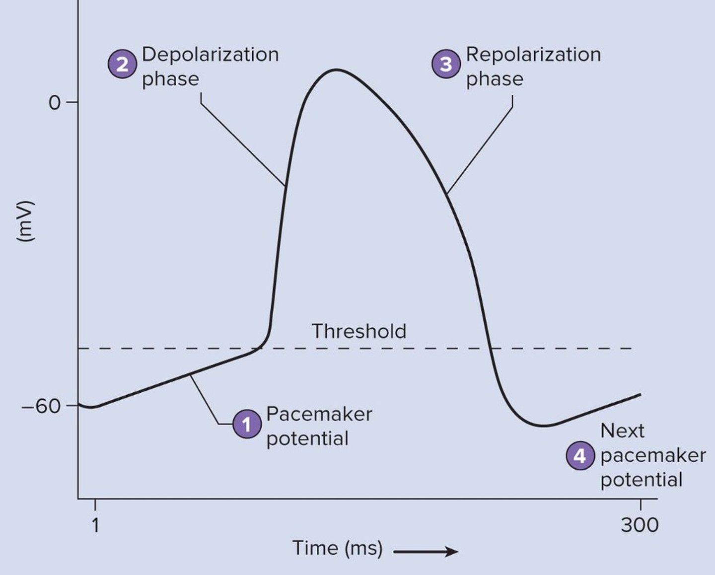
What occurs during the depolarization phase of the action potential in cardiac muscle?
The membrane potential rises sharply
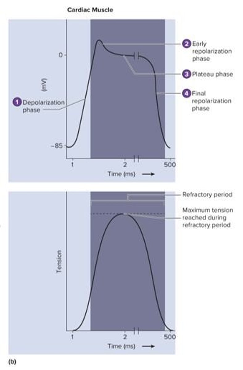
What is the plateau phase in cardiac action potential?
A phase where the membrane potential remains nearly constant at a slightly positive value
How does the action potential in cardiac muscle differ from skeletal muscle?
Cardiac muscle has a longer refractory period preventing tetanus
What is the role of intercalated disks in cardiac muscle?
To connect individual cardiac muscle cells and facilitate synchronized contraction
What happens during the final repolarization phase of cardiac action potential?
The membrane potential declines back toward the resting value
What is the function of the coronary arteries?
To supply blood to the heart tissue