Alteration in Perfusion: Atrial Rhythms
 Call Kai
 Learn
Learn Practice Test
 Spaced Repetition
 Match
Match Flashcards
 Knowt Play
1/38

There's no tags or description
Looks like no tags are added yet.
Name | Mastery | Learn | Test | Matching | Spaced | Call with Kai | Chat |
|---|
No analytics yet
Send a link to your students to track their progress
39 Terms
Atrial dysrhythmias occur when the
sinus node fails to fire or an irritable cell/cells in the atria decide to become the pacemaker.
Atrial Dysrhythmias
Changes are in the P wave (flattened, notched, irregular or peaked).
The QRS is Generally normal .12 sec or smaller
Wandering Pacemaker
The pacemaker site wanders between the sinus node, the atria, and the AV junction.
Although each impulse originates from a different focus, rate usually remains within a normal range, but it can be slower or faster.
Wandering Pacemaker conduction
Conduction through to the ventricles is normal.
Atrial Ps

Rules for Wandering Pacemaker
Regularity: Slightly irregular
Rate: Usually normal, 60–100 bpm
P Wave: Morphology changes from one complex to the next
PRI: Less than 0.20 second; may vary
QRS: Less than 0.12 second
Rarely causes symptoms

Mechanism of Premature Atrial Complex
The pacemaker is an irritable focus within the atrium that fires prematurely and produces a single ectopic beat.
Conduction through to the ventricles is normal.
Mechanism of Premature Atrial Complex: rhythm
This is a single beat, not an entire rhythm. The underlying rhythm also must be identified.
PAC may feel like an irregular pulse.
Rules for Premature Atrial Complex: Regularity, rate, P wave
Regularity: Depends on the underlying rhythm; regularity will be interrupted by the PAC
underlying rhythm__ with PAV
Rate: Depends on the underlying rhythm
P Wave: P wave of early beat differs from the sinus P waves; can be flattened or notched; may be lost in the preceding T wave
Rules for Premature Atrial Complex: PRI and QRS
PRI: 0.12–0.20 second; can exceed 0.20 second
QRS: Less than 0.12 second
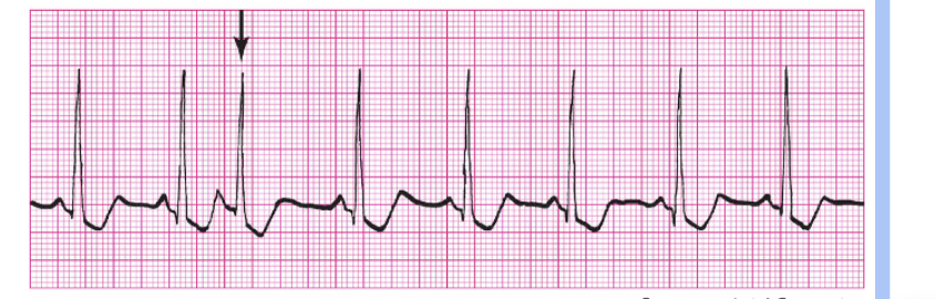
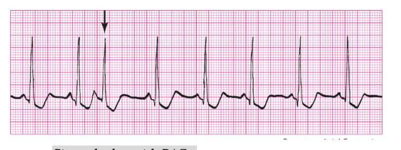
Rules for Premature Atrial Complex
Sinus rhythm with PAC
There is still a p before the qrs,
Count the pac when counting the rate
Premature Atrial Contraction: Significance
Usually benign
Can be early sign of HF
Can lead to atrial tachyarrhythmias
Premature Atrial Contraction: causes
fatigue, hypoxia, dig-toxicity, caffeine, ischemia, HF, alcohol, medications, amphetamines, heart disease and mitral prolapse
Premature Atrial Contraction: treatment and assessment
Assessment: if many occur may feel dizzy
Treat: Cause, B adrenergic blockers, monitor
Mechanism of Atrial Tachycardia
The pacemaker is a single irritable site within the atrium that fires repetitively at a very rapid rate. Conduction through to the ventricles is normal.
Rules for Atrial Tachycardia
Regularity: Regular
Rate: 150–250 bpm
P Wave: Atrial P wave; differs from sinus P wave; can be lost in T wave (P and T waves are smushed)
PRI: 0.12–0.20 second
QRS: Less than 0.12 second

Atrial Tachycardia significance
Very dangerous in AMI or heart disease
Commonly caused by dig-toxicity
Can also be called Paroxysmal (sudden) Atrial Tachycardia(PAT)
Atrial Tachycardia clincial picture
Rapid, regular pulse
May show signs/symptoms of drop in cardiac output
Can cause pulmonary edema, HF, shock (Immediately have the patient bear down to help lower HR)
Atrial tachycardia treatment
Tx: vagal maneuv, adenosine (rapid), Ca chnl blkr (diltiazem), B blkr (atenolol, propranolol), amiodarone, oxygen, cardiovert (low voltage)
Mechanism of Atrial Flutter
A single irritable focus within the atria issues an impulse that is conducted in a rapid, repetitive fashion.
Those that do get through are conducted normally.
Mechanism of Atrial Flutter: To protect the ventricles from receiving too many impulses, the
AV node blocks some of the impulses from being conducted through to the ventricles.
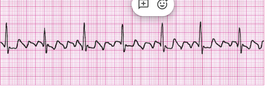
Rules for Atrial Flutter
Regularity: Atrial rhythm is usually regular; ventricular rhythm is usually regular but can be irregular if there is variable block
Rate: Atrial rate 250–350 bpm; ventricular rate varies
P Wave: Characteristic sawtooth pattern (F waves)
PRI: Unable to determine
QRS: Less than 0.12 second
Atrial Flutter significance
Rapid ventricular rate and loss of atrial kick (20%) can drop cardiac output
Risk of pulmonary and cerebral emboli
Can cause HF or myocardial ischemia
atrial flutter is seen in
CAD,
rheumatic heart disease,
alcoholism,
thyroid disease,
rheumatic heart disease
Atrial contraction forces
additional volumes of blood into the ventricles (often referred to as "atrial kick").
The atrial kick contributes a significant volume of blood toward ventricular preload (approximately 20%)
Atrial Flutter clinical picture
Pulse can be regular or irregular, fast or slow
Signs/symptoms of low cardiac output: SOB, palpitations, dizzy, chest pain
Atrial Flutter Tx
Oxygen 2 liters first thing you need is oxygen and then medication
Ca chnl: diltiazem, amlodipine
B blks: (olol), metoprolol, atenolol
Anticoagulants: warfarin, heparin, enoxaparin
Antiarrhythmics: amiodarone (brings it to be a reg rhythm)
Alternate Option: Cardioversion (synchronized) or Radiofrequency catheter ablation
first thing you need is oxygen and then medication
Mechanism of Atrial Fibrillation
The atria are so irritable that a multitude of foci initiate impulses, causing the atria to depolarize repeatedly in a fibrillatory manner.
The AV node blocks most of the impulses, allowing only a limited number through to the ventricles.
In atrial fib, the av node
holds the electricity for longer, there is a lot of p’s and then a qrs, we can use cardioversion
Conduction: AV nodes blocks some impulses but allow others to proceed normally through the ventricles
Rules for Atrial Fibrillation
Regularity: Grossly irregular
Rate: Atrial rate greater than 350 bpm; ventricular rate varies greatly
P Wave: No discernible P waves; atrial activity is referred to as fibrillatory waves (f waves)
we don’t know which one causes the qrs is because of that we can’t measure PRI
PRI: Unable to measure
QRS: Less than 0.12 second
Atrial Fibrillation image

Atrial Fibrillation controlled vs uncontrolled
Ventricular response irregularly, irregular
Goal to keep ventricular rate less than or equal to 100 to keep CO intact- controlled Atrial Fibrillation.
If HR greater than 100 – uncontrolled A fib. Sometimes noted as a fib with a rapid ventricular response.
Blood is sitting in the atrial for a extended time which leads to clots
Atrial Fibrillation cause and assessment
Cause: the same as A flutter
Assessment : dizziness, chest pain, anxiety, syncope, hypotension
Client at risk for mult. thrombi, stroke
If client has valvular disease, risk higher
Atrial Fibrillation treatment
Tx: if new onset and caught, then drugs same as A flutter.
If no response –Cardioversion or Radiofrequency catheter ablation
Take pulse for one full minute
Anticoagulants, Rate Controlling drug (ie. dig or lopressor) for life
Treatment: Radiofrequency Catheter Ablation Therapy
Electrode-tipped ablation catheter “burns” accessory pathways or ectopic sites in the atria, AV node, and ventricles.
Treatment: Radiofrequency Catheter Ablation Therapy Nonpharmacologic treatment for
AV nodal reentrant tachycardia
Reentrant tachycardia related to accessory bypass tracts
Control of ventricular response of certain tachydysrhythmias
The R wave has to be shocked→ if not leads to more serious problems
Treatment: Cardioversion
Synchronized counter shock (synchronized to R waves).
The shock depolarizes the myocardium simultaneously.
Firing on the patient’s t waves can lead to ventricular arrhythmias.
The shock stops abnormal rhythm and allows the sinus node to assume pacing
This is an elective procedure
Signed consent-Light anesthetic agent and sedative given.
Before Cardioversion, if not new…
If patient has been in atrial fibrillation for >48 hours, anticoagulation therapy with warfarin (Coumadin) is recommended for 3 to 4 weeks before cardioversion and for 4 to 6 weeks after successful cardioversion. Poss dislodge of clot.
Digitalis must be held for 48 hrs. prior to cardioversion.
Hold dig since it lower heart
Cardioversion Nursing Care
Check placement of “pads/paddles”
Shock is delivered while client is exhaling
Be sure all personnel are clear
Have crash cart ready
Start with 50-100 joules (lower volts)
Maintain patent airway
O2 needed
Assess client vital signs and LOC
Monitor for dysrhythmia
Document rhythm strips before, during, and after
Assess for chest burns
Provide emotional support for client
Complications: PE, CVA, MI