Cell Biology Ch. 1
 Call Kai
 Learn
Learn Practice Test
 Spaced Repetition
 Match
Match Flashcards
 Knowt Play
1/88

There's no tags or description
Looks like no tags are added yet.
Name | Mastery | Learn | Test | Matching | Spaced | Call with Kai | Chat |
|---|
No analytics yet
Send a link to your students to track their progress
89 Terms
basic unit of life and biology? what does this mean?
cell / smallest thing to exhibit all the characteristics of life
basic characteristics of cells
every organism consists of them, carry out necessary functions for life, are constantly changing and have capacity to grow, reproduce, and become specialized
3 parts of cell theory
all cells come from preexisting cells, all living things are made of cells, cell is basic unit of life
what is cell biology and what are the 3 areas of study that contribute to the overall field of cell bio
study of cell structure and function / cytology, biochemistry, and genetics
describe cytology
studies cellular structure, uses microscope, based on direct observations, easier
first to view and name cells? what type of cells?
Robert Hooke / dead cork
first to see living cells
antonie van leeuwenhoek
discovered and name nucleus
Robert Brown
said cells come from preexisting cells
Virchow
said all plants and animals are made of cells
Schleiden and Scwann
describe biochemistry
studies chemistry of living systems, function, isolating/separating (molecular level), and how the things we see work
describe genetics
study of behavior of genes and storage of hereditary information, PCR
define micro unit, value, and symbol
10^-6 m (um) / 1 millionth of a meter
define nano unit, value, and symbol
10^-9 m (nm) / 1 billionth of a meter
3 necessary parts of microscopy
illumination source, specimen, and lenses
light microscopy:
illumination source, lenses, mag limit, typically?
light or photons / glass / 1000X / compound
electron microscopy:
illumination source, lenses, mag limit
electrons / magnets / 10^6X
simple vs. compound microscope
simple using single convex lense to focus light while compound uses many lenses
identify 2 types of microscopy and the different types within those
light microscopy (brightfield, phase-contrast/differential interference contrast, fluorescence/confocal) and electron miscroscopy (transmission electron miscroscope TEM and scanning electron microscope SEM)
define magnification
increasing virtual size of sample (how large it looks)
define resolution
distinguishing between two points as separate
higher mag = (__) res
increased (but not always better); think beach ball and softball example with rope
t or f: monocular microscopes can be compound
t
how does a brightfield microscope work
light passes through specimen and glass lenses focus image onto eye
brightfield microscopes are usually (__) and can utilize chemical (__). background is (__) and has (__) look what can be seen without stains/dyes?
compound / dyes or stains / white / watercolor / nucleus, cell membrane, cytoplasm
phase contrast/differential interference contrast microscope has (__) background and (__) are visible. (__) appearance
white/gray / shadows and highlights / 3D
how does phase contrast/differential interference contrast microscope work?
converting small phase shifts in transmitted light into visible differences in image brightness and contrast.
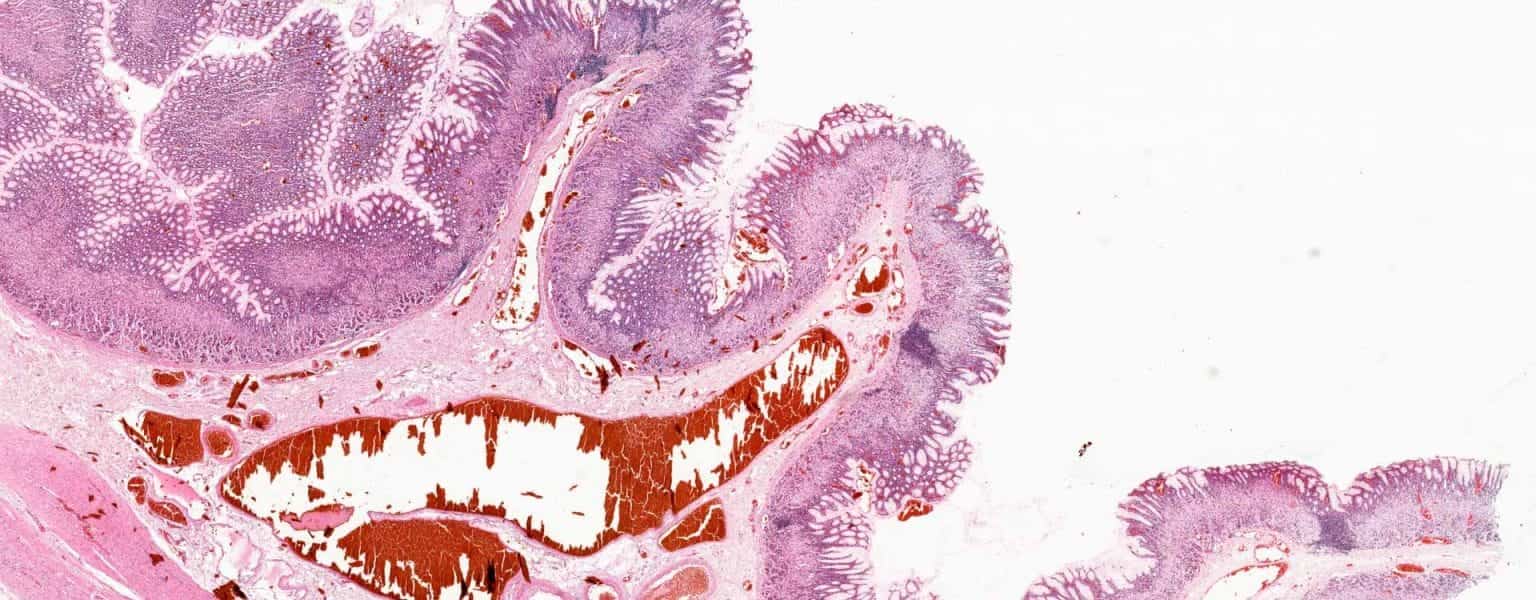
identify microscope used
brightfield
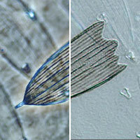
identify microscope used
phase contrast/ differential interference contrast
identify two types of immunofluorescence
primary (direct) and secondary (indirect)
describe basic antibody structure
protein made by B lymphocytes in immune system that binds a target antigen. it has a constant region (C) that is the same for all antibodies and variable (V) that are identical to each other but unique for each antibody and antigen-binding site.
explain how cytology is different from biochem in discipline of cell bio
cytology explores the structures while biochem exlains and clarifies the structures
list diff biochemical techniques used by cell biologists
centrifugation (subcellular fractionation), ultracentrigue, chromatograophy, electrophoresis, mass sepctrometry
differentiate between classical genetics and molecular genetics
classical genetics studied chromosomes, DNA, and how genetic information is passed. molecular genetics studies the chemical structure and function of genes
identify 3 parts of central dogma of molecular biology
DNA replication, transcription, and translation
differentiate between a hypothesis and null hypothesis
a hypothesis is a tentative explanation that can be tested experimentally or by observation; a null hypothesis is the opposite of the original hypothesis. failure to confirm null hypothesis verifies the hypothesis
what is bioinformatics
allows us to compare and analyze many genes or other molecules at the same time
what is chromatography
separates mixture into parts based on charge or affinity or size
chromosome theory of heredity
characteristics of organisms passed down generations results from inheritance of chromosomes carrying genes
CRISPR
genome editing
DNA sequencing
rapidly determines base sequences of DNA molecules
electrophoresis
electricity separates proteins, DNA, or RNA by size and electric charge
antibodies can be linked to an (___) to perform a chemical reaction resulting in a colored precipitation product that can be seen with standard microscope
enzyme
guide RNA
helps Cas9 target a specific place within the genome wehre Cas9 causes a double-stranded break in DNA
mass spectrometry
determines size and composition of individual proteins
recombinant DNA
contain DNA sequences from 2 different sources
subcellular fractionation
isolating structures based on size, shape, or density by ultracentrifugation. used to isolate organelles and speeding up pulls out smaller things
hypothesis
proposed explanation for a specific phenomenon that is testable
theory
predictive, repeatable, tested abundantly, not proven
explain how fluorescence/confocal microscopy works
light is projected onto the sample and emitted back to the detector
fluoresence/confocal microscopy is also called (__) microscopy and requires the use of (__). what do the images look like? what can it detect?
darkfield / fluorophores / dark background with very clear not blurry colorful images / specific molecules and ions
fluorophores (__) energy at shorter or (__) energy wavelengths. they (__) energy at longer or (__) energy wavelengths
absorb / high / release / low
fluorophores can be in stand-alone (__) and (__) and used to?
dyes / stains / tag antibodies for immunofluorescence microscopy
immnunofluorescence microscopy specifically identifies the location of (___) and uses (__). what are the two types
various molecules / fluorescently tagged antibodies / primary and secondary immunofluorescence
what is the epitope
specific part of foreign particle or antigen that the antibody binds to
how does primary/direct immunofluorescence work
antibodies are labeled with a fluorescent dye and then they bind directly to target antigen
specificity, background, and signal or primary/direct immunofluorescence
high specificity / low background / low signal
how does secondary/indirect immunoflourescence work
unlabeled primary antibody binds to the target antigen (it will not light up). then a secondary fluoro-tagged antibody binds to the primary antibodies
specificity, background, and signal of secondary/indirect immunoflourescence
lower specificity / higher background / high signal (greater ratio of binding)
fluorescent staining dye that binds to AT-rich regions of fixed cells in DNA
DAPI
fluorescent staining dye that binds to AT-rich regions of live cells in DNA
hoechst
fluorescent staining dye that intercalates with DNA
propidium iodide
fluorescent staining method for protein is (__). how does it work?
green fluorescent protein (GFP) / GFP amino sequence added to protein gene and then expresses green to show where protein is but may change how protein behaves
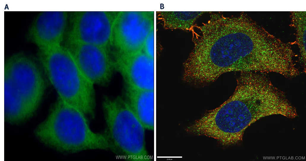
what type of microscope used
fluorescence/confocal microscopy
how does transmission electron microscope work
electron passed through specimen
how does scanning electron microscope work?
electrons projected onto specimen coated in gold and then bounce off
how to distinguish transmission vs. scanning EM and what can you see
transmission look like a more detailed brightfield image and you can see individual ribosomes and scanning are 3-D
for transmission EM, darker parts mean more
solid because electrons not passing through but bouncing back
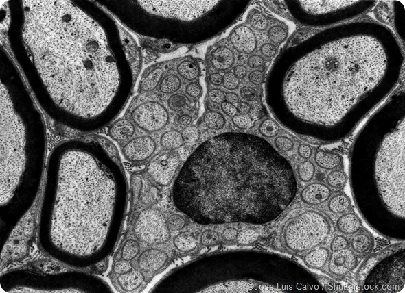
what microscope used
transmission electron microscope
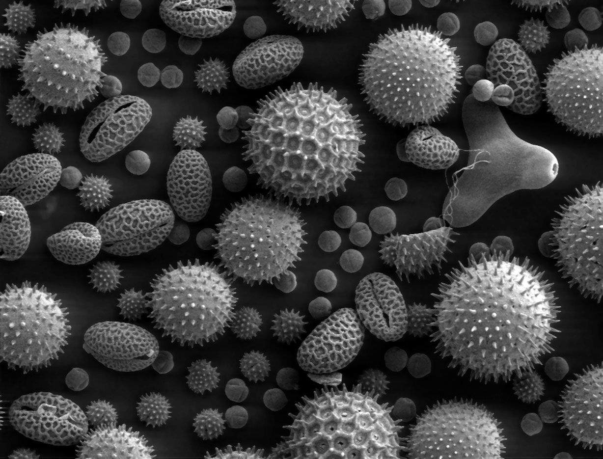
what type of microscope used
scanning electron microscope
how are antibodies used in EM
antibodies labeled with gold particles which show up as darker spots
how does centrifugation separate things
using centrifugal force to separate heavier things to bottom
how does immunoaffinity column chromatography work
beads coated with anti-specific antibodies and packed into a column. cell membranes are destroyed and mixture of molecules allowed to separate by gravity and antibodies bind to target protein but you also get non specific binding. Then washed to get rid of non specific binding proteins and then a final step to break off protein of interest from antibody
what type of gel is used in DNA electrophoresis
porous agarose
how does DNA electrophoresis work
smaller pieces go through gel more quickly and end up farther away from negative end (more towards positive end). thicker bands show that there is more DNA of that size there